검색 전체 메뉴
PDF
맨 위로
OA 학술지
Identification and Expression Analysis of Chloroplast p-psbB Gene Differentially Expressed in Wild Ginseng
  • 비영리 CC BY-NC
  • 비영리 CC BY-NC
ABSTRACT
Identification and Expression Analysis of Chloroplast p-psbB Gene Differentially Expressed in Wild Ginseng
KEYWORD
Cultivated ginseng , PCR , p-psbB gene , suppressive subtraction hybridization (SSH) , wild ginseng
  • 1. Introduction

    Ginseng, Panax ginseng C.A. Meyer, has been a commonly used herbal medicine in oriental countries, including China, Japan and Korea, for thousands of years. Ginseng is a deciduous perennial plant that belongs to the Araliaceae family. Currently, twelve species have been identified in the genus Panax. Ginseng is one of the most widely used herbal medicines in the world, which benefits to general health, including positive effects on the endocrine, cardiovascular, immune, and central nervous systems and preventing fatigue, oxidative damage, mutagenicity and cancer [1-5].

    Cultivated ginseng is cultivated artificially and accounts for the majority of ginseng in the current market. Mountain wild ginseng grows in natural environments, vegetating in deep mountains, while mountain cultivated wild ginseng is seeded and grown in forests and mountains and is considered as a mimicry of mountain wild ginseng. And they have been shown to contain higher levels of ginsenosides. On the other hand, the reported differences in total ginsenoside contents between wild and cultivated ginseng were minimal [6-7]. In both Korea and China, wild ginseng is widely accepted to be more active than cultivated ginseng in chemoprevention. However, because of its high cost and sparse distribution,few systematic studies on wild ginseng have been done, and little has actually been reported on the differences between wild ginseng and other types of ginseng. Also, the lack of quality control has led to chaos in market distribution [8-9]. Thus, our research team conducted a study to identify wild ginseng specific genes for standardization. We succeeded in identifying one novel clone, the NRT2 gene which is a high-affinity nitrate transporter [10]. In addition, we searched for another novel gene that wild ginseng to be distinguished from cultivated ginseng, and found the p-psbB gene, which is manifested in wild ginseng.

    Light-induced photosynthetic water oxidation and plastoquinone reduction takes place in the thylakoids of plants. These redox-mediated reactions are catalyzed by a multi-subunit membrane complex designated as photosystem II [11]. The minimum subcomplex that can evolve oxygen and release protons is referred to as the PSII core complex. More than twenty five different protein subunits make up the photosystem II complex of oxygenic photosynthetic organisms [12].

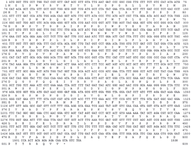
    At the heart of this complex is the reaction center consisting of the D1 and the D2 proteins, where primary charge separation occurs [13]. Closely associated with the D1 and the D2 proteins are two similar chlorophyll a-binding proteins, CP43 and CP47(product of the [14]. These proteins serve as an"inner antennae" system that is linked to a secondary light-harvesting system.

    Here, we have cloned the p-psbB gene encoding CP47, a chlorophyll -binding inner antenna protein, as a candidate target gene of the wild ginseng-specific genes using suppressive subtraction hybridization (SSH). We have further analyzed the differentially expressed levels of the p-psbB gene between cultivated ginseng and mountain cultivated wild ginseng by means of real-time quantitative PCR.

    2. Materials and methods

       2.1. Various ginsengs for RNA isolation

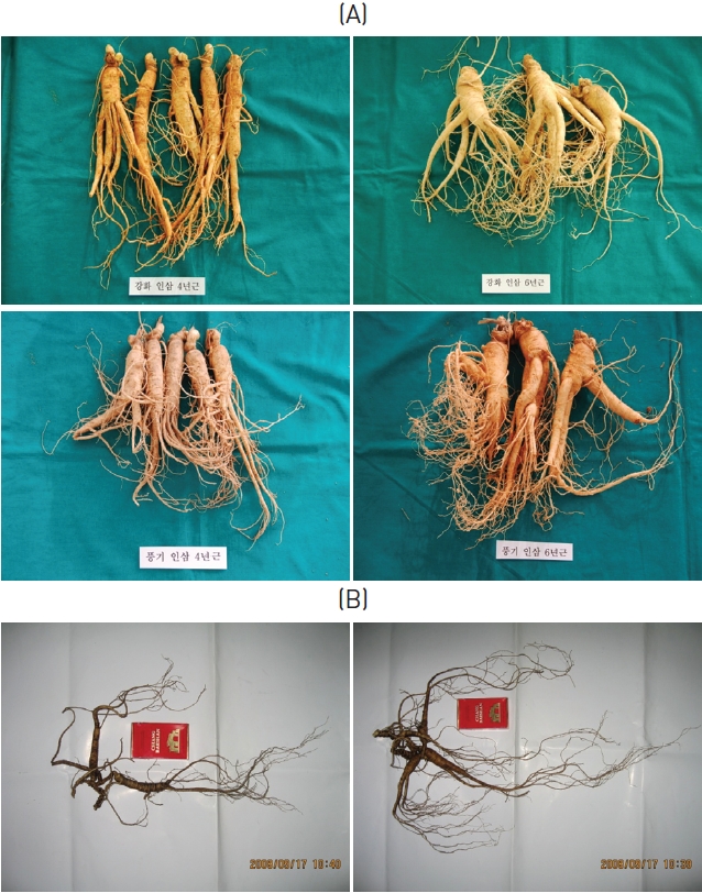
    The cultivated ginsengs (CGs) used in this experiment were 4 and 6 years of age and from various region in Korea. The wild ginsengs (WGs) used in this experiment were collected from Changbai Mt. in 2008, and were about 20 to 40 cm long, with masses of about 20 to 30 g and approximate ages of 30 to 50 years (Fig.1 ).

       2.2. Total RNA isolation and mRNA purification

    Ginseng was ground in liquid nitrogen by using a mortar and pestle, and RNA was isolated using the RNeasy Plant RNA Isolation Kit (Qiagen). The concentration of isolated RNA was estimated by measuring its absorbance at 260 nm. An aliquot of the RNA extract was treated with DNase-I (Invitrogen) prior to cDNA synthesis by using Superscript III reverse transcriptase(Invitrogen) and random hexamers according to the manu-facturer|s protocol.

       2.3 Suppressive subtractive hybridization

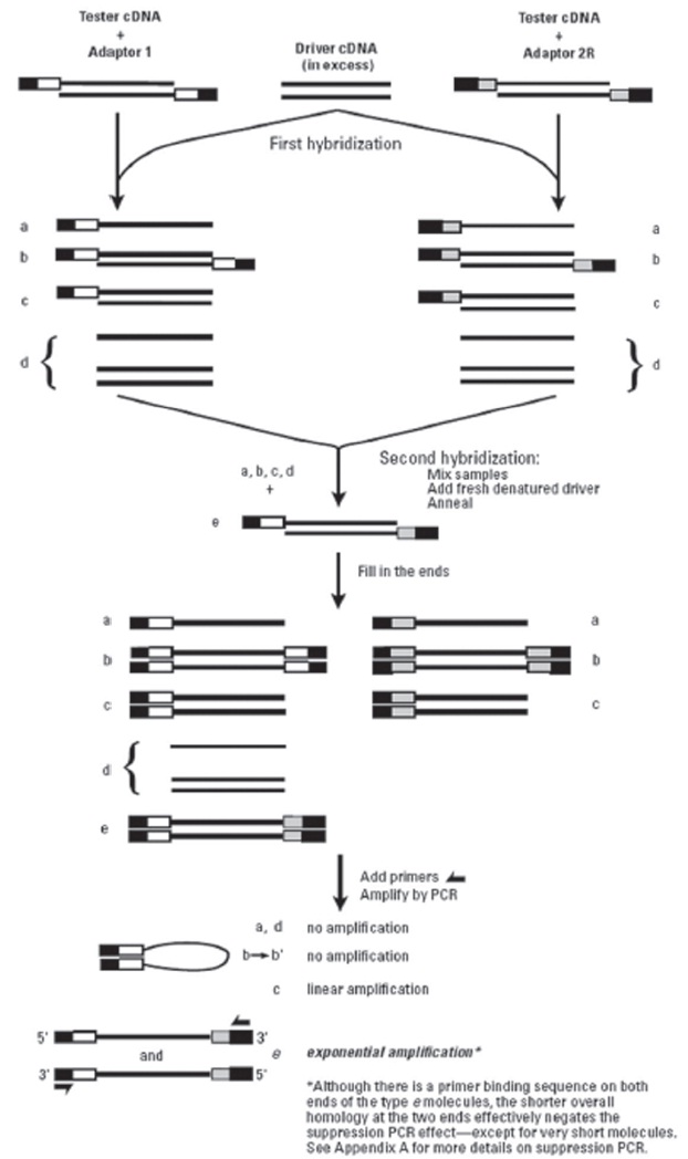
    Suppressive subtractive hybridization (SSH) was performed using Clontech PCR-SelectTM cDNA Subtraction Kit (Clontech)according to the manufacturer’s protocol. SSH method includes several steps (cDNA synthesis, RsaI digestion and adaptor ligation, two rounds of hybridization and PCR) for isolating differentially expressed genes.

    The cDNA fragments, derived from SSH forward subtractive library (tester: mountain cultivated wild ginseng; driver:ginseng), were cloned into pEC-T vector (KOMA Co., Seoul, Korea). The positive clones containing inserted fragments were identified by using the colony-PCR method.

       2.4. RT-PCR Assay

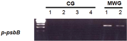
    Semi-quantitative RT-PCR was performed to compare the differential expression of the genes in the SSH library by using gene-specific primers. Total RNA (2 μg) was used for cDNA synthesis according to the First Strand cDNA Synthesis Kit(Invitrogen), and 1.0 μl of cDNAs was used as a template for PCR. PCR amplification was performed under the following conditions: 95°C for 5 min, 30 cycles at 95°C for 45 s, 54°C for 30 s, and 72°C for 60 s. The final incubation was done at 72°C for 5 min. PCR products were electrophoresed in a 2% agarose gel.

       2.5. Quantitative Real-time quantitative RT-PCR

    Real-time quantitative RT-PCR detection was performed with a StepOne machine and Fast SYBR Green Master Mix (Applied Biosystem, USA) and were measured in a 96-well plate. For each well, the 20 μl reaction involved 10 μl of the 2 X Fast SYBR Green Master Mix, 0.5 μM each of forward and reverse primer, 2.75 μl of DNase-free H2O, 2 μl of cDNA templates. PCR reactions were performed using the following parameters: 8 min at 95°C and 40 cycles of 45 s at 95°C, 45 s at 56°C and 45 s at 72°C. PCR products were melted by gradually increasing the temperature from 60°C to 95°C in 0.5°C steps.

    The identities of the amplicons and the specificity of the reaction were verified by using a melting curve analysis. Normalization of the cDNA templates was achieved by using 18S quantification. The primers presented in Table 1 were used to analyze
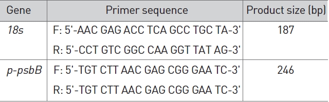
 p-psbB gene expressions.

    [Table 1] Primer for RT-PCR.

    label

    Primer for RT-PCR.

       2.6. Sequencing and homology analysis

    PCR products were cloned into the pEC-T vector (KOMA Co., Ltd, Seoul, Korea) and then sequenced by using the ABI 3700 DNA sequencers (PerkinElmer Applied Biosystems). The sequence analysis was performed using Chromas sequence analysis software. BLASTn was used to study similar nucleotide sequences.

    3. Results

       3.1. Isolation of differentially expressed genes in wild ginseng

    To identify wild ginseng-specific genes, wild ginseng cDNAs were subtracted from a pool of cultivated-ginseng cDNAs (Fig.1). The subtraction was expected to significantly reduce common cDNAs and to enrich for wild-ginseng-specific cDNAs.More than 100 transformants were obtained from the library, and the recombinant efficiency detected by using colony-PCR was about 90%.

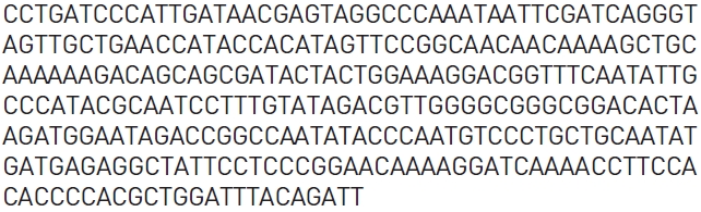
    One hundred positive clones confirmed by PCR amplification were randomly selected, from which, 16 significantly different clones were sequenced. Because the suppression subtractive hybridization procedure includes a restriction enzyme digestion of the cDNAs produced, none of the clones obtained from the resulting libraries were full length.

    Among the novel cDNAs identified here as putative wild-ginseng-specific genes is a putative chloroplast p-psbB, designated as p-psbB (Fig.2 ). The open reading frame contained in the p-psbB cDNA encodes a protein with 509 amino acids with a predicted molecular mass of 56,364 Da (Fig.3 ).

       3.2. RT-PCR analysis

    To confirm the differential expression of the p-psbB gene, we employed the RT-PCR analysis was employed. Total cellular RNA from the four cultivated ginsengs, and two mountain wild ginsengs were used for the RT-PCR analysis. The p-psbB gen-especific primers were designed to amplify both the cDNA from the cultivated ginsengs and from the wild ginsengs. The number of PCR cycles was optimized to ensure that the comparison of the levels of expressions of the p-psbB gene was within the linear phase of amplification.

    As shown in Fig. 4, all of the p-psbB transcripts derived from mountain wild ginsengs reveal on upper band whereas all of the p-psbB transcripts derived from the cultivated ginsengs showed lower bands. Thus, these results suggest that p-psbB mRNA is specifically expressed in wild ginsengs.

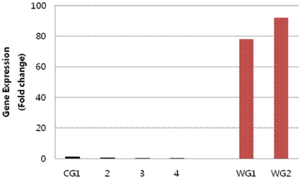
       3.3. Real-time RT-PCR analysis

    4. Discussion

    P. ginseng is categorized as either cultivated (in the farm) or wild (in the mountain) according to its different nurturing methods. Cultivated ginseng is systematically farmed on an open land and is harvested after a 4 to 6 year of cultivation period. On the other hand, wild ginseng is planted through seeding in a deep mountain. Wild ginseng is slower in growth and more sensitive to environmental changes than cultivated ginseng, showing a preference for areas with fluctuating daily temperatures and less exposure to direct sunlight. These differences may result in a variation of active compounds between cultivated and wild ginseng. In both Korea and China,wild ginseng is widely accepted to produce more potent medicinal activity than cultivated ginseng. However, few studies have been conducted to compare the food components and pharmacological activities between wild and cultivated ginseng.

    In the present study, to identify a wild ginseng-specific gene, we subtracted cDNAs expressed in wild ginsengs from those in cultivated ginsengs by using the SSH technique [15]. The technique of SSH is believed to generate an equalized representation of differentially expressed genes and to provide a high enrichment of differentially expressed mRNA. SSH overcomes the limitations of other gene analysis methods for differential expression. Its PCR-based approach allows for the effective removal of common genes from the RNA population prior to creating the library and has the advantage that reverse transcriptions are amplified efficiently [16].

    We isolated a novel gene, p-psbB (Panax ginseng chloroplast p-psbB;). Sequence analysis revealed that p-psbB possessed significant homology to p-psbB sequences reported from other plant species. p-psbB mRNA is differentially expressed in wild ginsengs. Thus, p-psbB may be one of the important markers of wild ginseng.

    The p-psbB encoded CP47 protein, a chlorophyll binding inner antenna protein in the photosystem II complex is located in the lipid matrix of the thylakoid membrane. The p-psbB has a light-harvesting function; it absorbs light and transfers the excitation energy to the reaction center of photosystem II [14]. Even more importantly, it also accepts excitation energy from the peripheral antenna and transfers it to the reaction center as well. However, although the mechanism by which p-psbB is up-regulated in wild ginseng is not clear, we suppose that it may an important marker of wild ginseng.

참고문헌
  • 1. Zhang D, Yasuda T, Yu Y, Zheng P, Kawabata T, Ma Y et al. 1996 Ginseng extract scavenges hydroxyl radical and protectsunsaturated fatty acids from decomposition caused by iron-mediatedlipid peroxidation. [Free Radic Biol Med.] Vol.20 P.145-50 google cross ref
  • 2. Yun TK, Lee YS, Lee YH, Kim SI, Yun HY. 2001 Anticarcinogenic effect of Panax ginseng C.A. Meyer and identification of acti-ve compounds. [J Korean Med Sci.] Vol.16 P.S6-18 google
  • 3. Joo S, Won TJ, Lee DI. 2005 Reciprocal activity of ginsenosides in the production of proinflammatory repertoire, and their po-tential roles in neuroprotection in vitro. [Planta Med.] Vol.71 P.476-81 google cross ref
  • 4. Kwon KR, Yoon HC, Kim HH. 2006 Anti-cancer effects of cultivated wild ginseng herbal acupuncture in C57BL/6 mice injected with B16/F10 cells reproductive toxicity by doxorubicin. [TheJouranl of Korean Acupuncture and Moxibustion society.] Vol.23 P.105-20 google
  • 5. Jang HY, Park HS, Kwon KR, Rhim TJ. 2008 A study on the compa-rison of antioxidant effects among wild ginseng, cultivated wild ginseng, and cultivated ginseng extracts. [Journal of Pharmacopuncture.] Vol.11 P.67-78 google cross ref
  • 6. Lui JH, S EJ. 1980 The Ginsenosides of Various Ginseng Plants and Selected Products. [J Nat Prod.] Vol.43 P.340-6 google cross ref
  • 7. Lim W, Mudge KW, Vermeylen F. 2005 Effects of population, age,and cultivation methods on ginsenoside content of wild Ame-rican ginseng (Panax quinquefolium). [J Agric Food Chem.] Vol.53 P.8498-505 google cross ref
  • 8. Shin SS, Kim KC, Choi YH, Lee YT, Eom HS, Kim CS. 2001 Critic st-andardization and objectivity of mountain grown ginseng. [Research Institute of Oriental Medicine Dong-Eui University.] Vol.5 P.107-14 google
  • 9. Kwon KR, Seo JC 2004 Genetical Identification of Korean Wild Ginseng and American Wild Ginseng by using Pyrose-quencing Method. [Kor J Herbology.] Vol.19 P.45-50 google
  • 10. Kwon KR, Park WP, Kang WM, Jeon EY, Jang JH. 2011 Identific-ation and analysis of differentially expressed genes in mou-ntain cultivated ginseng and mountain wild ginseng. [Journal of Acupuncture and Meridian Studies.] Vol.4 P.123-8 google cross ref
  • 11. Debus RJ. 1992 The manganese and calcium ions of photosynthe-ticoxygen evolution. [Biochim Biophys Acta.] Vol.1102 P.269-352 google cross ref
  • 12. Biochim Biophys Acta. B, Barber J., Boekema EJ. 1997 Structure and Membr-ane Organization of Photosystem Ii in Green Plants. [AnnuRev Plant Physiol Plant Mol Biol.] Vol.48 P.641-71 google cross ref
  • 13. Nanba O, Satoh K. 1987 Isolation of a photosystem II reaction center consisting of D-1 and D-2 polypeptides and cytochro-me b-559. [Proc Natl Acad Sci USA.] Vol.84 P.109-12 google cross ref
  • 14. Bricker TM, Frankel LK. 2002 The structure and function of CP47 and CP43 in photosystem II. [Photosynth Res.] Vol.72 P.131-46 google cross ref
  • 15. Diatchenko L, Lau YF, Campbell AP, Chenchik A, Moqadam F, Huang B, et al. 1996 Suppression subtractive hybridization: a method for generating differentially regulated or tissue-specific cDNA probes and libraries. [Proc Natl Acad Sci USA.] Vol.93 P.6025-30 google cross ref
  • 16. Diatchenko L, Diatchenko S, Lau YF, Siebert PD. 1999 Suppression subtractive hybridization: a versatile method for identifying differentially expressed genes. [Methods Enzymol.] Vol.303 P.349-80 google cross ref
OAK XML 통계
이미지 / 테이블
  • [ Figure 1 ]  Several cultivated ginsengs (A) and wild ginsengs (B) were used for RNA isolation.
    Several cultivated ginsengs (A) and wild ginsengs (B) were used for RNA isolation.
  • [ Table 1 ]  Primer for RT-PCR.
    Primer for RT-PCR.
  • [ Figure 2 ]  Overview of suppressive subtraction hybridization..
    Overview of suppressive subtraction hybridization..
  • [ Figure 3 ]  Determined partial DNA sequence of putative Panax ginseng Chloroplast p-psbB gene.
    Determined partial DNA sequence of putative Panax ginseng Chloroplast p-psbB gene.
  • [ Figure 4 ]  Nucleotide and predicted amino acid sequences of Panax ginseng p-psbB. The deduced amino acid sequence of the p-psbB is shown in one-letter symbols below the nucleotide sequence. Amino acid residues are numbered beginning with the first methionine, and the translation termination codon is denoted by an asterisk. Numbers to the right of the sequence correspond to amino acids (lower) and nucleotid-es (upper).
    Nucleotide and predicted amino acid sequences of Panax ginseng p-psbB. The deduced amino acid sequence of the p-psbB is shown in one-letter symbols below the nucleotide sequence. Amino acid residues are numbered beginning with the first methionine, and the translation termination codon is denoted by an asterisk. Numbers to the right of the sequence correspond to amino acids (lower) and nucleotid-es (upper).
  • [ Figure 5 ]  RT-PCR analysis of differential expression of p-psbB genes. Total RNAs (2.0 g) from four cultivated ginsengs (CG) and two wild ginsengs (MWG) were used for RT-PCR using the specific primers p-psbB.
    RT-PCR analysis of differential expression of p-psbB genes. Total RNAs (2.0 g) from four cultivated ginsengs (CG) and two wild ginsengs (MWG) were used for RT-PCR using the specific primers p-psbB.
  • [ Figure 6 ]  Quantitative real-time RT-PCR analysis of p-psbB transcripts. Total RNA extracted from the cells (2 μg) was reverse-transcribed to cDNA (40 μl), and aliquots (1.5 μl) were applied to real-time PCR (20 μl) with each primer (0.4mM). Values represented relative expression of p-psbB gene (calculated with threshold cycle number, CT) of two wild ginsengs (WG) compared with that of four cultivated ginsengs (CG). Each value was adjusted with CT of internal control (18s). All reations were performed in triplicate and resulting S.E. values are also given.
    Quantitative real-time RT-PCR analysis of p-psbB transcripts. Total RNA extracted from the cells (2 μg) was reverse-transcribed to cDNA (40 μl), and aliquots (1.5 μl) were applied to real-time PCR (20 μl) with each primer (0.4mM). Values represented relative expression of p-psbB gene (calculated with threshold cycle number, CT) of two wild ginsengs (WG) compared with that of four cultivated ginsengs (CG). Each value was adjusted with CT of internal control (18s). All reations were performed in triplicate and resulting S.E. values are also given.
(우)06579 서울시 서초구 반포대로 201(반포동)
Tel. 02-537-6389 | Fax. 02-590-0571 | 문의 : oak2014@korea.kr
Copyright(c) National Library of Korea. All rights reserved.