검색 전체 메뉴
PDF
맨 위로
OA 학술지
Cytoskeletal changes during nuclear and cell division in the freshwater alga Zygnema cruciatum (Chlorophyta, Zygnematales)
  • 비영리 CC BY-NC
  • 비영리 CC BY-NC
ABSTRACT
Cytoskeletal changes during nuclear and cell division in the freshwater alga Zygnema cruciatum (Chlorophyta, Zygnematales)
KEYWORD
anti-α-tubulin , microfilament , microtubule , nuclear division , phallacidin , Zygnema
  • INTRODUCTION

    Since the first observation of microfilaments in a freshwater alga, Nitella sp. (Nagai and Rebhun 1966), many papers have been published on the organization and function of actin filaments in interphase and other cell cycles in higher plants, fungi, green algae (Grolig 1990),red algae (Suzuki et al. 1995, Garbary and McDonald 1996a, 1996b), and brown algae (Karyophyllis et al. 2000).

    Microfilaments are thought to play a key role in cytokinesis furrowing, while not being involved in nuclear division. Sampson and Pickett-Heaps (2001) observed microfilaments associated with chromosomes, often in paired spots, in Oedogonium sp., a green alga, and suggested that actin filaments may be a functional component of the spindle. Similar results have been reported regarding F-actin organization in Sphacelaria, a brown alga (Karyophyllis et al. 2000). Karyophyllis et al. (2000)observed actin filaments aligned with spindle microtubules during nuclear division and posited the existence of organized actin flilament-kinetochore bundles. They also suggested that actin filaments might be involved in chromosome movement as well as in cytokinesis furrowing.In the Zygnematales, cytokinesis starts at the center of the cell and moves toward the periphery in the phragmoplast and furrowing with a persistent spindle. The organization and function of actin filaments, and the relationship between actin filaments and microtubules during nuclear division has never been studied in this group.

    In this study, we observed the cytoskeletal changes during cell division of the green alga Zygnema cruciatum (Vauch.) Agardh to elucidate the coordinate involvement of actin filaments and microtubules in nuclear division and chromosome movement.

    MATERIALS AND METHODS

      >  Plant material and laboratory culture

    Algal materials were collected from ponds in Kongju, Korea, from December, 2004 to January, 2005. The plants were washed three times with Bolds basal medium (Bischoff and Bold 1963) and kept in the same medium at 4°C, using a 12 : 12 hour light : dark cycle with cool-white fluorescent lighting (> 20 ㎛ol photons m-2s-1). Germanium dioxide was added to the medium (final concentration 1 mg L-1) for 2 weeks to eliminate diatoms.

      >  Fluorescence localization of actin and DNA

    To visualize the actin cytoskeleton, Zygnema filaments were fixed for 30 min in 3.7% (w/v) formaldehyde diluted in microfilament stabilizing buffer (MFSB) consisting of 10 mM EGTA, 5 mM MgSO4, 100 mM PIPES-KOH, pH 6.9 (Traas et al. 1987). Zygnema filaments were rinsed three times with MFSB, and then placed for 30 min in 0.5% Triton X-100 diluted in MFSB. The Zygnema filaments were then washed three times with MFSB before being placed in the staining solution of Bodipy FL phallacidin (Molecular Probes, Eugene, OR, USA) (Wieland et al. 1983) for 3-6 h in the dark at 4°C. Bodipy FL phallacidin was prepared as a stock solution of 300 units mL-1 in methanol and was stored at -20°C in the dark. The stock was diluted with MFSB to a final concentration of 1.5 units mL-1. For double staining of F-actin and nuclear DNA, Bodipy FL phallacidin samples were placed in a solution of 2.5 μg mL-1 4’-6 diamidino-2-phenolindole (DAPI) for 10 s, washed with MFSB and mounted on slide glass with MFSB. Double-stained materials were observed with a model BX-50 fluorescence microscope (Olympus, Tokyo, Japan) under blue (excitation = 470 nm) and ultraviolet (excitation = 356 nm) filters for F-actin and nuclear DNA staining, respectively.

      >  Fluorescence localization of microtubules

    Zygnema filaments were fixed in 3.7% formaldehyde diluted with MFSB for 30 min and then rinsed three times in the same buffer. For microtubule staining, Zygnema filaments were cut with razor blade into small pieces (0.5-1 mm in length) and incubated overnight in the dark with monoclonal anti-α-tubulin antibody conjugated to fluorescein isothiocyanate (Sigma-Aldrich, St. Louis, MO, USA), diluted 1 : 40 in phosphate buffered saline (PBS; 8 mM Na2HPO4, 2 mM NaH2PO4, 140 mM NaCl, pH 7.4). For double staining of microtubules and nuclear DNA, the sample stained with the flourescein isothiocynate (FITC)-conjugated antibody were placed in a solution of 2.5 μg mL-1 DAPI for 10 s and washed with PBS. Double-stained materials were microscopically examined as described above.

      >  Time-lapse microscopy

    For time-lapse video-microscopy, the filaments were placed on a glass slide and a coverslip was applied and sealed with a 1 : 1 : 1 mixture of Vaseline, lanolin and paraffin that had been prepared by melting on a hot plate at 70oC. The slide preparations were examined using an Olympus BX-51 microscope under the oil immersion ×20 objective lens, and the images were recorded using a Digital Imaging Time-Lapse Recorder (TCS Korea, Daejeon, Korea).

    RESULTS

      >  Observation of cell division using time-lapse video-microscopy

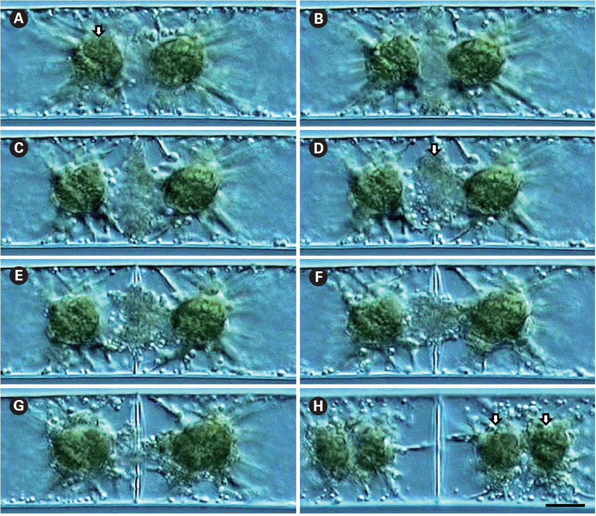
    Cell division was observed using time-lapse video-microscopy. Cells were uninucleate and possessed two star-shaped chloroplasts (Fig. 1A). Cell division always proceeded simultaneously with nuclear division, with the cell wall first discerned at the cell periphery, with development towards the cell center (Fig. 1B). Vesicles involved in cell plate formation decreased with the development of the cell wall (Fig. 1C-G). Usually, chloroplast division followed nuclear division and cell plate formation (Fig. 1H).

    Fluorescence localization of actin and DNA

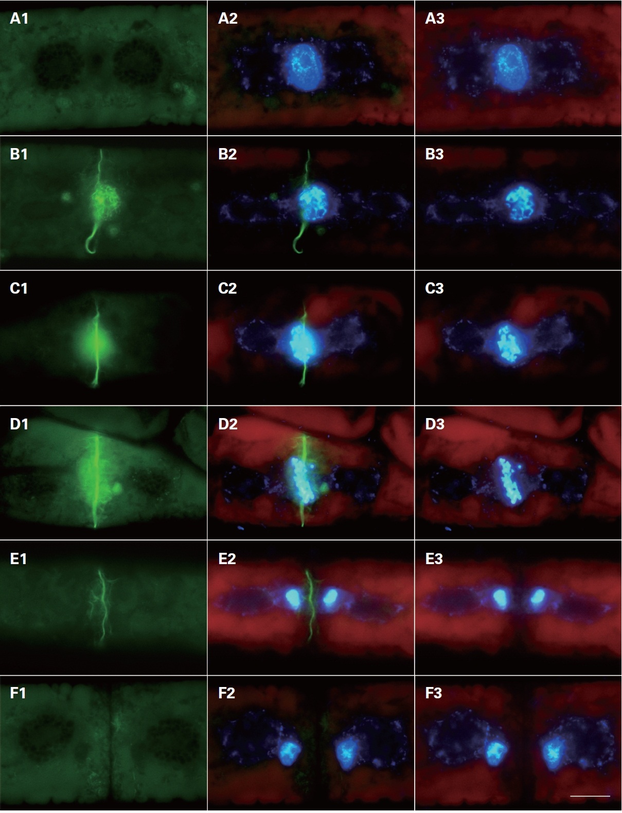
    Cytoskeletal changes were observed during cell division using FITC-phallacidin for F-actin staining. Microfilaments began to appear at the center of the division plate when nuclear division started (Fig. 2). These filaments were not visible in control cells pretreated with non-fluorescent phallacidin (data not shown). Therefore, filaments revealed using FITC-phallacidin presumably represented cytoskeletal microfilament bundles composed of F-actin.

    In interphase cells, F-actin was randomly distributed in the cytoplasm near the cell surface. Normally, the actin filaments were not observed in the centers of the cells (Fig. 2A1). In prophase, the nuclear envelope was broken down; two typical forms of staining were observed in different locations of the cell. The first type of staining was a fibrous web around the nucleus, in which F-actin tightly surrounded the prophase nucleus forming a finely interwoven net-like cage as apparent in the surface view (Fig. 2B1). In the other type of staining, the F-actin was evident at the middle of the cells forming a fibril ring situated just beneath the plasma membrane (Fig. 2C1 & D1). A faint trace of protoplast-furrowing was visible in mid-

    prophase cells. These protoplast-furrowing structures were tightly associated with the fibril ring (Fig. 2C1).

    In metaphase cells, the chromosomes were aligned at the cell equator, and the net-like cage of F-actins ran nearly parallel with the individual chromosomes, but ultimately most formed a spindle located on the individual chromosomes (Fig. 2D1, 3A1 & B1). In this stage, the fibril rings were similar in appearance to those in prophase cells (Fig. 2B1 & C1). The diameter of the fibril ring

    continuously decreased with the development of the furrow, until each cell divided into two daughter cells (Fig. 2D1-F1).

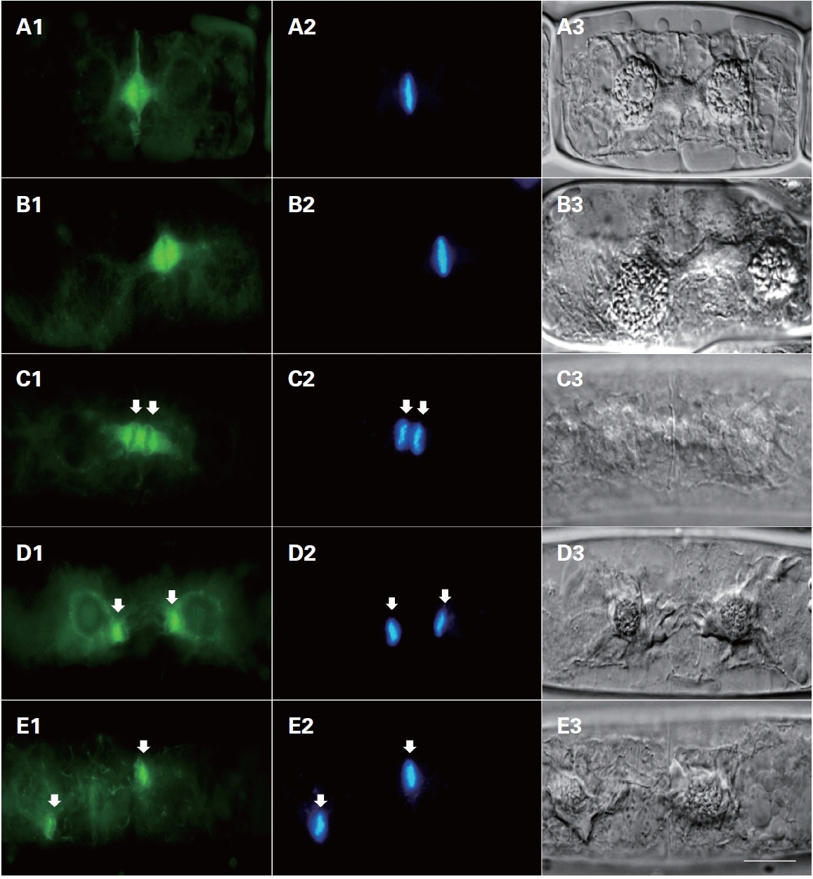
    In anaphase cells, the duplicated chromosomes separated, and the spindle form of F-actins associated with the daughter chromosomes moved from the equator of the cell to spindle poles (Fig. 3C1 & D1). The spindle form of F-actins was similar in appearance to those in metaphase cells (Fig. 3E1). The F-actins were localized at the spindle pole side in late anaphase. Furrowing of the protoplast was most conspicuous at this stage. A fibril ring was not detected in the same area at anaphase.

    In telophase cells, the daughter chromosomes reached to the mitotic poles, and actin filaments were observed on the daughter nuclei (Fig. 3E1 & E2). These F-actins tightly surrounded the telophase nucleus and continuously decreased with the completion of cell division. Different types of F-actins were evident in the cytoplasm near the surface of the cell, most of them running irregularly (Fig. 3E1).

      >  Fluorescence localization of microtubules and DNA

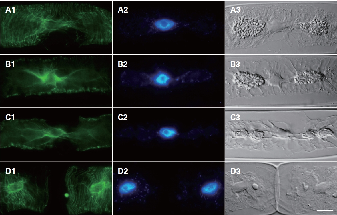
    Cytoskeletal changes were observed during cell division using FITC-anti-α-tubulin to stain microtubules. In interphase cells, the microtubules were arranged beneath the protoplasm of the cells, most of them running nearly parallel to the cross-wall surrounding the nucleus (Fig. 4). In cells progressing from prophase to meta-

    phase, microtubules were localized at the nuclear region at prophase (Fig. 4B1) but became an organized mitotic spindle at metaphase (Fig. 4B1). Interestingly, microtubules appeared between dividing chloroplasts (Fig. 4C1 & C3). Finally, after forming the new cell wall (Fig. 4D3), microtubules were re-arranged beneath the cell wall (Fig. 4D1).

    DISCUSSION

    The present study recorded the following observations. During interphase, actin filaments were located along the cell surface running parallel to the long axis of the cell. During prophase, a fibrous web of F-actin appeared around the nucleus and a fibril ring was formed at the division center just beneath the plasma membrane. The F-actins of metaphase cells co-aligned with spindle microtubules to contact individual chromosomes. The spindle form of the F-actin was associated with daughter chromosomes moving from the equator of the cell to spindle poles during anaphase. Finally, during telophase, F-actins tightly surrounded the nucleus. The amount of F-actin in the center of cell continuously decreased with the completion of cell division. These data provided the first comprehensive evidence suggesting a coordinate involvement of microfilaments and microtubules during nuclear division in Z. cruciatum.

    The F-actins apparent during interphase appeared in the cytoplasm near the surface of the cell, and disappeared with the progress of cytokinesis. They might be involved in vesicle and organelle transport, either alone or with the microtubules. They could also be responsible for cytoplasmic streaming, although this function was not evident in Zygnema. The latter activity is usually generated by the sub-cortical actin filaments (Shimmen and Yokota 1994). Similar roles have been reported for F-actin systems in other green algae (Menzel and Schliwa 1986, Goto and Ueda 1988, Grolig 1990), in some red algae (Garbary and McDonald 1996a) and in some brown algae (Karyophyllis et al. 2000). Kim et al. (2005) reported phototaxis of the filamentous green algae Spirogyra spp. and suggested that coordination of actin filaments with microtubules on cell surface might be the mechanical effector of the filament movement.

    Four types of microfilaments have been reported in Spirogyra spp. (Goto and Ueda 1988). Each type displays a unique behavior during the cell cycle: dispersed in the cytoplasm near the cell surface, located beneath the plasmalemma running parallel to the cell's long axis, located at the edges of the chloroplasts, and surrounding the nucleus. Among them, types 2 and 4 are similar to the F-actin type in Zygnema and in Oedogonium spp. (Sampson and Pickett-Heaps 2001). F-actins surrounding the nucleus were also presently associated with spindle forming microtubules in the prophase stage, which might suggest that the actin filament helps in the formation of mitotic spindles as well as the attachment of spindle fibers to the chromosomes in prophase. In many higher plant cells, as well as in some algae, the actin filaments show a particular organization during mitosis and cytokinesis, similar to that of the microtubule spindle (Panteris et al. 1992, Czaban and Forer 1994). It is important to note that actin has never been reported in the spindle of vegetative meristematic angiosperm cells (McCurdy and Gunning 1990, Liu and Palevitz 1992, Hepler et al. 1993, Cleary and Mathesius 1996). On the contrary, the actin filaments seem to participate in the spindle in the cultured callus cells of higher plants (Seagull et al. 1987, Traas et al. 1987), cells of the pteridophyte Adiantum (Panteris et al. 1992), and cells of Haemanthus endosperm (Schmit and Lambert 1987, 1990). Although similar F-actins type have been reported in brown algae Sphacelaria (Karyophyllis et al. 2000), this is the first report of F-actins in the metaphase stage of Zygnematales.

    The organization of F-actin in the spindle of Zygnema cells persisted through all stages of mitosis, suggesting that F-actins may be necessary for spindle organization. The prophase actin filament cage may be involved in the maintenance of the nuclear materials after the degeneration of the nuclear envelope during the first mitotic stages. F-actins might also be involved in daughter chromosome separation, because these spindle forms of F-actin associated with daughter chromosomes move from the equator of the cell to the spindle poles during anaphase stage. Although the evidence against the involvement of actin filaments in mitosis is considerable, several studies showed the involvement of actin filaments in chromosome movement (e.g., Sampson and Pickett-Heaps 2001). Sampson et al. (1996) showed that treatment of the freshwater algae Oedogonium spp. with the actin inhibitor cytochalasin D blocks chromosomal attachment to the spindle, which prevents cells from entering anaphase. Our data also provide evidence that actin filaments might be involved in nuclear division and chromosome movement in Z. cruciatum.

참고문헌
  • 1. Bischoff H. W, Bold H. C 1963 Phycological studies IV. Some soil algae from enchanted rock and related algal species P.95 google
  • 2. Cleary A. L, Mathesius U 1996 Rearrangements of F-actin during stomatogenesis visualised by confocal microscopy in fixed and permeabilised Tradescentia leaf epidermis [Bot. Acta] Vol.109 P.15-24 google
  • 3. Czaban B. B, Forer A 1994 Rhodamine-phalloidin and anti-tubulin antibody staining of spindle fibres that were irradiated with an ultraviolet microbeam [Protoplasma] Vol.178 P.18-27 google cross ref
  • 4. Garbary D. J, McDonald A. R 1996a Actin rings in cytokinesis of apical cells in red algae [Can. J. Bot] Vol.74 P.971-974 google cross ref
  • 5. Garbary D. J, McDonald A. R 1996b Fluorescent labelling of the cytoskeleton in Ceramium strictum (Rhodophyta) [J. Phycol] Vol.32 P.85-93 google cross ref
  • 6. Goto Y, Ueda K 1988. Microfilament bundles of F-actin in Spirogyra observed by fluorescence microscopy [Planta] Vol.173 P.442-446 google cross ref
  • 7. Grolig F 1990 Actin-based organelle movements in interphase Spirogyra [Protoplasma] Vol.155 P.29-42 google cross ref
  • 8. Hepler P. K, Cleary A. L, Gunning B. E. S, Wadsworth P, Wasteneys G. O, Zhang D. H 1993 Cytoskeletal dynamics in living plant cells. [Cell Biol. Int] Vol.17 P.127-142 google cross ref
  • 9. Karyophyllis D, Katsaros C, Galatis B 2000 F-actin involvement in apical cell morphogenesis of Sphacelaria rigidula (Phaeophyceae): mutual alignment between cortical actin filaments and cellulose microfibrils [Eur. J. Phycol] Vol.35 P.195-203 google
  • 10. Kim G. H, Yoon M, Klotchkova T. A 2005. A moving mat: phototaxis in the filamentous green algae Spirogyra (Chlorophyta Zygnemataceae) [J. Phycol] Vol.41 P.232-237 google cross ref
  • 11. Liu B, Palevitz B. A 1992 Organization of cortical microfilaments in dividing root cells [Cell Motil. Cytoskelet] Vol.23 P.252-264 google cross ref
  • 12. McCurdy D. W, Gunning B. E. S 1990 Reorganization of cortical actin microfilaments and microtubules at preprophase and mitosis in wheat root-tip cells: a double label immunofluorescence study [Cell Motil. Cytoskelet] Vol.15 P.76-87 google cross ref
  • 13. Menzel D, Schliwa M 1986 Motility in the siphonous green alga Bryopsis. II. Chloroplast movement requires organized arrays of both microtubules and actin filaments [Eur. J. Cell Biol] Vol.40 P.286-295 google
  • 14. Nagai R, Rebhun L. I 1966 Cytoplasmic microfilaments in streaming Nitella cells [J. Ultrastruct. Res] Vol.14 P.571-589 google cross ref
  • 15. Panteris E, Apostolakos P, Galatis B 1992 The organization of F-actin in root tip cells of Adiantum capillus veneris throughout the cell cycle [Protoplasma] Vol.170 P.128-137 google cross ref
  • 16. Sampson K, Pickett-Heaps J. D 2001 Phallacidin stains the kinetochore region in the mitotic spindle of the green alga Oedogonium spp [Protoplasma] Vol.217 P.166-176 google cross ref
  • 17. Sampson K, Pickett-Heaps J. D, Forer A 1996 Cytochalasin D blocks chromosomal attachment to the spindle in the green alga Oedogonium [Protoplasma] Vol.192 P.130-144 google cross ref
  • 18. Schmit A. C, Lambert A. M 1987 Characterization and dynamics of cytoplasmic F-actin in higher plant endosperm cells during interphase mitosis and cytokinesis [J. Cell Biol] Vol.105 P.2157-2166 google cross ref
  • 19. Schmit A. C, Lambert A. M 1990 Microinjected fluorescent phalloidin in vivo reveals the F-actin dynamics and assembly in higher plant mitotic cells [Plant Cell] Vol.2 P.129-138 google cross ref
  • 20. Seagull R. W, Falconer M. M, Weerdenburg C. A 1987 Microfilaments: dynamic arrays in higher plant cells [J. Cell Biol] Vol.104 P.995-1004 google cross ref
  • 21. Shimmen T, Yokota E 1994 Physiological and biochemical aspects of cytoplasmic streaming [Int. Rev. Cytol] Vol.155 P.97-139 google cross ref
  • 22. Suzuki H, Oiwa K, Yamada A, Sakakibara H, Nakayama H, Mashiko S 1995 Linear arrangement of motor protein on a mechanically deposited fluoropolymer thin film [Jpn. J. Appl. Phys] Vol.34 P.3937-3941 google cross ref
  • 23. Traas J. A, Doonan J. H, Rawlins D. J, Shaw P. J, Watts J, Lloyd C. W 1987 An actin network is present in the cytoplasm throughout the cell cycle of carrot cells and associated with the dividing nucleus [J. Cell Biol] Vol.105 P.387-395 google cross ref
  • 24. Wieland T, Miura T, Seeliger A 1983 1983. Analogs of phalloidin: D-Abu2-Lys7-phalloin an F-actin binding analog its rhodamine conjugate (RLP) a novel fluorescent F-actin-probe and D-Ala2-Leu7-phalloin an inert peptide. Int [J. Pept. Protein Res] Vol.21 P.3-10 google cross ref
OAK XML 통계
이미지 / 테이블
  • [ Fig. 1. ]  Time-lapse images of cell division process in Zygnema cruciatum. (A) Beginning of cell wall formation. Arrow shows the nucleus between chloroplasts. (B) Two stellate chloroplasts associated with the nucleus and the peripheral protoplasm. (C) Formation of vesicles at the center of cell. (D) New cell wall begins to develop. Arrow shows phragmoplast the new cell wall and dividing chloroplasts. (E) Developing wall. (F) Developing wall with reduced number of vesicles. (G) Number of vesicles reduced abruptly. (H) Chloroplast division starts (arrows). Scale bar represents: 10 ㎛.
    Time-lapse images of cell division process in Zygnema cruciatum. (A) Beginning of cell wall formation. Arrow shows the nucleus between chloroplasts. (B) Two stellate chloroplasts associated with the nucleus and the peripheral protoplasm. (C) Formation of vesicles at the center of cell. (D) New cell wall begins to develop. Arrow shows phragmoplast the new cell wall and dividing chloroplasts. (E) Developing wall. (F) Developing wall with reduced number of vesicles. (G) Number of vesicles reduced abruptly. (H) Chloroplast division starts (arrows). Scale bar represents: 10 ㎛.
  • [ Fig. 2. ]  Phallacidin staining of Zygnema cruciatum actin during cell division. (A) Interphase. (B) Prophase. (C) Prometaphase. (D) Metaphase. (E) Anaphase. (F) Telophase. (A1-F1) Phallacidin stained actin filaments. (A2-F2) Merged pictures of Phallacidin stained actin filaments and DAPI stained nucleus. (A3-F3) DAPI stained nuclei. Scale bar represents: 10 ㎛.
    Phallacidin staining of Zygnema cruciatum actin during cell division. (A) Interphase. (B) Prophase. (C) Prometaphase. (D) Metaphase. (E) Anaphase. (F) Telophase. (A1-F1) Phallacidin stained actin filaments. (A2-F2) Merged pictures of Phallacidin stained actin filaments and DAPI stained nucleus. (A3-F3) DAPI stained nuclei. Scale bar represents: 10 ㎛.
  • [ Fig. 3. ]  Phallacidin staining of Zygnema cruciatum actin during cell division. (A& B) Metaphsae. (C & D) Anaphase. The spindle form of F-actins associated with the daughter chromosomes (arrows). (E) Telophase. The F-actins tightly surrounded the telophase nucleus (arrows). (A1->E1) Phallacidin stained actin filaments. (A2?E2) DAPI stained nucleus. (A3-E3) Differential interference microscopic image. Scale bar represents: 10 ㎛.
    Phallacidin staining of Zygnema cruciatum actin during cell division. (A& B) Metaphsae. (C & D) Anaphase. The spindle form of F-actins associated with the daughter chromosomes (arrows). (E) Telophase. The F-actins tightly surrounded the telophase nucleus (arrows). (A1->E1) Phallacidin stained actin filaments. (A2?E2) DAPI stained nucleus. (A3-E3) Differential interference microscopic image. Scale bar represents: 10 ㎛.
  • [ Fig. 4. ]  Fluorescein isothiocynate (FITC)-anti-α-tubulin stained microtubulin of Zygnema cruciatum during cell division. (A) Interphase. (B) Prophase. (C) Metaphase. (D) Telophase. (A1-D1) FITC-anti-α-tubulin stained microtubulin. (A2-D2) DAPI stained nucleus. (A3-D3) Differential interference microscopic image. Scale bar represents: 10 ㎛.
    Fluorescein isothiocynate (FITC)-anti-α-tubulin stained microtubulin of Zygnema cruciatum during cell division. (A) Interphase. (B) Prophase. (C) Metaphase. (D) Telophase. (A1-D1) FITC-anti-α-tubulin stained microtubulin. (A2-D2) DAPI stained nucleus. (A3-D3) Differential interference microscopic image. Scale bar represents: 10 ㎛.
(우)06579 서울시 서초구 반포대로 201(반포동)
Tel. 02-537-6389 | Fax. 02-590-0571 | 문의 : oak2014@korea.kr
Copyright(c) National Library of Korea. All rights reserved.