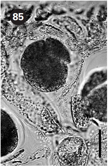
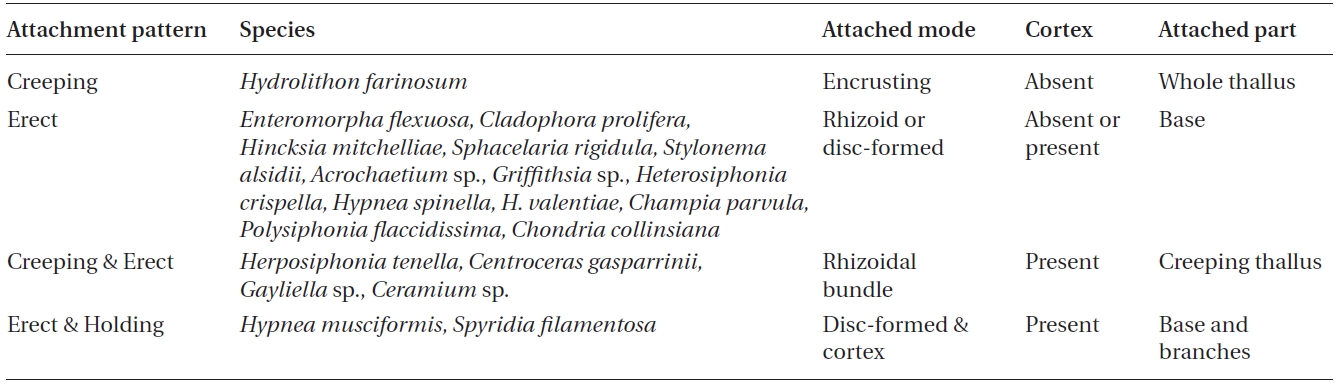
Seagrass epiphyte blooms potentially have important economic and ecological consequences in Tampa Bay, one of the Gulf of Mexico’s largest estuaries. As part of a Tampa Bay pilot study to monitor the impact of environmental stresses,precise characterization of epiphyte diversity is required for efficient management of affected resources. Thus, epiphyte diversity may be used as a rational basis for assessment of ecosystem health. In May 2001, epiphytic species encompassing green, brown and red macroalgae were manually collected from dense and sparse seagrass beds of Thalassia testudinum and Syringodium filiforme. A total of 20 macroalgal epiphytes, 2 Chlorophyta, 2 Phaeophyta, and 16 Rhodophyta,were found on T. testudinum and S. filiforme seagrass at the four sampling sites (Bishop Harbor, Cockroach Bay, Feather Sound, and Mariposa Key). The Rhodophyta, represented by 16 species, dominated the numbers of species. Among them, the thin-crusted Hydrolithon farinosum was the most commonly found epiphyte on seagrass leaves. Species number, as well as species frequency of epiphytes, is higher at dense seagrass sites than sparse seagrass sites. Four attachment patterns of epiphytes can be classified according to cortex and rhizoid development: 1) creeping, 2) erect,3) creeping & erect, and 4) erect & holding. The creeping type is characterized by an encrusting thallus without a rhizoid or holdfast base. Characteristics of the erect type include a filamentous thallus with or without a cortex, and a rhizoid or holdfast base. The creeping and erect type is characterized by a filamentous thallus with a cortex and rhizoid. A filamentous thallus with a cortex, holdfast base, and host holding branch is characteristics of the erect and holdfast attachment type. This study characterized each species found on the seagrass for epiphyte identification.
Seagrass meadows are very productive ecosystems of which a large proportion is often attributed to epiphytes(Heijs 1984, Leliaert et al. 2001). Seven seagrass species occur in Florida:
Seagrass epiphytes are very important components of the meadows. At least 113 epiphytes and up to 120 macroalgal species have been identified from Florida seagrass blades and communities, respectively (Dawes 1987). Although lists and ecological studies about epiphytes on
This paper characterizes macroalgal epiphytes and determines attachment patterns on seagrass blades of
During the spring of 2002, seagrass shoots of
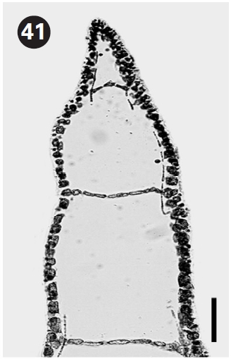
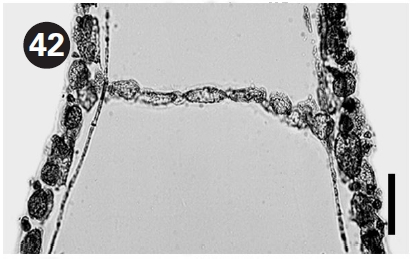
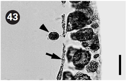
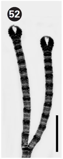
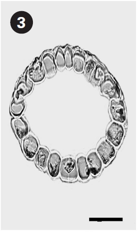
Comparison of epiphyte attachment patterns on Syringodium filiforme and Thalassia testudinum
abundance of epiphytes between sparse and dense sites.
>
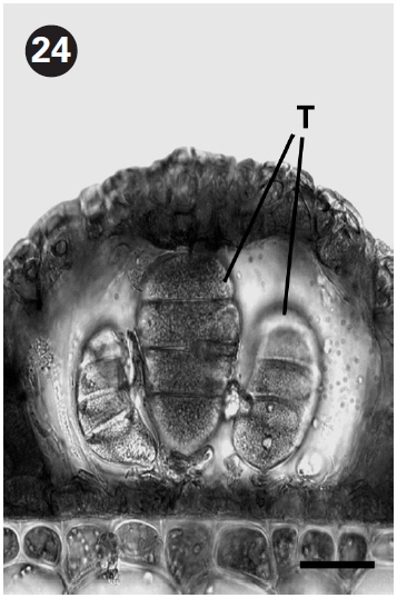
Epiphytic species composition, species abundance,and attachment pattern
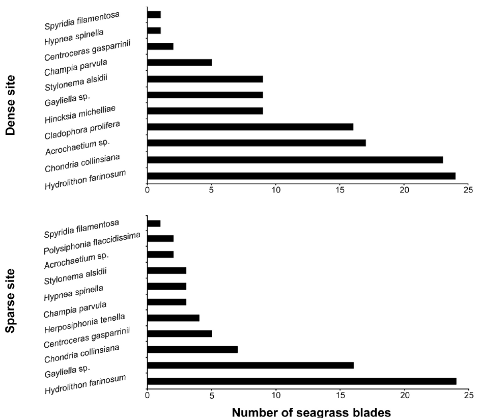
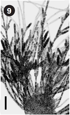
As shown in Table 1, a total of 20 macroalgal epiphytes (2 Chlorophyta, 2 Phaeophyta, and 16 Rhodophyta) are found in
Rhodophyta exceeds 80% at the total species number. Of them, the thin-crusted
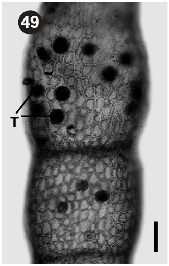
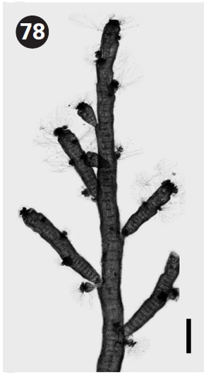
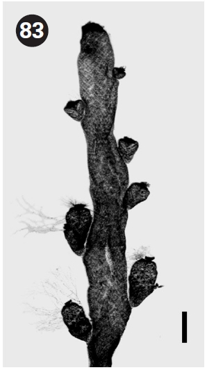
The total species number of epiphytes on each narrow
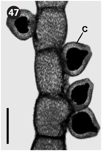
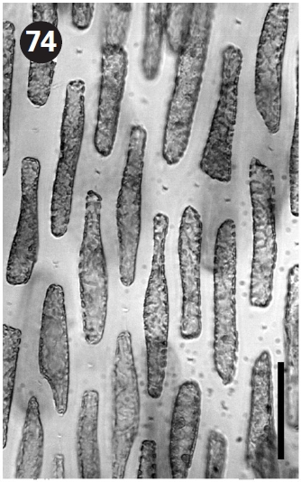
Species number, as well as species frequency, of epiphytes is higher at dense seagrass sites than sparse seagrass sites. Fourteen epiphytes were identified from dense sites of
ity of attachment of macroalgal epiphytes to seagrass blades, a larger number of epiphytes may occur in dense sites. Species frequency of epiphytes on each blade of
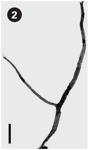
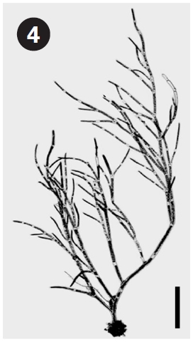
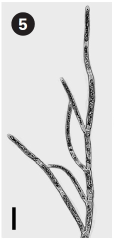
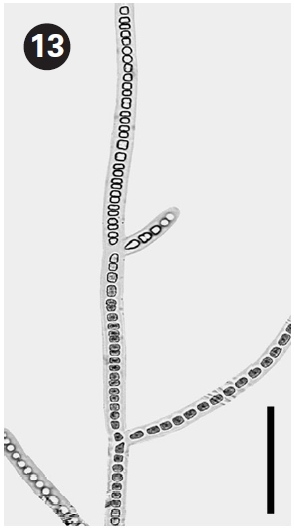
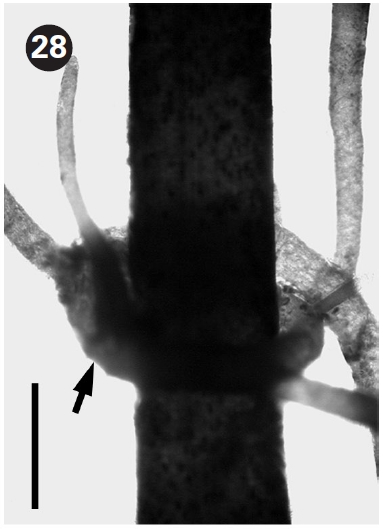
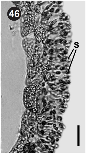
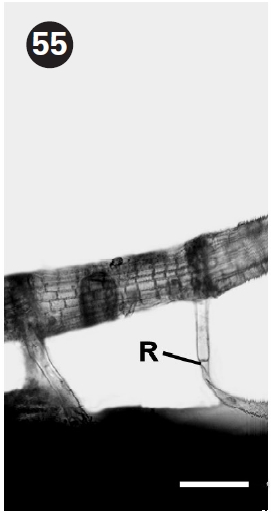
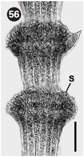
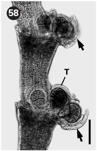
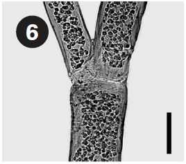
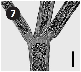
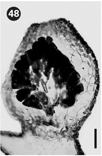
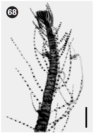
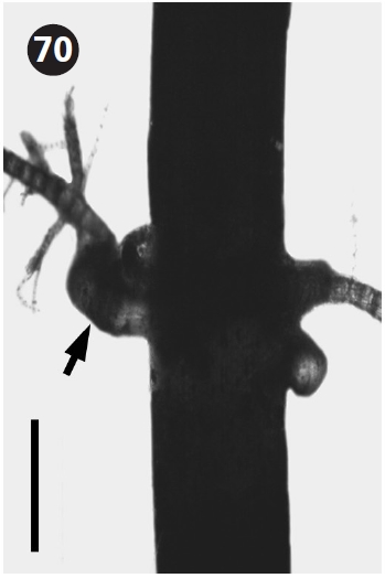
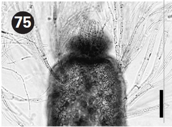
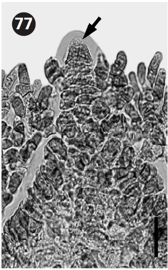
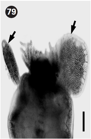
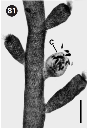
As summarized in Table 1, four attachment patterns of epiphytes can be classified according to development of cortex and rhizoid: 1) creeping, 2) erect, 3) creeping & erect, and 4) erect & holding. The creeping type is characterized by an encrusting thallus without a rhizoid or holdfast base. This type is found in
>
List and characterization of epiphytes
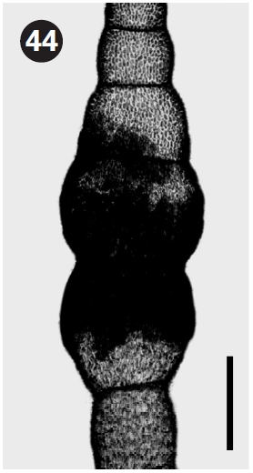
Although most of these epiphytic species have previously been reported from Florida (Dawes 1987, Littler and Littler 2000), we characterize each species with detailed morphology.
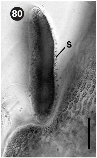
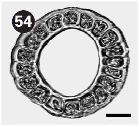
The thallus is slender, erect, and about 1 cm high. Blades taper toward base and are cylindrical and hollow. Rhizoids form a tightly knit basal pad.
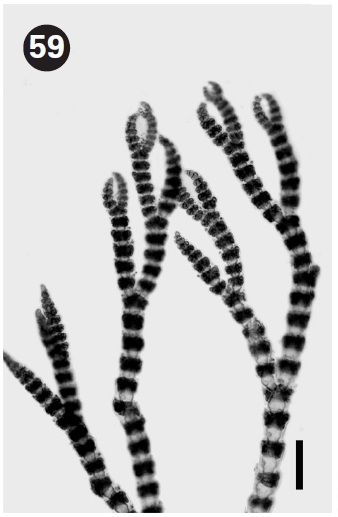
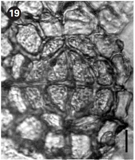
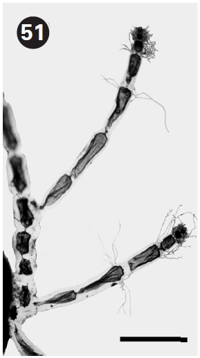
The thallus is filamentous, pseudo-dichotomous or pseudo-trichotomous, branching, erect, and about 1 cm high. Filaments are straight to slightly curved. Rhizoids are formed from basal cells.
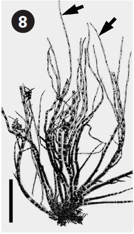
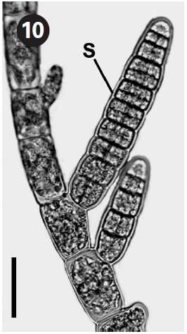
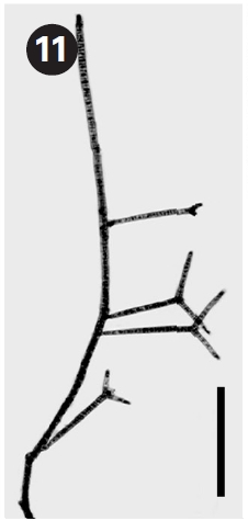
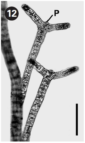
Hincksia mitchelliae (Harvey) P. C. Silva in Silva et al. 1987 (Figs 8-10)
Basionym:
The thallus is filamentous tufts or mats, erect, and 0.5 cm high. Filaments are irregularly branched, and taper toward apices. Plurilocular sporangia are cylindrical, rarely stalked, and lateral on filaments.
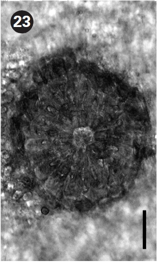
The thallus is filamentous, erect, and 0.3 cm high. Filaments are straight and cylindrical. Propagules have 2-3 cylindrical arms.
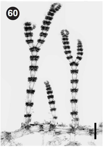
The thallus is erect, pseudodichotomously branched, and 0.2-0.3 cm high. Cells are discoid to ellipsoid.
The thallus is filamentous, erect, and 0.3-0.5 cm high. Cells are cylindrical or rod-shaped. Monosporangia are basal in lateral clusters and develop adaxially at the upper part of the cell.
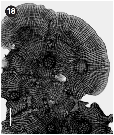
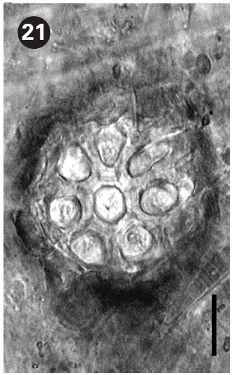
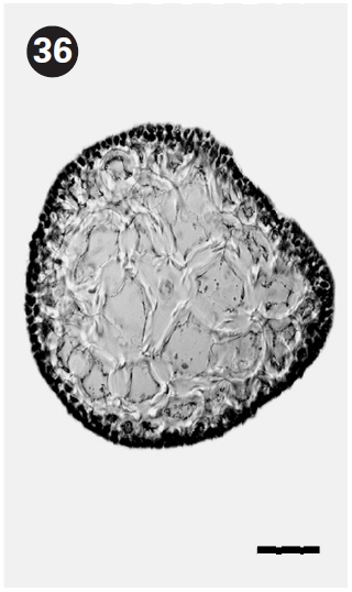
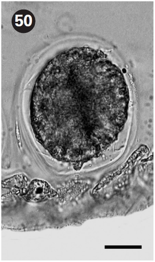
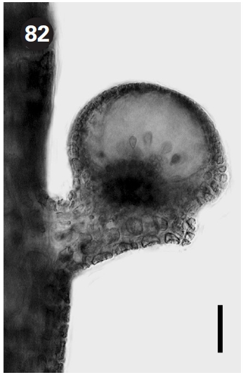
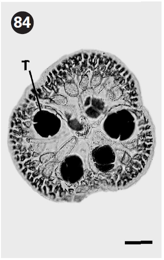
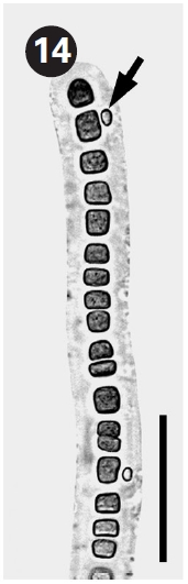
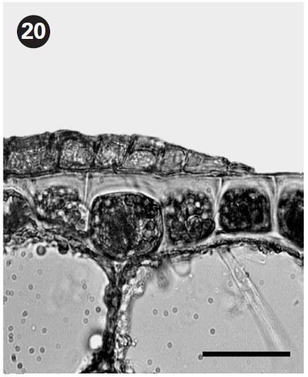
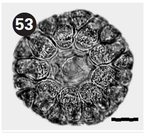
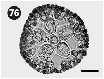
The thallus is prostrate, thin, crusts, develops from an initial four-celled structure, and measures 0.3-0.5 cm diam. Tetrasporangial conceptacles are hemispherical and tetrasporangia are zonately divided.
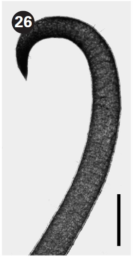
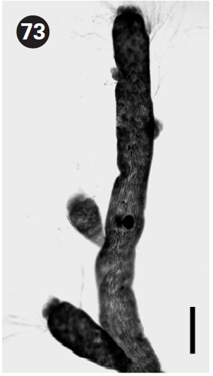
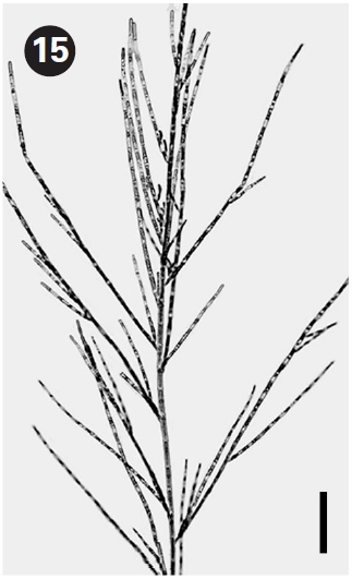
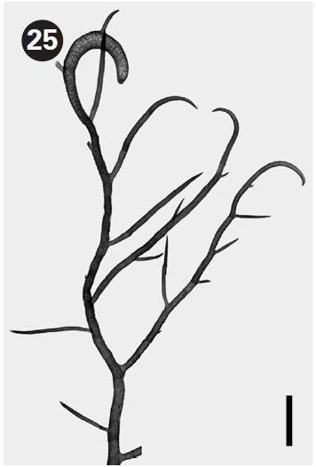
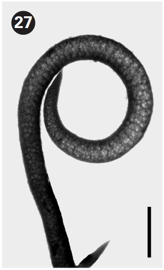
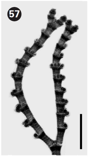
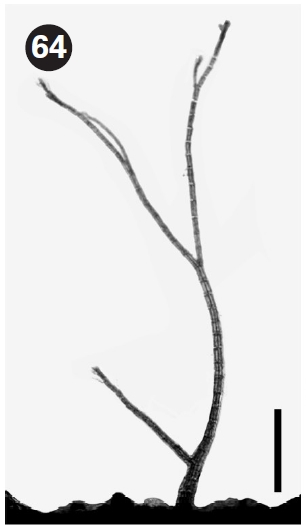
The thallus is tangled, wiry, erect, then coiled, and about 10-15 cm high. Apices are slightly upcurved, flattened hooks. Holdfast is disc-like, becoming more tangled by the coiled apex.
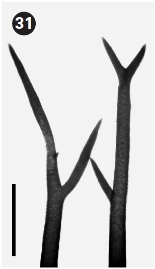
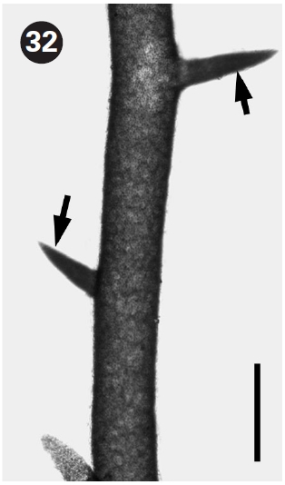
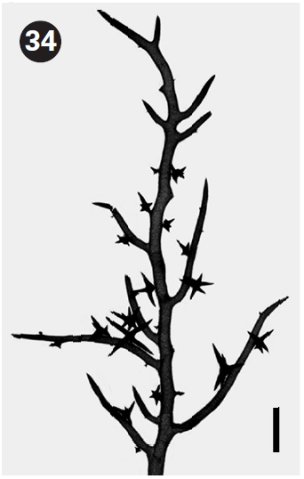
The thallus is wiry, erect, and 5-6 cm. Apices are tapering and pointed, but not upcurved. Branchlets are spine-like and numerous. Holdfast is disc-like.
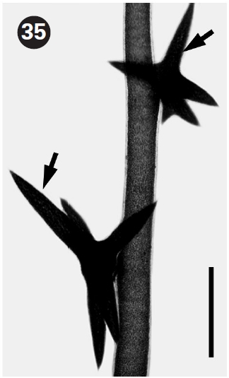
The thallus is tough, wiry, erect, and 7-8 cm high. Apices are tapering and pointed, but not upcurved. Branchlets are spine-like and star-shaped with up to six points. Holdfast is disc-like.
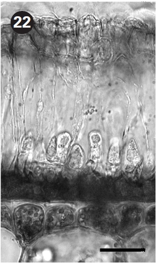
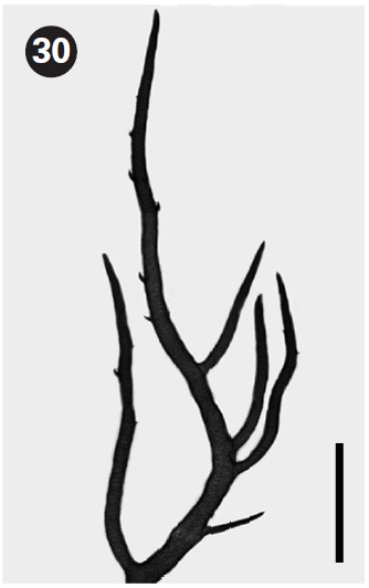
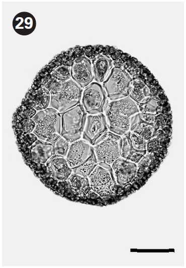
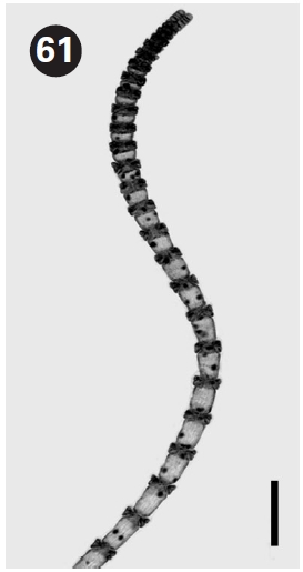
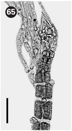
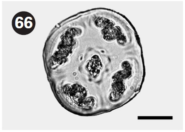
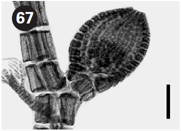
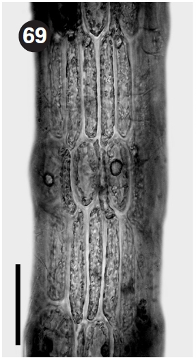
The thallus is gelatinous, alternately branching, erect, and about 3-5 cm high. Branches are cylindrical to slightly flattened. Apices are bluntly pointed. Segments are swollen or barrel-shaped. The inner wall is lined with faint longitudinal filaments with sparsely scattered and oval gland cells. Spermatangia are in swollen spermatangial sori and produced from cortical cells. Cystocarps are protuberant with wide ostioles. Tetrasporangia are spherical, tetrahedrally divided, and produced on the inner side of cortical cell.
The thallus is monosiphonous, dichotomous, erect, and 1 cm high. Sterile filaments are whorled at upper ends of segments and trichotomously branched.
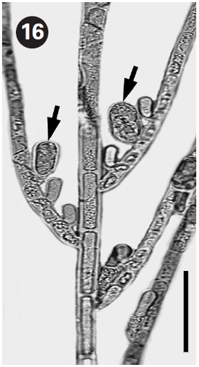
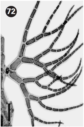
The thallus is filamentous, dichotomous, creeping and erect, and 2-4 cm high. Apices are incurved. The cortex is complete and has whorled spines. Spermatangia are in the terminal clusters of the node. Tetrasporangia are spherical, produced from periaxial cells, and protected by involucral branchlets. Recently, Won et al. (2009) resurrected this species based on morphological and molecular evidence.
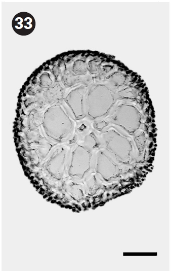
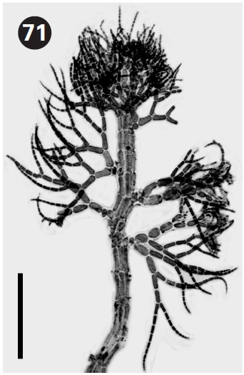
The thallus consists of prostrate axes giving rise to erect axes, and is 0.2-0.3 cm high. The axis has four periaxial cells. Three cortical initials are produced per periaxial cell. Of them, basipetal cortical cells are produced horizontally and grow basipetally. This species is similar to
The thallus is simple, filamentous, pseudo-dichotomous, creeping and erect, and 0.5 cm high. Cortication is incomplete. Two cortical cells are acropetally produced from a peraxial cell.
The thallus is tangled, prostrate, creeping and erect, and 0.5 cm high. Branching is irregularly alternate. Rhizoids arise from each node.
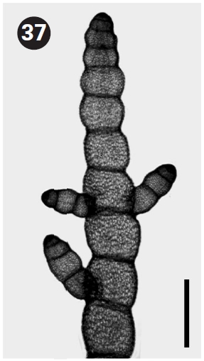
The thallus is filamentous, erect, and 0.3 cm high. Branching is irregularly alternate with four pericentral cells. Scar cells are common between segments and apical filaments are highly branched. Cystocarps are spherical and on short stalk.
The thallus is filamentous, erect and then coiled, and about 7 cm high. Branchlets are delicate and unbranched, with incomplete cortication.
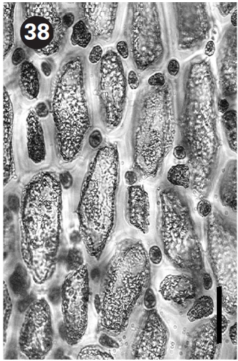
The thallus is delicate, erect, not corticated, and 0.4 cm high. Branchlets are deciduous, and dichotomously to alternately branched. Our material is at a young plant stage.
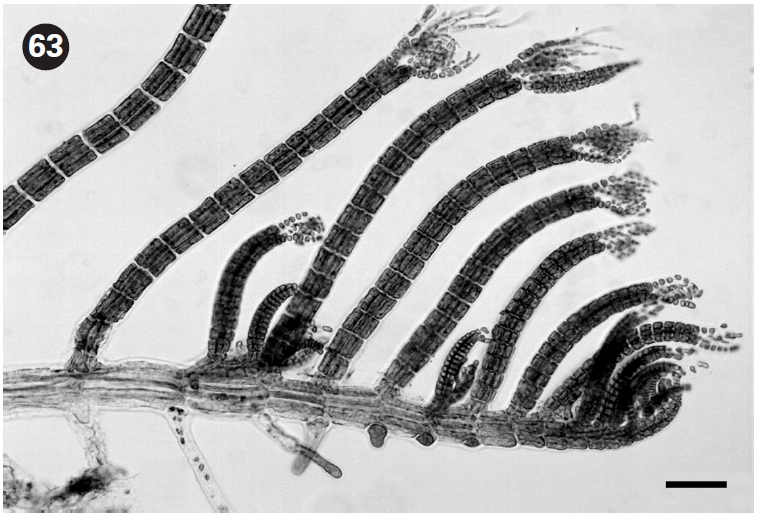
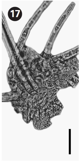
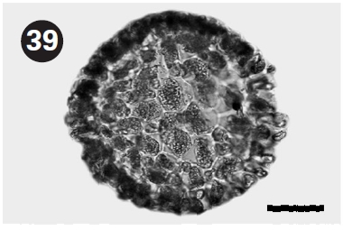
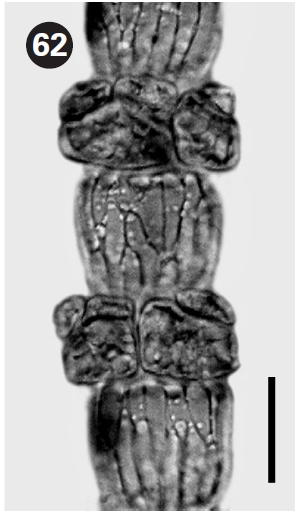
The thallus is solitary, erect, and 0.8-1.2 cm high. There are 5-6 pericentral cells. Apices are truncate to slightly rounded and tufted with dichotomously branched fila-
ments. Tetrasporangia are spherical, tetrahedrally divided, and produced on branchlets. Spermatangial sori are disc-shaped, circular to oval, flat, and form at the base of apical filaments. Cystocarps are on the short stalk and spherical to oval.