검색 전체 메뉴
PDF
맨 위로
OA 학술지
Preparation and pH-Sensitive Release Behavior of Alginate/Activated Carbon Composite Magnetic Hydrogels
  • 비영리 CC BY-NC
ABSTRACT
The alginate-based hydrogel was prepared as a pH-sensitive drug delivery system. To enhance the drug loading capacity,activated carbon was introduced as a drug absorbent. The iron oxide was incorporated into the alginate matrix for the magnetic transferring to the target organ. The activated carbon and iron-oxide were dispersed uniformly in the alginate hydrogel. The drug release from the alginate/activated carbon composite hydrogel was carried out in various pH conditions with vitamin B12 and Lactobacillus lamnosers as model drugs. The fast and sustainable release of drug was observed in the basic condition due to the pH-sensitive solubility of alginate. The novel drug delivery system having pH-sensitive release property and magnetic movement to target place was developed by using the alginate/activated carbon composite magnetic hydrogels.
KEYWORD
pH-sensitive release , Alginate , Activated carbon , Iron oxide , Hydrogel , Microcapsule
  • 1. Introduction

    There is a growing interest in the use of magnetic nanosized particles in various biomedical applications. In particular, iron-oxide nanoparticles have been used as magneticresonance imaging contrast agents for the targeted drug delivery [1-3]. Recently, the fabrication and magnetic characterization of iron-oxide (γ-Fe2O3) nanoparticles precipitated inside the alginate beads have been reported [4].Hydrogels are promising materials used for biomedical applications due to their bio-similarity, aqueous environment,porous structure, and facility of conjugation with various biological macromolecules [5-9]. Alginate is a hydrogelforming polysaccharide consisting of (1-4) linked β-Dmannuronate (M) and its C-5 epimer α-L-guluronate (G) residues [10]. One of the attractive properties of alginate is the versatility of gel formation simply induced by various divalent cations. Alginate hydrogel beads cross-linked by Ca2+ are the most typical one and have been widely utilized in tissue engineering and drug delivery [11-14]. The other attractive property of alginate is the pH-dependent solubility due to the carboxyl groups in the backbone. Therefore, alginate has the properties of shrinking at lower pH and getting dissolved at higher pH [15]. The alginate hydrogel beads could protect the acid-sensitive drug from the gastric fluid and the drug could be delivered safely to the intestine [16-17].

    Activated carbon (AC) is one of the most effective adsorbents for several organic compounds because of their extended surface area, high adsorption capacity, micro porous structure, and specific surface activity [18-19]. Coconut shell-based AC is an effective candidate for drug adsorbent because it has high specific surface area and well-developed pore structure.

    Vitamin B12 (VB12), a model drug, combines with a substance called gastric intrinsic factor (IF). This complex can then be absorbed by the intestinal tract. IF is secreted by the stomach lining and it tightly binds VB12 and helps it pass through the intestinal lining and into the blood. VB12 is needed for normal nerve cell activity, DNA replication, and blood cell production. However, VB12 deficiency can potentially cause severe and irreversible damage, especially to the brain and nervous system. At levels only slightly lower than normal, a range of symptoms such as fatigue, depression,and poor memory may be experienced. Lactobacillus lamnosers also need the protection from the gastric fluid to maximize their survival ability in the intestine.

    The objective of this study is to protect the loaded drugs from the gastric condition and to deliver effectively to the target intestinal area by using the alginate/AC composite beads loaded with Lactobacillus lamnosers and VB12 as drugs. The alginate/AC composite beads were modified to contain the poly(vinyl alcohol) (PVA)/maghemite (γ-Fe2O3) particles to impart the superparamagnetic property for the targeting delivery. The effects of pH on the drug release from alginate/AC composite beads were studied in terms of the pH-dependent release behavior and the survivability of Lactobacillus lamnosers.

    2. Experimental

       2.1. Materials

    AC was purchased from Dong Yang Tanso Co. (Korea). Alginic acid sodium salt (from brown algae, viscosity of 2% solution at 25℃ : ~250 cps), γ-Fe2O3, calcium chloride (CaCl2), PVA, glutaraldehyde (GA), span 80, and n-hexane were obtained from Sigma Chemical Company. VB12, the model drug, was purchased from Samchun Chemical. Lactobacillus lamnosers was obtained from Bio-shield.

       2.2. Preparation of γ -Fe2O3 microcapsules

    The γ-Fe2O3 microcapsules having shell of PVA and core of γ-Fe2O3 were synthesized by emulsion polymerization method. A mixed aqueous solution of PVA (Mw 31,000-50,000, 10 wt%, 20 ml) and γ-Fe2O3 (0.2 g) was slowly added to n-hexane as oil phase (150 ml) containing span 80 as emulsifier (7 ml) with stirring at 1000 rpm for 1 h at 50 to form the water-in-oil emulsion. GA as crosslinking agent (25 wt%, 1 ml) was slowly added to the emulsion with stirring at 1300 rpm for 3 h at 50℃. The γ-Fe2O3 microcapsules were formed and purified by multiple washing with distilled water and petroleum ether, respectively, followed by solvent evaporation under vacuum at 50℃.

       2.3. Preparation of vitamin B12-loaded activated carbon

    AC was pretreated with washing at 100℃ for 2 h using mixed acids of sulfuric (35 wt%) and nitric (20 wt%) acids with weight ratio of 1:1. The treated AC was loaded with VB12 by immersing in the aqueous solution of VB12.

       2.4. Preparation of alginate/AC composite hydrogel magnetic beads

    The microorganism solution was prepared by mixing the aqueous solution of sodium alginate (1.5 wt%) and Lactobacillus lamnosers with volume ratio of 7:3. γ-Fe2O3 microcapsules and AC were introduced in the mixture solution to form the dispersion. Then, 20 ml of this dispersion was added drop wise to 200 ml of calcium chloride solution (0.2 M) by means of a peristaltic pump equipped with Tygon® tubes. As a mixture solution is added to a calcium chloride solution, the gel beads were formed immediately upon crosslinking. The sodium ions of alginate are exchanged with calcium ions and the polymers become crosslinked to form the composite hydrogel beads. The composite hydrogel beads were spherical and black-colored due to the presence of AC. The composite hydrogel beads were washed with distilled water several times to remove the excess calcium ions.

       2.5. Characterization of alginate/AC composite hydrogel magnetic beads

    2.5.1. Morphology

    The morphology of γ-Fe2O3 microcapsules was monitored by the optical microscope (Olympus, CH-2). The surface morphology of alginate/AC composite magnetic beads andγ-Fe2O3 microcapsules was evaluated by JSM-7500F (Jeol, Japan) scanning electron microscopy. As a pretreatment, the composite beads were vacuumed up to 10-3 Pa and sputtered using Pt. γ-Fe2O3 microcapsules were sputtered using Os.

    2.5.2. Magnetic characteristic

    The magnetization curves of the dried alginate/AC composite hydrogel magnetic beads were determined with 2900-02 AGIM vibrating sample magnetomer (PMC Co.).

    2.5.3. Drug release behavior

    The release of VB12 from alginate/AC composite beads was carried out in each of the various buffer media (pH 2, 7, and 10) at 25 ± 1℃. Accurately weighed beads were placed in vials containing 25 ml of the buffer medium and maintained in a thermostat bath. At each predetermined time interval, 0.5 ml of buffer was collected from the release medium. The released amounts of VB12 were measured by UV/VIS spectrophotometer (Optizen 2120 UV, Mecasus, Korea) at 361 nm. The cumulative release was calculated according to the following equation:

    Cumulative amount released (%) = (Mt/M)× 100

    where Mt is the amount of drug released from the composite beads at time t and M is the amount of VB12 pre-loaded in AC. All the release experiments were carried out three times.

    2.5.4. Survivability of Lactobacillus lamnosers

    Survivability of Lactobacillus lamnosers in the composite beads was evaluated by culturing in the customized agar medium. The survival of Lactobacillus lamnosers was studied under different pH conditions simulating the gastrointestinal environment. The dried composite beads were introduced to the falcon tube containing 20 ml of the simulated gastric fluid (pH 2) at 25 ± 1℃ of shaking incubator for 30 min. And 1.0 ml aliquot was extracted from the pH 2 fluid. After then, the composite beads were moved to the other falcon tube containing 20 ml of the simulated intestinal fluid (pH 7) at 25 ± 1℃ of shaking incubator. At intervals of 30, 60, 90, 120, and 150 min, 1.0 ml aliquot was extracted from the pH 7 fluid. The extracts were diluted with pure water and 1 ml was plated on the agar medium. The inoculated agar plates were incubated at 35 ± 1℃ for 72 h.

    3. Results and Discussion

       3.1. Morphology of γ -Fe2O3 microcapsules

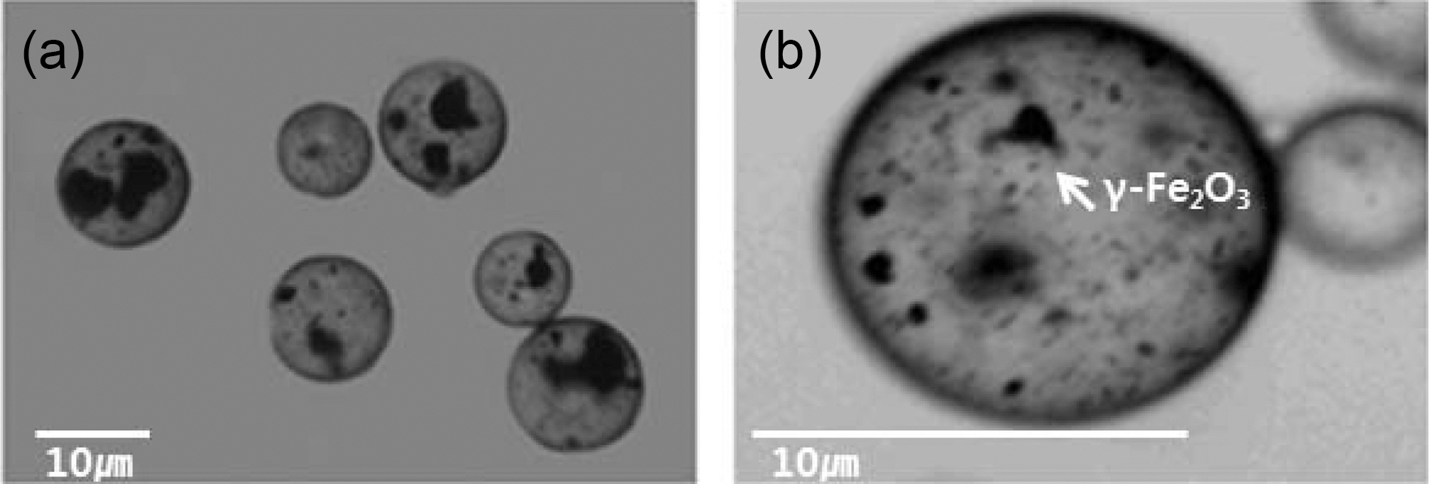
    The optical microscope images of the γ-Fe2O3 microcapsules are presented in Fig. 1. Within each microcapsule, the black-colored γ-Fe2O3 particles are uniformly embedded in the microcapsules although some agglomeration was observed. Fig.

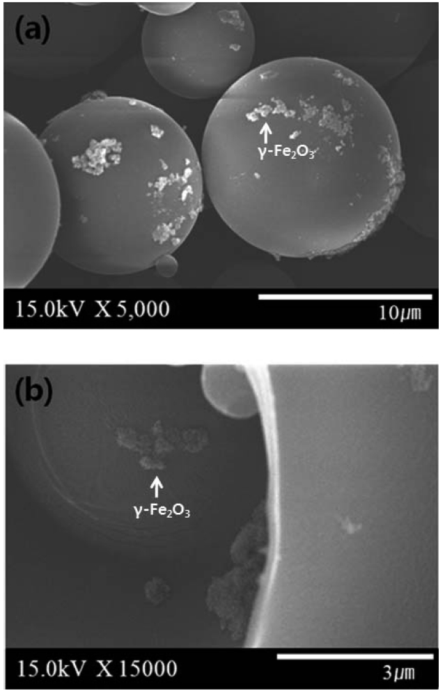
    2 shows the SEM microphotographs of γ-Fe2O3 microcapsules. γ-Fe2O3 microcapsules were spherical with a their mean diameter of about 10 ㎛. The fractured microcapsules showed the γ-Fe2O3 contained in the core of microcapsules.

       3.2. Magnetic characteristics of alginate/AC composite hydrogel magnetic beads

    The composite hydrogel beads were spherical and blackcolored due to the presence of AC and γ-Fe2O3 inside the alginate matrix. AC and γ-Fe2O3 microcapsules were dispersed well in the composite hydrogel beads. The beads had the wet size of 3.00 mm and the dry size of 1.70 mm. Fig. 3 showed the magnetic properties of composite hydrogel beads. The saturation magnetization of composite hydrogel beads reached to 0.01 emu and the ferromagnetic nature of composite hydrogel

    beads. The saturation magnetization of composite hydrogel beads reached to 0.01 emu and the ferromagnetic nature of composite hydrogel beads was seen from the hysteresis curve.The ferromagnetic properties are known useful in magnetic resonance imaging and therapeutic agent because of their inherent biocompatibility and relative stability of chemical and magnetic characteristics in ambient conditions [20]. The alginate/AC composite hydrogel magnetic beads in aqueous fluid were easily attracted under an external magnetic field as shown in Fig. 4. The composite beads exhibited a clear magnetic response because γ-Fe2O3 particles were immobilized uniformly in the composite beads. The composite hydrogel beads could be easily moved in the aqueous phase with an external magnet.

       3.3. pH-sensitive release behavior of alginate/AC composite hydrogel magnetic beads in gastrointestinal tract conditions

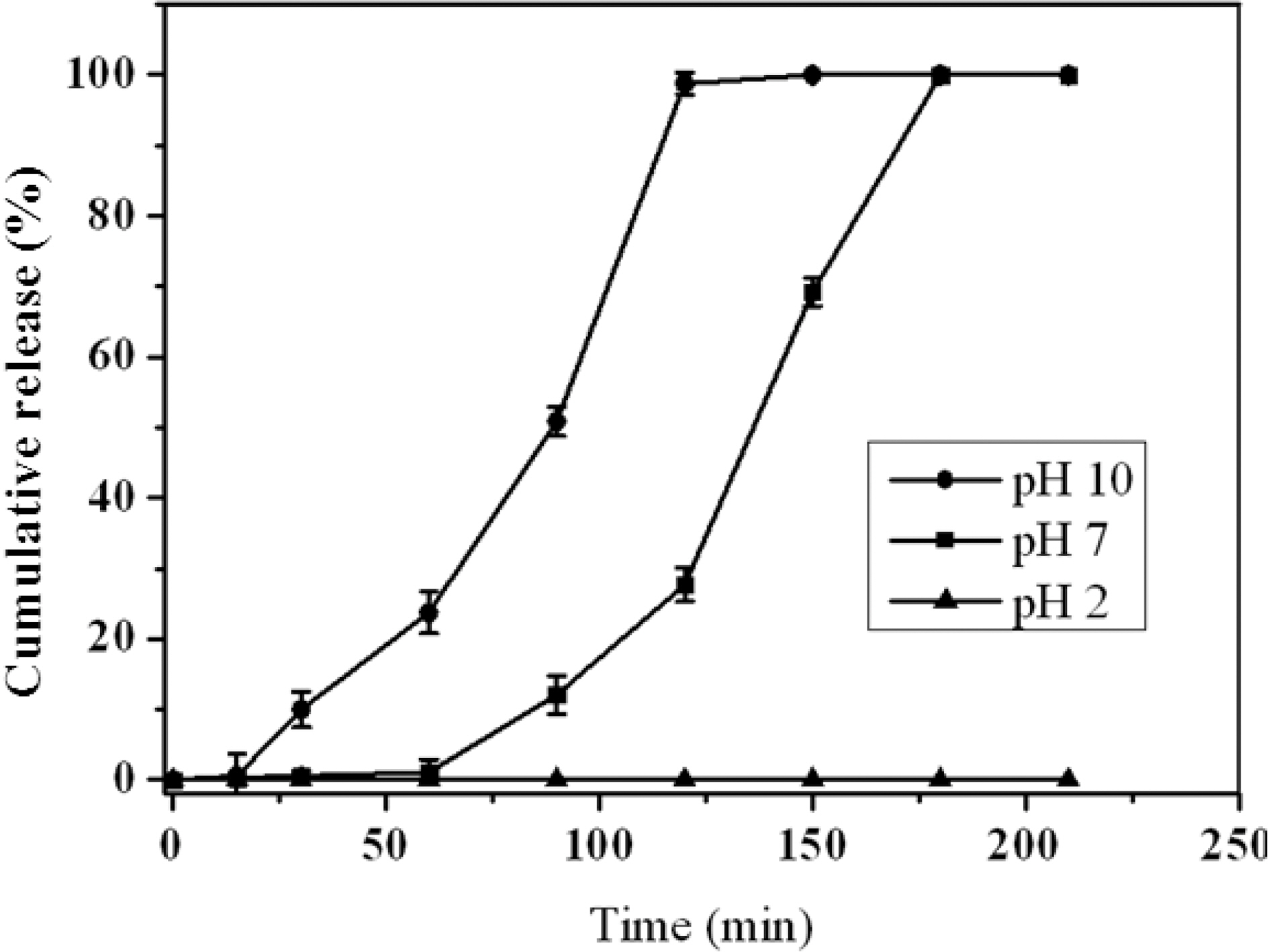
    The release behavior was investigated for the alginate/AC composite beads containing VB12 in several different buffer media at room temperature. The composite beads containing VB12 were placed in the buffer media from pH 2 to 10 and the released amounts were measured depending on time. The pH-sensitive release behavior of the alginate/AC composite beads is well presented in Fig. 5. The released amount of drug from the composite beads increased as the pH increased into the basic condition due to the selective dissolution of alginate matrix. The alginate/AC composite beads could control the release of VB12 successfully depending on the degree of selective dissolution of alginate matrix by changing the pH of the release media. Therefore, the alginate/AC composite beads were able to protect the loaded drug effectively from the acidic gastric condition.

       3.4. Survivability of Lactobacillus lamnosers loaded in alginate/AC composite hydrogel magnetic beads

    The effects of pH on the survivability and the protection of Lactobacillus lamnosers loaded in the alginate/AC composite hydrogel beads were studied by changing the pH of releasing medium from 2 to 7. Fig. 6 shows the colony formation on agar plate inoculated with the releasing medium containing the alginate/AC composite hydrogel beads under simulated gastrointestinal tract conditions. There was no colony formed on agar plates under the acidic condition because the alginate matrix protected the loaded Lactobacillus lamnosers completely. On the other hand, many colonies of Lactobacillus lamnosers were grown successfully under the basic condition because the intact Lactobacillus lamnosers could be released from the composite beads to the medium. The number of colonies increased as the release time increased in the basic condition because more amounts of alginate were dissolved in the basic condition. These survival results showed the protection from the acidic gastric condition by encapsulation and the proper delivery to the basic intestinal condition.

    4. Conclusions

    The alginate/AC composite hydrogel beads containing γ-Fe2O3 microcapsules were prepared as a pH-sensitive drug delivery system to protect and deliver the loaded drug effectively. The AC and γ-Fe2O3 clusters were uniformly dispersed in the alginate hydrogel matrix. The alginate/AC composite hydrogel beads could be easy moved to the target by using external magnet due to the γ-Fe2O3 clusters in alginate matrix. VB12 was released from the composite beads with sustained and pH-sensitive manner. The drug was released fast in the basic condition based on the pH-sensitive solubility of alginate hydrogel. The Lactobacillus lamnosers could be protected effectively from the acidic gastric condition by encapsulation with the alginate/AC composite hydrogel and the Lactobacillus lamnosers could be delivered intact to the basic intestinal condition. The novel drug delivery system having pH-sensitive property, sustained release, and easy magnetic movement to target place was obtained by using alginate/AC composite hydrogel beads.
참고문헌
  • 1. Lacava L. M, Lacava Z. G. M, Da Silva M. F, Silva O, Chaves S. B, Azevedo R. B, Pelegrini F, Gansau C, Buske N, Sabolovic D, Morais P. C 2001 [Biophys. J.] Vol.80 P.2483
  • 2. Babes L, Denzoit B, Tonguy G, Jueune J. J, Jalet P 1999 [J. Colloid Interface Sci.] Vol.212 P.474
  • 3. Berry C. C, Wells S, Charles S, Curtis A. S. G 2003 [Biomaterials] Vol.24 P.4551
  • 4. Naik R, Senaratne U, Powell N, Buc E. C, Tsoi G. M, Naik V. M, Vaishnava P. P, Wenger L. E 2005 [J. Appl. Phys.] Vol.97 P.10J313
  • 5. Tabata Y, Ikada Y 1998 [Adv. Drug Deliver. Rev.] Vol.31 P.287
  • 6. Hoffman A.S 2002 [Adv. Drug Deliver. Rev.] Vol.354 P.3
  • 7. Omidian H, Rocca J. G, Park K 2005 [J. Control. Release] Vol.102 P.3
  • 8. Neves J, Bahia M. F 2006 [Int. J. Pharm.] Vol.318 P.1
  • 9. Wang C. Y, Liu H. X, Gao Q. X, Liu X. X, Tong Z 2007 [Chem. Phys. Chem.] Vol.8 P.1157
  • 10. Liew C. V, Chan L. W, Ching A. L, Heng P. W. S 2006 [Int. J. Pharm.] Vol.309 P.25
  • 11. Chretien C, Chaumeil J. C 2005 [Int. J. Pharm.] Vol.304 P.18
  • 12. Pasparakis G, Bouropoulos G 2006 [N. Int. J. Pharm.] Vol.323 P.34
  • 13. Sriamornsak P, Kennedy R. A 2006 [Int. J. Pharm.] Vol.323 P.72
  • 14. Pongjanyakul T, Puttipipatkhachorn S 2007 [Int. J. Pharm.] Vol.331 P.61
  • 15. George M, Abraham T. E 2006 [J. Control. Release] Vol.114 P.1
  • 16. Hwang S. J, Rhee G. J, Lee K. M, Oh K. H, Kim C. K 1995 [K. Int. J. Pharm.] Vol.116 P.125
  • 17. Anson M, Marchese J, Garis E, Ochoa N, Pagliero C 2004 [Membrane Sci.] Vol.243 P.19
  • 18. Fernandez-Hervas M. J, Holgado M. A, Fini A, Fell J. T 1998 [Int. J. Pharm.] Vol.163 P.23
  • 19. Ballinas L, Torras C, Fierro V, Garcia-Valls R 2004 [J. Phy.Chem. Solids] Vol.65 P.633
  • 20. Choi J. H, Lee S, Kang H. J, Lee J. Y, Kim J, Yoo H. S, Stratton T. R, Applegate B. M, Youngblood J. P, Kim H. J, Ryu K. N 2010 [Macromolecular Research] Vol.18 P.504
이미지 / 테이블
  • [ Fig. 1. ]  Optical microscope images of (a) γ-Fe2O3 microcapsules and (b) γ-Fe2O3 microcapsules with higher magnification.
    Optical microscope images of (a) γ-Fe2O3 microcapsules and (b) γ-Fe2O3 microcapsules with higher magnification.
  • [ Fig. 2. ]  SEM microphotographs of the γ-Fe2O3 microcapsules; (a) external structure and (b) internal structure.
    SEM microphotographs of the γ-Fe2O3 microcapsules; (a) external structure and (b) internal structure.
  • [ Fig. 3. ]  Magnetic hysteresis curve of alginate/AC composite hydrogel magnetic beads.
    Magnetic hysteresis curve of alginate/AC composite hydrogel magnetic beads.
  • [ Fig. 4. ]  Photographs of (a) the alginate/AC composite hydrogel magnetic beads and (b) the alginate/AC composite hydrogel magnetic beads attracted by an external magnet.
    Photographs of (a) the alginate/AC composite hydrogel magnetic beads and (b) the alginate/AC composite hydrogel magnetic beads attracted by an external magnet.
  • [ Fig. 5. ]  pH-sensitive release behavior of the alginate/AC composite hydrogel magnetic beads containing VB12 at several different pHs.
    pH-sensitive release behavior of the alginate/AC composite hydrogel magnetic beads containing VB12 at several different pHs.
  • [ Fig. 6. ]  Colony formation of Lactobacillus lamnosers protected by alginate/AC composite hydrogel magnetic beads after staying for (a)30 min at pH 2 (b) 30 min at pH 7 (c) 60 min at pH 7 (d) 90 min at pH 7 (e) 120 min at pH 7 and (f) 150 min at pH 7.
    Colony formation of Lactobacillus lamnosers protected by alginate/AC composite hydrogel magnetic beads after staying for (a)30 min at pH 2 (b) 30 min at pH 7 (c) 60 min at pH 7 (d) 90 min at pH 7 (e) 120 min at pH 7 and (f) 150 min at pH 7.
(우)06579 서울시 서초구 반포대로 201(반포동)
Tel. 02-537-6389 | Fax. 02-590-0571 | 문의 : oak2014@korea.kr
Copyright(c) National Library of Korea. All rights reserved.