검색 전체 메뉴
PDF
맨 위로
OA 학술지
Geno- and Ecotoxicity Evaluation of Silver Nanoparticles in Freshwater Crustacean Daphnia magna
  • 비영리 CC BY-NC
  • 비영리 CC BY-NC
ABSTRACT
Geno- and Ecotoxicity Evaluation of Silver Nanoparticles in Freshwater Crustacean Daphnia magna
KEYWORD
Daphnia magna , DNA damage , Ecotoxicity , Genotoxicity , Silver nanoparticles
  • 1. Introduction

    Silver nanoparticles (AgNPs) have a wide range of current and potential future applications, including spectrally selective coatings for solar energy absorption, chemical catalysts, surface-enhanced Raman scattering for imaging and; in particular, antimicrobial sterilization, which has made them one of the most commonly used nanomaterials[1-5]. Widely used NPs, such as AgNPs, will most likely enter the environment, and may produce a physiological response in certain organisms, possibly altering their fitness, and might ultimately change their populations or community densities. Research and literature regarding the ecotoxicity of NPs is still emerging, and gaps still exist in our knowledge of this area.

    Despite the dramatic increase in the use of such nanomaterials, little information is available on their potential harmful effects on the environment. Most current literature on the toxicity of nanoparticles; however, comes from mammalian studies that have focused on respiratory exposure, or from in vitro assays using mammalian cells[6-11]. Ecotoxicological studies on nanoparticles are more limited, with only a few reports on the acute toxic effects of nanoparticles on aquatic organisms[12-16]. Few ecotoxicity studies on aquatic organisms have been performed that include genotoxic endpoints. However, the presence of genotoxic and potentially carcinogenic compounds in aquatic environments is of major concern with respect to the health of aquatic media biota[17-19]. The potential genotoxic effects of emerging nanomaterials, such as AgNPs, on aquatic systems should be identified to allow for their safe use.

    Genotoxic assessments of AgNPs were conducted on aquatic sentinel species, the freshwater crustacean Daphnia magna. The small-sized freshwater crustacean, D. magna, holds an important position in the aquatic food chain, respond to many pollutants, are easy to culture and have short life cycles; thus, are considered suitable species for aquatic biomonitoring[20, 21]. Conventional ecotoxicity tests were also conducted on the Daphnia systems, as they may provide insights to the potential toxic effects of AgNPs on aquatic environments. Given the importance of D. magna in aquatic ecosystems, information concerning the geno- and ecotoxicity of widely used nanomaterials on these species could be valuable in relation to aquatic nanoecotoxicology. To compare the toxicity of AgNPs to that of Ag ions, the toxicity of Ag ions was also investigated in D. magna using the same toxic endpoints as used in the AgNPs toxicity assay.

    2. Experimental Methods

       2.1. Organism Culture, AgNPs, and Ag Ion Preparation and Exposure to D. magna

    Using an original strain provided by the Korea Institute of Toxicology (Daejeon, Korea), D. magna were obtained in our laboratory from adults reared using M4-media, as described previously[22, 23]. AgNPs (size <100 nm, Sigma-Aldrich Chemical, St. Louis, MO, USA) were homogenously dispersed in deionized water by sonication for 13 hours (Branson-5210 sonicator, Branson Inc., Danbury, CT, USA), stirring for 7 days, and filtering through a cellulose membrane (pore size 100 nm, Advantec, Toyo Toshi Kaisha, Tokyo, Japan) to remove NP aggregations. To compare the toxicities of AgNPs and Ag ions, aqueous AgNO3 (AG002, Next Chimica, Centurion, Republic of South Africa), in deionized water, was used, with the final concentrations of AgNPs and AgNO3 estimated using a Multitype Inductively Coupled Plasma Emission Spectrometer (ICPE-9000, Shimadzu, Tokyo, Japan). The concentrations for AgNPs and AgNO3 had equivalent Ag masses. From stock solutions (4 mg/L), experimental concentrations of AgNPs and AgNO3 were prepared in M4-media. Neonates, aged less than 24 hours, were used for Comet assays and ecotoxicity tests.

       2.2. Characterization of AgNPs

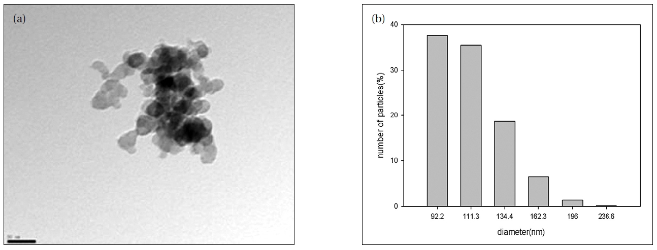
    Energy filtering transmission electron microscopy (TEM) was used to examine the particle shape and size of the AgNPs. Twenty μL of the particle suspension were dried onto a 400 mesh carboncoated copper grid and imaged with a LIBRA 120 TEM (Carl Zeiss, Oberkochen, Baden-W rttemberg, Germany) at 80-120 kV. The size distribution of the AgNPs was evaluated using a photal dynamic light scattering (DLS) spectrometer, DLS-7000 (Otsuka Electronics Co., Inc., Osaka, Japan).

       2.3. Mortality, Growth, Reproduction Assays

    For the mortality test, 10 individuals, less than 24 hours, were exposed to AgNPs and Ag ions for 24 hours, with live and dead individuals then counted[23]. For the growth test, 20 individuals, less than 24 hours, were incubated with AgNPs and Ag ions for 96 hours, with the fresh weights measured immediately after exposure. The body dry weight was evaluated after drying Daphnia at 105℃ for 24 hours. Daphnia reproduction tests were conducted according to the OECD guidelines[24]. Ten individuals, less than 24 hours, were exposed to various concentrations of the test chemicals, and then observed and fed daily for 21 days. Three replicates were prepared for each concentration, with jars filled with 100 mL of test media. Each jar was provided with Chlorella as the food source, at a concentration of 5×10⁵ cells/mL daily. Test animals were transferred to new medium every 2 days. Neonates were removed from the jar daily, and the numbers of neonates counted.

       2.4. Comet Assay

    To prepare Daphnia, a total of 150 neonates were collected from the control and experimental tanks after 24 hours exposure to nanoparticles, and pooled for the Comet assay. Organisms were placed in 1 mL of phosphate-buffered saline (PBS), containing 20 mM ethylene diamine tetra acetic acid (EDTA) and 10% dimethyl sulfoxide (DMSO), and disintegrated mechanically by mincing. An alkaline comet assay was performed based on the method of Singh et al.[25], with adaptation for Daphnia, as described previously[22]. Briefly, about 50 cells per slide (3 slides per treatment) were analyzed using a fluorescence microscope (Nikon, Kanagawa, Japan), equipped with an excitation filter with a BP 546/12 nm and 590 nm barrier filter at 400×magnification. DNA damage was expressed as the olive tail moment (OTM) using an image analysis computerized method (Komet 5.5, Kinetic Imaging Limited, Nottingham, UK).

       2.5. Data Analysis

    The genotoxic- and ecotoxic assays results were tested for significance using an analysis of variance (ANOVA) test, employing the Dunnett's multiple comparison test. All statistical tests were performed using SPSS version 12.0 (SPSS Inc., Chicago, IL, USA).

    3. Results and Discussion

    The AgNPs used for the toxicity assays were characterized using TEM and DLS methods (Fig. 1). The TEM provided information on the size and shape of the nanoparticles, and showed sizes mainly <50 nm (Fig. 1a); however, it could not provide information on whether the nanoparticles existed in single or aggregated forms in the test medium, as the nanoparticles form aggregates when dried on the microscopic observation slide. The results of the DLS suggested that the AgNPs did not exist as single particles, but tended to aggregate in the test medium, as the main nanoparticle sizes distributed in the test medium were about 100 nm (Fig. 1b). In relation to nanotoxicity, it is often expected that the smaller the size, the stronger the toxicity exerted[26]. However, the relationship between the physico-chemical properties of nanoparticle and their toxicities seems to be much more complicated than just being related to their size and surface area (i.e. shape, charge, concentration, etc.); there is still much on going debate[8, 27, 28]. Many studies have failed to show any clear relationship between toxicity and the size of nanoparticles [8, 29]. The TEM images of the nanoparticles from the test medium showed the size of the nanoparticles tested. However, the line of evidence provided from the present study is rather limited; therefore, to identify key properties of nanoparticles with respect to causing ecotoxicity, toxic responses of a broad range of physico-chemical properties to various classes of nanoparticles may be investigated in various environmental relevant species.

    [Table 1.] Acute toxicity of silver nano particles estimated in Daphnia magna

    label

    Acute toxicity of silver nano particles estimated in Daphnia magna

    To find the range of sublethal concentrations for geno-and ecotoxicity tests, an acute toxicity test was performed on D. magna exposed to AgNPs, using mortality as an endpoint (Table 1). A broad range of AgNPs concentrations was tested (data not shown from 1 to 4 mg/L). Beyond exposure to 2 μg/L of AgNPs, complete mortality was observed. A steep concentrationresponse relationship was observed (i.e. 0% mortality at 1 μg/L and 100% mortality at 2 μg/L of AgNPs exposure). Based on the acute toxicity test, 0.5, 1, and 1.5 μg/L of AgNPs were selected as concentrations for geno- and ecotoxicity tests. To compare the toxicity of AgNPs to that of Ag ions, the toxicity of Ag ions was also investigated in D. magna using the same concentrations and toxic endpoints as used in the AgNPs toxicity assay.

    DNA damage, particularly DNA strand breaks, was measured using the Comet assay to evaluate whether AgNPs induced any genetic toxicity in D. magna (Table 2). AgNPs and Ag ions may exert genotoxic effects on D. magna, given that DNA strand breaks (OTMs) increased in D. magna exposed to AgNPs and Ag ions. A statistically significant increase in OTMs was observed in D. magna exposed to 1 and 1.5 μg/L of AgNPs and Ag ions. However, the degree of increase in OTM was more consequential in AgNPs exposed D. magna than in those exposed to Ag ions. Moreover, exposure concentration dependant DNA damage was observed in AgNPs exposed D. magna. Even though genotoxicity tests with the Comet assay are widely used in aquatic environmental monitoring, most Comet assays have been performed on in vitro systems of aquatic species, mostly using fish-driven cell lines[19, 30]. The measurement of genotoxic effects of emerging nanomaterials, using in vivo genotoxicity biomarker in aquatic invertebrates, could be a useful tool for monitoring aquatic toxicity due to nanoparticles. AgNPs may influence the genetic constitution of populations by directly damaging DNA molecules within the individual cell nucleus, but the ecological relevance of changes in single cells within some tissues of certain individual organisms is extremely difficult to assess.

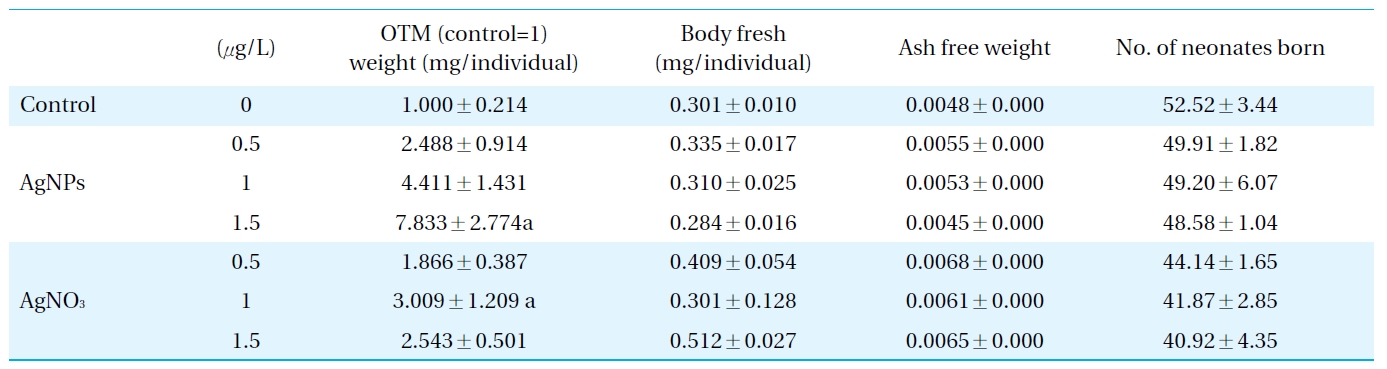
    Therefore, conventional ecotoxicity tests, using mortality, growth and reproduction as endpoints, were subsequently conducted to validate the ecotoxicological relevance of the response of DNA to damage in D. magna exposed to AgNPs and Ag ions (Table 2). The response of Daphnia to AgNPs in terms of their mortality, growth and reproduction may explain the higher biological-level consequences of the observed DNA damage. Aquatic toxicity tests may provide insights to the relative sensitivity of D. magna to AgNPs, which may also provide information on the impact of nanoparticles on water systems, as these species hold important positions in aquatic ecosystems[24, 31-33]. A significant increase in mortality was observed in D. magna exposed to 1.5 ㎕/L of AgNPs; whereas, no significant alteration was observed in growth and reproduction. It seemed Ag ion exposure leads to a slight increase in mortality, but decrease in reproduction potential; however, those alterations were not statistically significant. An increase in DNA strand breaks occurred concomitantly with an increase in mortality in D. magna exposed to 1.5 ㎕/L of AgNPs, which suggests DNA alteration induced by AgNPs might provoke higher level consequences. As mortality is the most obvious sign of progression of serious toxicity at the organism level, the impairment of survival due to AgNPs exposure may be considered a consequence of a serious progression of sub-organism level toxicities, such as the increased DNA damage in Daphnia. The relationships between the responses of the genotoxic biomarker and the physiological /individual/population effects are complicated due to the compensatory mechanisms regulating the physiological/ individual fitness and population dynamics in a natural system. As the mere presence of genotoxic compounds, which are potentially carcinogenic, is of major concern in human and ecosystem health, the sensitive and rapid detection of the genotoxic properties of aquatic systems themselves is considered important, although does not necessarily include alteration at a higher level of biological organization. Especially for the nanomaterials concerned, despite the dramatic increase in the use of nanomaterials and; hence, their ubiquitous distribution in aquatic environments, little information is available on their potential genotoxicity on aquatic organisms. Considering the potential of D. magna as a bioindicator species, and the importance of the genotoxicity of nanoparticles in ecotoxicity monitoring, the measurement of the DNA damage in these species after exposure to nanoparticles could provide useful information for freshwater monitoring. There have been discussions regarding the comparative toxicity of AgNPs and Ag ions[34, 35], with the latter’s bactericidal action having been studied previously[36, 37]. Our previous ecotoxicity study using Caenorhabditis elegans, comparing the toxicity of AgNPs and Ag ions, suggested that AgNPs were slightly more toxic than Ag ions in terms of their effect on reproduction potential, and it also appeared that different mechanisms exerted the toxicity of AgNPs and with Ag ions[38]. Results of the geno- and ecotoxicities (Table 2) in D. magna exposed to AgNPs and Ag ions also suggest that AgNPs are slightly more toxic than Ag ions. However, as it appeared that the biocidal effects of AgNPs might be partially due to Ag ion generation, further studies on this aspect of toxicity are required.

    [Table 2.] Growth, reproduction parameters investigated in Daphnia magna exposed to silver nanoparticles and AgNO3, and DNA damage (as OTM obtained by comet assay) measured in D. magna exposed to silver nanoparticles and AgNO3

    label

    Growth, reproduction parameters investigated in Daphnia magna exposed to silver nanoparticles and AgNO3, and DNA damage (as OTM obtained by comet assay) measured in D. magna exposed to silver nanoparticles and AgNO3

    In this study, the geno- and ecotoxicities of AgNPs on D. magnawere evaluated. The results suggested that AgNPs may havegenotoxic potential toward Daphnia, and AgNPs-induced DNAdamage might provoke higher-level consequences, which couldcomprise a contribution to the knowledge on the aquatic toxicityof AgNPs on aquatic ecosystems, for which little data areavailable. However, further studies on the mechanism behindAgNPs-induced DNA damage and mortality are needed to betterexplain the ecotoxicity of AgNPs in D. magna.

    Acknowledgements

    This research was supported by the International Research & Development Program of the National Research Foundation of Korea(NRF) funded by the Ministry of Education, Science and Technology(MEST) of Korea(Grant number: K20912000002-09B1300-00210) and the Korean Ministry of Environment through the Ecotechnopia 21 project

참고문헌
  • 1. Rand BP, Peumans P, Forrest SP 2004 Long-range absorption enhancement in organic tandem thin-film solar cells containing silver nanoclusters [J Appl Phys] Vol.96 P.7519-7526 google cross ref
  • 2. Zhai HJ, Sun DW, Wang HS 2006 Catalytic properties of silica/silver nanocomposites [J Nanosci Nanotechnol] Vol.6 P.1968-1972 google cross ref
  • 3. Yamamoto S, Watarai H 2006 Surface-enhanced Raman spectroscopy of dodecanethiol-bound silver nanoparticles at the liquid/liquid interface [Langmuir] Vol.22 P.6562-6569 google cross ref
  • 4. Pal S, Tak YK, Song JM 2007 Does the antibacterial activity of silver nanoparticles depend on the shape of the nanoparticle? A study of the gram-negative bacterium Escherichia coli [Appl Environ Microbiol] Vol.73 P.1712-1720 google cross ref
  • 5. Maynard A, Michelson E 2010 The Nanotechnology Consumer Product Inventory Project on Emerging Nanotechnology google
  • 6. Lam CW, James JT, McCluskey R, Hunter RL 2004 Pulmonary toxicity of single-wall carbon nanotubes in mice 7 and 90 days after intratracheal instillation [Toxicol Sci] Vol.77 P.126-134 google
  • 7. Braydich-Stolle L, Hussain S, Schlager JJ, Hofmann MC 2005 In vitro cytotoxicity of nanoparticles in mammalian germline stem cells [Toxicol Sci] Vol.88 P.412-419 google cross ref
  • 8. Hussain SM, Hess KL, Gearhart JM, Geiss KT, Schlager JJ 2005 In vitro toxicity of nanoparticles in BRL 3A rat liver cells [Toxicol In Vitro] Vol.19 P.975-983 google cross ref
  • 9. Monteiro-Riviere NA, Nemanich RJ, Inman AO, Wang YYY, Riviere JE 2005 Multi-walled carbon nanotube interactions with human epidermal keratinocytes [Toxicol Lett] Vol.155 P.377-384 google cross ref
  • 10. Limbach LK, Wick P, Manser P, Grass RN, Bruinink A, Stark WJ 2007 Exposure of engineered nanoparticles to human lung epithelial cells: Influence of chemical composition and catalytic activity on oxidative stress [Environ Sci Technol] Vol.41 P.4158-4163 google cross ref
  • 11. Eom HJ, Choi J 2009 Oxidative stress of CeO2 nanoparticles via p38-Nrf-2 signaling pathway in human bronchial epithelial cell [Beas-2B Toxicol Lett] Vol.187 P.77-83 google cross ref
  • 12. Hund-Rinke K, Simon M 2006 Ecotoxic effect of photocatalytic active nanoparticles TiO2 on algae and daphnids [Environ Sci Pollut Res] Vol.13 P.225-232 google cross ref
  • 13. Lovern SB, Klaper R 2006 Daphnia magna mortality when exposed to titanium dioxide and fullerene (C-60) nanoparticles [Environ Toxicol Chem] Vol.25 P.1132-1137 google cross ref
  • 14. Handy RD, Shaw BJ 2007 Ecotoxicity of nanomaterials to fish:challenges for ecotoxicity testing [Integr Environ AssessManag] Vol.3 P.458-460 google
  • 15. Lovern SB, Strickler JR, Klaper R 2007 Behavioral and physiological changes in Daphnia magna when exposed to nanoparticle suspensions (titanium dioxide nano-C-60 and C(60)HxC (70)Hx) [Environ Sci Technol] Vol.41 P.4465-4470 google cross ref
  • 16. Houk VS, Waters MD 1996 Genetic toxicology and risk assessment of complex environmental mixtures [Drug Chem Toxicol] Vol.19 P.187-219 google cross ref
  • 17. Ohe T, Watanabe T, Wakabayashi K 2004 Wakabayashi K Mutagens in surface waters: a review [Mutat Res-Rev Mut Res] Vol.567 P.109-149 google
  • 18. Nehls S, Segner H 2005 Comet assay with the fish cell line rainbow trout gonad-2 for in vitro genotoxicity testing of xenobiotics and surface waters [Environ Toxicol Chem] Vol.24 P.2078-2087 google cross ref
  • 19. Giesy JP, Graney RL, Newsted JL 1988 Comparison of three sediment bioassay methods using detroit river sediments [Environ Toxicol Chem] Vol.7 P.483-498 google cross ref
  • 20. Atienzar FA, Cheung VV, Jha AN, Depledge MH 2001 Fitness parameters and DNA effects are sensitive indicators of copper-induced toxicity in Daphnia magna [Toxicol Sci] Vol.59 P.241-250 google cross ref
  • 21. Park SY, Choi J 2007 Cytotoxicity genotoxicity and ecotoxicity assay using human cell and environmental species for the screening of the risk from pollutant exposure [Environ Int] Vol.3 P.817-822 google
  • 22. Lee SW, Park K, Hong J, Choi J 2008 Ecotoxicological evaluation of octachlorostyrene in fourth instar larvae of Chironomus riparius (Diptera Chironomidae) [Environ Toxicol Chem] Vol.27 P.1118-1127 google cross ref
  • 23. 1984 Effects on biotic systems Daphnia magna acute immobilization test 202 google
  • 24. 1998 Effects on biotic systems Daphnia magna reproduction test 211 google
  • 25. Singh NP, McCoy MT, Tice RR, Schneider EL 1988 A simple technique for quantitation of low-levels of dna damage in individual cells [Exp Cell Res] Vol.175 P.184-191 google cross ref
  • 26. Oberdorster G, Oberdorster E, Oberdorster J 2005 Nanotoxicology: an emerging discipline evolving from studies of ultrafine particles [Environ Health Perspect] Vol.113 P.823-839 google
  • 27. Sayes CM, Wahi R, Kurian PA 2006 Correlating nanoscale titania structure with toxicity: A cytotoxicity and inflammatory response study with human dermal fibroblasts and human lung epithelial cells [Toxicol Sci] Vol.92 P.174-185 google cross ref
  • 28. Fujiwara K, Suematsu H, Kiyomiya E, Aoki M, Sato M, Moritoki N 2008 Size-dependent toxicity of silica nano-particles to Chlorella kessleri [J Environ Sci Health A Tox Hazard Subst Environ Eng] Vol.43 P.1167-1173 google
  • 29. Yin H, Too HP, Chow GM 2005 The effects of particle size and surface coating on the cytotoxicity of nickel ferrite [Biomaterials] Vol.26 P.5818-5826 google cross ref
  • 30. Cotelle S, Ferard JF 1999 Comet assay in genetic ecotoxicology: A review [Environ Mol Mutagen] Vol.34 P.246-255 google cross ref
  • 31. Okamura H, Omori M, Luo R, Aoyama I, Liu D 1999 Application of short-term bioassay guided chemical analysis for water quality of agricultural land run-off [Sci Total Environ] Vol.234 P.223-231 google cross ref
  • 32. Kikuchi M, Sasaki Y, Wakabayashi M 2000 Screening of organophosphate insecticide pollution in water by using Daphnia magna [Ecotoxicol Environ Saf] Vol.47 P.239-245 google cross ref
  • 33. Lee SB, Choi J 2006 Multilevel evaluation of nonylphenol toxicity in fourth-instar larvae of Chironomus riparius (Diptera Chironomidae) [Environ Toxicol Chem] Vol.25 P.3006-3014 google cross ref
  • 34. Ji JH, Jung JH, Kim SS 2007 Twenty-eight-day inhalation toxicity study of silver nanoparticles in Sprague-Dawley rats [Inhal Toxicol] Vol.19 P.857-871 google cross ref
  • 35. Hidalgo E, Dominguez C 1998 Study of cytotoxicity mechanisms of silver nitrate in human dermal fibroblasts [Toxicol Lett] Vol.98 P.169-179 google cross ref
  • 36. Clement JL, Jarrett PS 1994 Antibacterial silver [Met Based Drugs] Vol.1 P.467-482 google cross ref
  • 37. Silver S 1996 Bacterial resistances to toxic metal ions - A review [Gene] Vol.179 P.9-19 google cross ref
  • 38. Roh JY, Sim SJ, Yi J 2009 Ecotoxicity of Silver Nanoparticles on the Soil Nematode Caenorhabditis elegans Using Functional Ecotoxicogenomics [Environ Sci Technol] Vol.43 P.3933-3940 google cross ref
OAK XML 통계
이미지 / 테이블
  • [ Fig. 1. ]  Images of AgNPs in the test media using transmission electron microscopy (a) and dynamic light scattering spectrometers (b).
    Images of AgNPs in the test media using transmission electron microscopy (a) and dynamic light scattering spectrometers (b).
  • [ Table 1. ]  Acute toxicity of silver nano particles estimated in Daphnia magna
    Acute toxicity of silver nano particles estimated in Daphnia magna
  • [ Table 2. ]  Growth, reproduction parameters investigated in Daphnia magna exposed to silver nanoparticles and AgNO3, and DNA damage (as OTM obtained by comet assay) measured in D. magna exposed to silver nanoparticles and AgNO3
    Growth, reproduction parameters investigated in Daphnia magna exposed to silver nanoparticles and AgNO3, and DNA damage (as OTM obtained by comet assay) measured in D. magna exposed to silver nanoparticles and AgNO3
(우)06579 서울시 서초구 반포대로 201(반포동)
Tel. 02-537-6389 | Fax. 02-590-0571 | 문의 : oak2014@korea.kr
Copyright(c) National Library of Korea. All rights reserved.