Echiuran worms (phylum Annelida: class Polychaeta: subclass Echiura) comprise about 165 described species, most of which live in burrows in soft sediments (Goto et al., 2013). This animal group is poorly known in terms of their copepod associates. Currently, only four species of copepods have been found in association with echiurans (Anker et al., 2005).
Echiuran hosts were dug out with a shovel on the tidal flat during low tide. The collected echiuran samples were placed in plastic bags and fixed with ethanol. In the laboratory, echiurans were agitated in water and copepods were sorted out from the resulting residues. Prior to microscopic observation and dissection, copepod specimens were immersed in lactic acid for more than 10 min. Mounting was done following the reversed slide method (Humes and Gooding, 1964). All illustrations were drawn with the aid of a drawing tube mounted on an Olympus BH microscope. The terminology for the caudal ramus setae follows that of Huys and Boxshall (1991). The intact type specimens have been deposited in the National Institute of Biological Resources (NIBR), Incheon, Korea. Descriptions of species were made on the basis of the dissected and figured paratypes. In the species descriptions body length was measured from the anterior apex of the cephalothorax to the posterior margin of the caudal rami, excluding caudal setae. In the formula for the armature of antenna and legs 1-4, Roman numerals indicate spines and Arabic numerals represent setae.
Diagnosis. Body cyclopiform. Prosome flattened, consisting of cephalothorax and 3 metasomites. Urosome 5-segmented in female and 6-segmented in male. Fifth pedigerous somite of female with distinct epimera. Female genital area located ventrally. Dorsal suture between last 2 abdominal somites obscure. Caudal ramus elongate, with 7 setae, including well developed outer proximal seta (seta I). Rostrum lammelliform or not well defined. Antennule 6-segmented; in female, first segment with claw-like anterior process, with identical armature in both sexes. Antenna consisting of coxo-basis and 3-segmented endopod; basis and first endopodal segment each with 1 seta; second endopodal segment with 1 seta and 1 pectinate claw in female, or 1 pectinate claw and 1 large setiform element bearing 1 large and several smaller sucking discs in male; terminal endopodal segment with 2 setae and 4 distally pectinate claws. Labrum with concave posterior margin bearing a pair of linguiform processes. Mandible with 1 thick, spiniform element distally. Maxillule with 3 setae. Female maxilla 2-segmented; syncoxa extended posterodistally forming strong horse-head shaped process; allobasis tipped with 3 small setae. Male maxilla 1-segmented, lammelliform. Female maxilliped represented by sclerotized rudiment. Male maxilliped 4-segmented; terminal segment represented by claw. Legs 1-4 with 3-segmented rami; coxae without inner seta; second endopodal segment with inner seta. Number of setae and spines on third exopodal and endopodal segments of legs 1-4: 7, 8, 6, 6, and 4, 4, 3, 2, respectively. No sexual dimorphism in legs 1-4. Leg 5 consisting of well defined protopod and elongate exopod; exopod with total of 4 setae.
Type species.
Etymology. The generic name
Remarks. The new genus is closely related to
[Table 1.] Differences between the genera Goidelia Embleton and Echiuricopus n. gen.
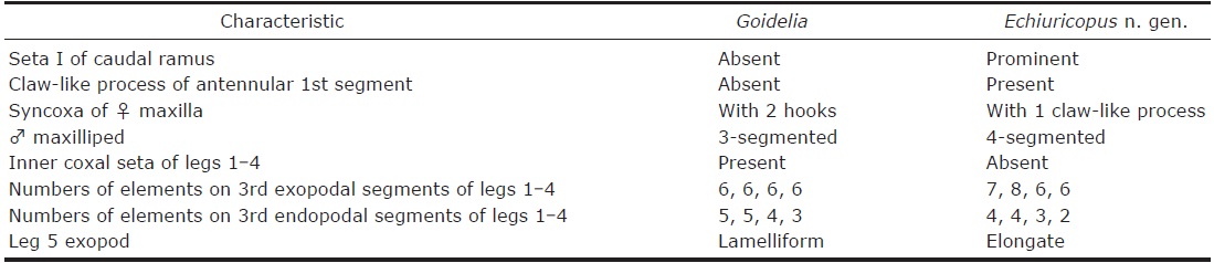
Differences between the genera Goidelia Embleton and Echiuricopus n. gen.
The familial assignment of the genus
>
Echiuricopus aprilis n. sp. (
Material examined. Three ♀♀ and 3♂♂ from an unidentified echiuran, collected from an intertidal mudflat (37°28ʹ 40ʺN, 126°22ʹ46ʺE) on Yeongjong Island off Incheon, Korean coast of the Yellow Sea, on 20 Apr 2015, by Kim IH. Holotype (♀, NIBRIV0000330540), allotype (♂, NIBRIV 0000325915), and paratypes (1♀, 1♂, NIBRIV0000330541) have been deposited in the NIBR, Incheon, Korea. Dissected paratypes (1♀, 1♂) are retained in the collection of the author.
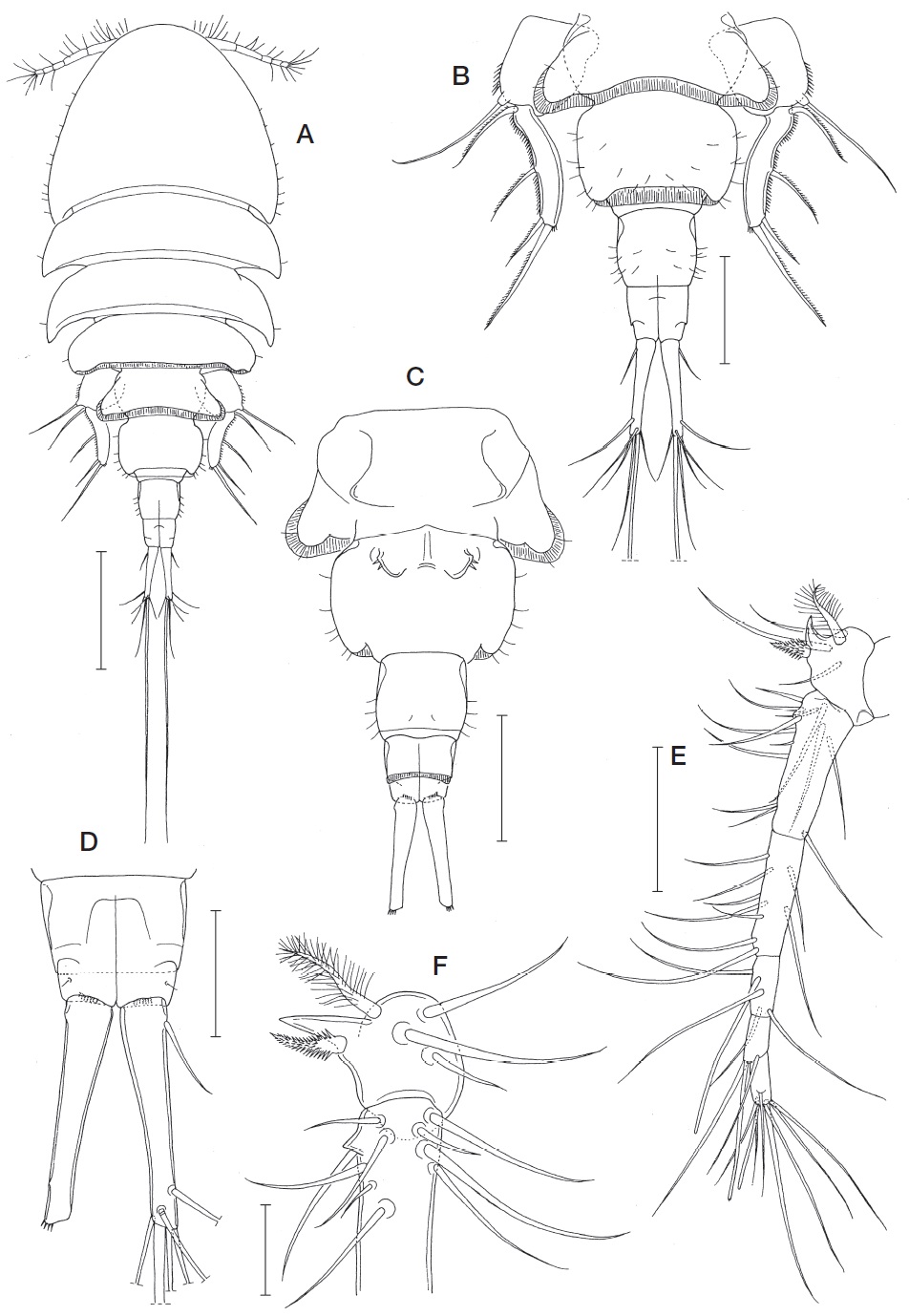
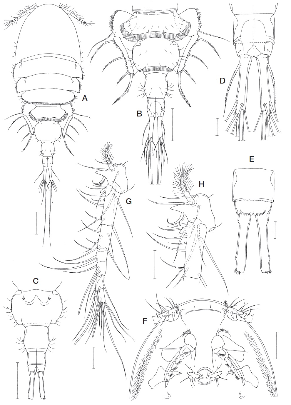
Female. Body (Fig. 1A) with flattened prosome and cylindrical urosome. Body length of dissected and figured specimen 990 μm. Other 2 examined specimens 1,018 and 1,061 μm, respectively. Prosome consisting of cephalothorax and 3 metasomites, 589 μm long. Cephalothorax nearly hemicircular and 287×393 μm, with longitudinal rows of setules and spinules on each side of ventral surface, as in next species. First to third metasomites (second to fourth pedigerous somites) 113×407, 98×389, and 95×324 μm, respectively. Second and third metasomites with posterolaterally projected, tapering epimera. Third metasomite with blunt lateral margins and rimmed with membrane along posterodorsal margin. Urosome (Fig. 1B, C) 5-segmented. First urosomite (fifth pedigerous somite) broad, 78×209 μm, with posterolaterally expanded, tapering epimera, and rimmed with membrane along posterodorsal margin and distal part of lateral margins. Genital double-somite sub-rectangular, 109×148 μm, wider than long, with rounded corners and membrane along posterodorsal margin; genital area located ventrally on somite (Fig. 3C). Three abdominal somites markedly narrower than genital double-somite, 70×75, 34×57, and 16×48 μm, respectively. Dorsal boundary obscure between last 2 abdominal somites (Fig. 1D). Second abdominal somite with membrane along posteroventral margin. Last abdominal somite with row of several spinules near base of caudal ramus. Caudal ramus slender, 91×18 μm (ratio of length to width 5.06 : 1), gradually tapering distally, with 4 spinules on outer distal corner, and armed with 7 naked setae; outer proximal seta (seta I) prominent but not longer than half length of caudal ramus; other 6 setae located distally or subdistally; seta V longest among setae, 436 μm long; seta IV second longest, 105 μm long.
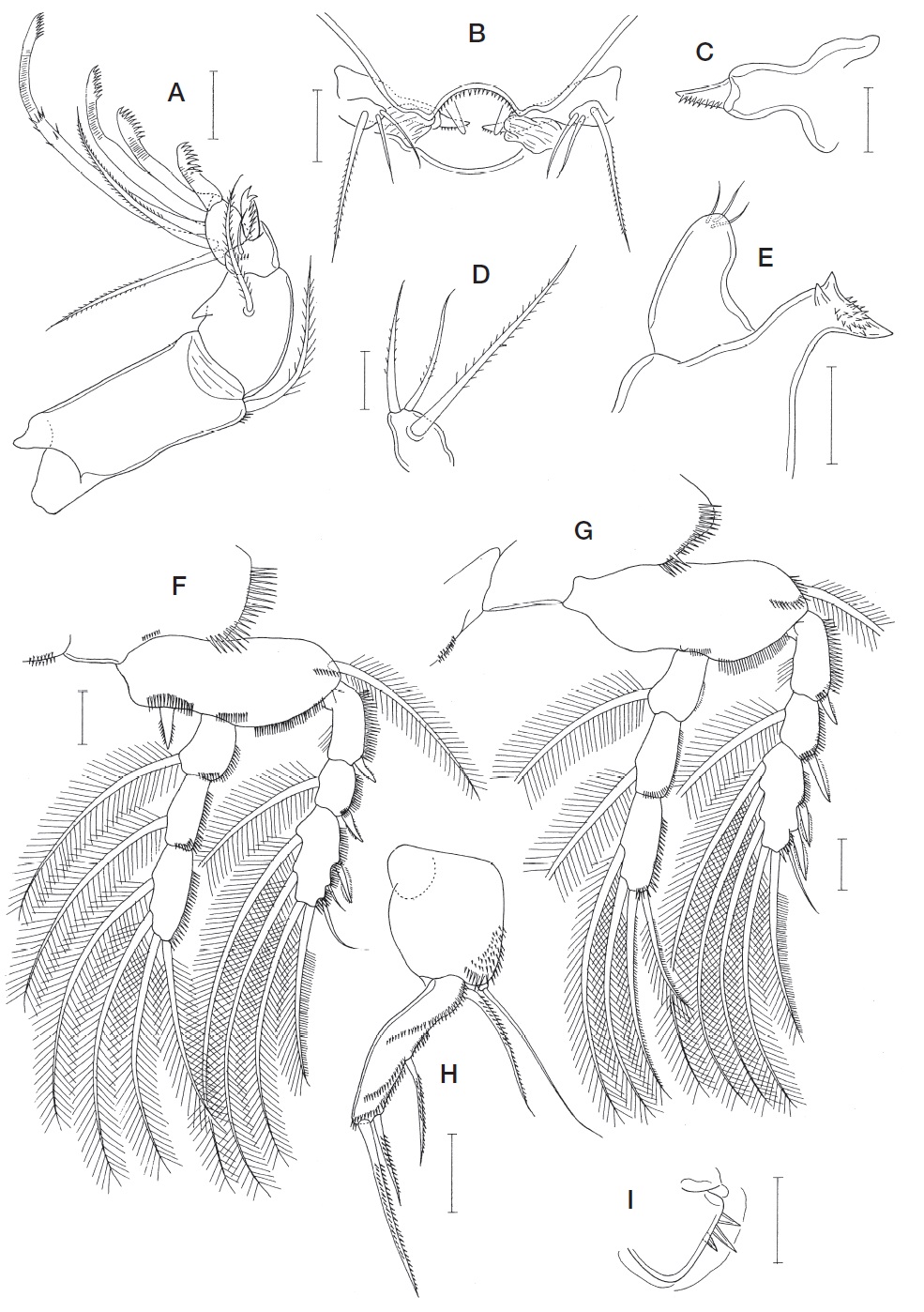
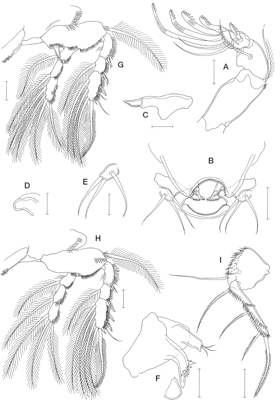
Rostrum absent. Antennule (Fig. 1E) 6-segmented and 169 μm long; armature formula 4+I, 13, 9, 4+aesthetasc, 2+ aesthetasc, and 7+aesthetasc; first segment with 1 plumose seta, 3 simple setae, 1 spine bearing spinules all around its shaft, and 1 backwardly recurved spinous process arising from the anterior margin; spine on first segment inserted on posterior proximal region of anterior process (Fig. 1F); second segment with small dentiform process proximally on anterior margin (Fig. 1F). Antenna (Fig. 2A) consisting of coxo-basis and 3-segmented endopod; coxo-basis longer than endopod, with 1 inner distal seta; first endopodal segment with 1 seta and 1 dentiform process on outer margin; second endopodal segment with 1 claw bearing row of 8 spinous processes and 1 small seta; terminal endopodal segment with 2 setae and 4 claws; these claws unequal in length and armed each with 7, 9, 9, and 8 spinous processes from inner to outer, respectively.
Labrum (Fig. 2B) tapering abruptly, with roundly concave posterior margin bearing row of spinules and 1 pair of transparent linguiform processes. Mandible (Fig. 2C) with 1 thick, spinulose spine distally. Paragnaths weakly developed, represented by transparent, smooth lobes (Fig. 2B). Maxillule (Fig. 2D) represented by a small lobe bearing 2 smaller inner and 1 longer outer setae, all of which being sparsely plumose. Maxilla (Fig. 2E) 2-segmented; syncoxa sclerotized, with pronounced, horse-head-shaped posterodistal outgrowth bearing 2 large dentiform processes and about 20 spinules; allobasis gradually tapering distally, with rounded distal end, and tipped with 3 small setae. Maxilliped represented by sclerotized vestige.
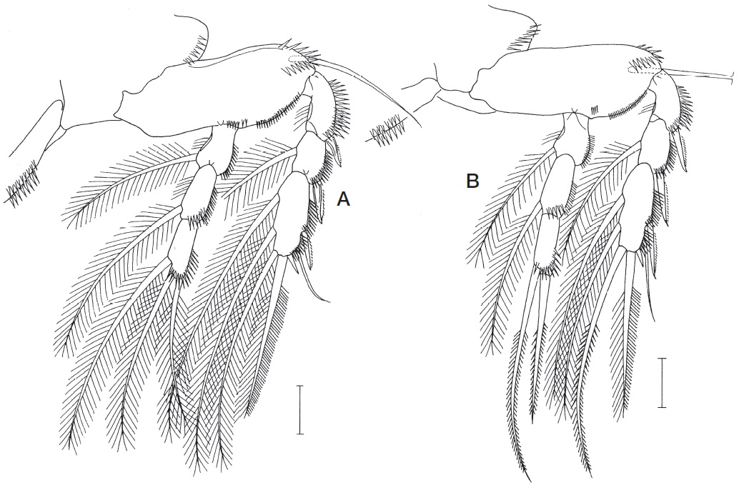
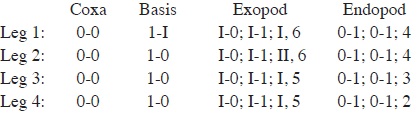
Legs 1-4 (Figs. 2F, G, 3A, B) with 3-segmented slender rami; outer margin of rami spinulose; coxae spinulose along outer margin and lacking inner seta. Inner distal spine on basis 19 μm long. Armature formula of legs 1-4 as follows:
Leg 5 (Fig. 2H) consisting of protopod and exopod. Protopod nearly rectangular, about 83×76 μm, clearly defined at base, with fine spinules on outer distal surface and 1 long, slender outer distal seta. Exopod 115×27 μm (ratio 4.26 : 1), broadest in middle, with 3 obliquely longitudinal rows of spinules and 4 spinulose setae (proximal, middle, subdistal, and distal ones); lengths of these setae 98, 73, 62, and 124 μm, from proximal to distal, respectively. Leg 6 (Fig. 2I) represented by 3 small spiniform elements on genital operculum.
Male. Body (Fig. 3C) tapering posteriorly and 630 μm long. Prosome 354 μm long. Cephalothorax 197×255 μm. Three metasomites similar in length. First metasomite as wide as cephalothorax. Urosome (Fig. 3D) 6-segmented. First urosomite (fifth pedigerous somite) 48×121 μm, much wider than other urosomites. Genital somite 40×82 μm, with row of fine spinules along genital operculum and 1 naked seta posterolaterally. First to last abdominal somites 36×56, 39×46, 27×40, and 9×34 μm, respectively. Caudal ramus 69×11 μm (ratio 6.27 : 1).
Rostrum absent as in female. Antennule with same number of armature elements, but first segment lacking anterior process (Fig. 3E). Antenna (Fig. 3F) different from that of female; second endopodal segment with 1 claw bearing 5 spinous processes and 1 long, spiniform element bearing proximal articulation, 1 large sucking disc and row of 7 or 8 small sucking discs; claws on terminal endopodal segment each with 4, 6, 5, and 5 distal spinous processes, from inner to outer, respectively.
Labrum (Fig. 4A) spiniferous on ventral surface. Mandible, paragnath and maxillule as in female. Maxilla (Fig. 4B) broad, lamella-like, with spinulose posterior margin and accompanied by transversely elongate anterolateral patch of spinules. Maxilliped (Fig. 4C) 4-segmented; first segment with 1 seta subdistally; second segment nearly fusiform, with 2 similar setae and 3 longitudinal rows of spinules on inner surface; third segment small and unarmed; terminal segment as strong claw bearing 3 unequal setae proximally and row of several denticles in distal region.
Leg 1 with weakly sigmoid inner distal spine on basis, otherwise as in female. Legs 2-4 as in female. Leg 5 (Fig. 4D) represented by 1 naked dorsolateral (basal) seta on fifth pedigerous somite and free exopod; exopod 28×10 μm and armed with 4 setae; these setae 42, 46, 59, and 81 μm long, respectively, from proximal to distal; largest distalmost seta plumose, adjacent seta sparsely plumose, and other 2 proximal setae naked. Leg 6 represented by 1 posterolateral seta on genital operculum (Fig. 3D).
Etymology. The specific name is derived from the Latin
>
Echiuricopus tenuipes n. sp. (
Material examined. Four ♀♀ from an unidentified echiuran (host species different from that of
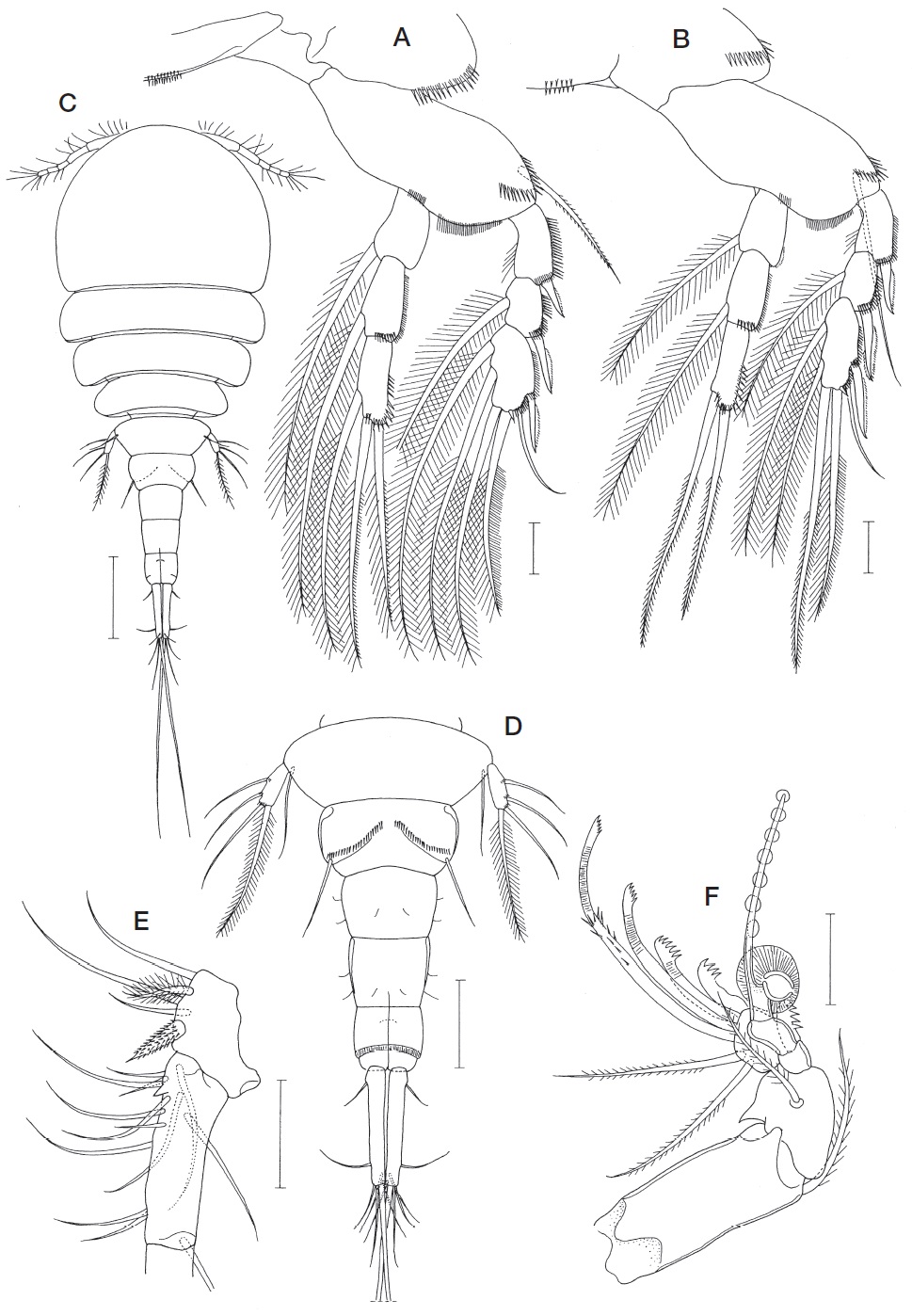
Female. Body (Fig. 5A) similar in form to
Rostrum obscure (Fig. 5F). Antennule (Fig. 5G) 6-segmented; armature formula 4+I, 13, 9, 4+aesthetasc, 2+ aesthetasc, and 7+aesthetasc; first segment with 1 broad plumose seta, 4 setae, and 1 pointed anterior process (Fig. 5H); one of setae inserted on posterior proximal region of anterior process and spinulose along posterior margin; second segment with beak-like process proximally on anterior margin. Antenna (Fig. 6A) similar to that of
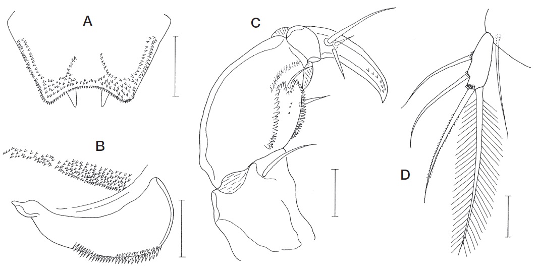
Labrum (Fig. 6B) similar to that of
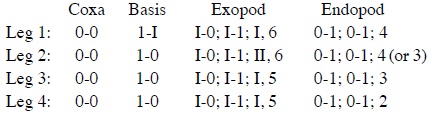
Legs 1-4 (Figs. 6G, H, 7A, B) with 3-segmented slender rami; outer margin of rami and posterior margin basis densely spinulose; coxa of these legs spinulose along outer margin and lacking inner seta. Inner distal spine on basis 16 μm long. Third exopodal segment of legs 1-4 with large spinule at outer distal corner. Third endopodal segment of leg 2 with 3 or 4 setae (4 being typical condition). Armature formula of legs 1-4 as follows:
Leg 5 (Fig. 6I) consisting of protopod and exopod. Protopod nearly rectangular, about 75×64 μm, clearly defined at base, with spinules on outer surface and 1 long, slender, naked outer distal seta. Exopod 114×19 μm (ratio 7.58 : 1), narrowest about halfway its length, with 3 obliquely longitudinal rows of spinules and 4 spinulose setae; lengths of these setae 101, 75, 60, and 116 μm, from proximal to distal, respectively. Leg 6 (Fig. 5C) represented by 3 small spiniform elements on genital operculum.
Male. Unknown.
Etymology. The specific epithet is derived from a combination of the Latin words
Remarks.
[Table 2.] Differences between females of Echiuricopus aprilis n. sp. and E. tenuipes n. sp.
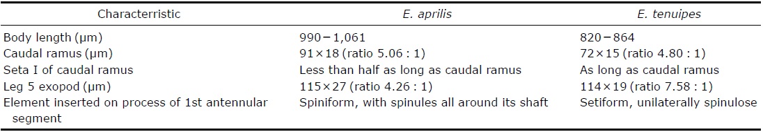
Differences between females of Echiuricopus aprilis n. sp. and E. tenuipes n. sp.