The centric diatom genus
Morphometric analysis is very useful for characterpoor species (Pappas et al. 2001). Shape analysis has proven to be an especially useful method, because shape is commonly the first distinguishing characteristic of many species, such as diatoms (Pappas et al. 2001). Many previous studies of diatoms have involved morphometric analysis of their fine structure (e.g., valve outline, spine, striae, and rimoportulae). Some studies have measured the simple shape, such as valve outlines, which, although they are not reciprocal, can nevertheless be highly effective in separating semi-cryptic or cryptic species, e.g., of the genus
The aim of this study was three-fold. First, the present study provides a detailed survey of the genus
To compare specimens that previous studies identified as
Specimens were observed under LM and SEM. The organic matters in water samples were removed following the protocol developed by Hasle and Fryxell (1970) and Simonsen (1974), with some modifications. The permanent slides were examined under LM (Axioskop 40; Carl Zeiss, Oberkochen, Germany). LM micrographs were taken using an Axiocam MRc5 digital camera (Carl Zeiss). Some cleaned materials were examined using SEM (JSM- 5600LV; Jeol, Tokyo, Japan). The dimensions of frustules in the LM and SEM images were measured using ImageJ 1.32 software (Schneider et al. 2012).
We identified initial definitions to guide the first morphological features and biometric data-gathering phase, then reviewed and confirmed them on the basis of the available data. The characteristics of
1) The ratio of the pervalvar axis to the diameter. Defined as the change in the length of the pervalvar axis due to a change in diameter.
2) The ratio of internal linking spines to diameter. Defined as the change in the internal linking spines within 10 μm due to a change in diameter.
3) The ratio of marginal linking spines to diameter. Defined as the change in the marginal linking spines within 10 μm due to a change in diameter.
4) The ratio of fenestrae to diameter. Defined as the change in the fenestrae within 10 μm due to a change in diameter.
Terminology used follows the general proposals of Crawford (1979), Round et al. (1990), and MacGillivary and Kaczmarska (2015).
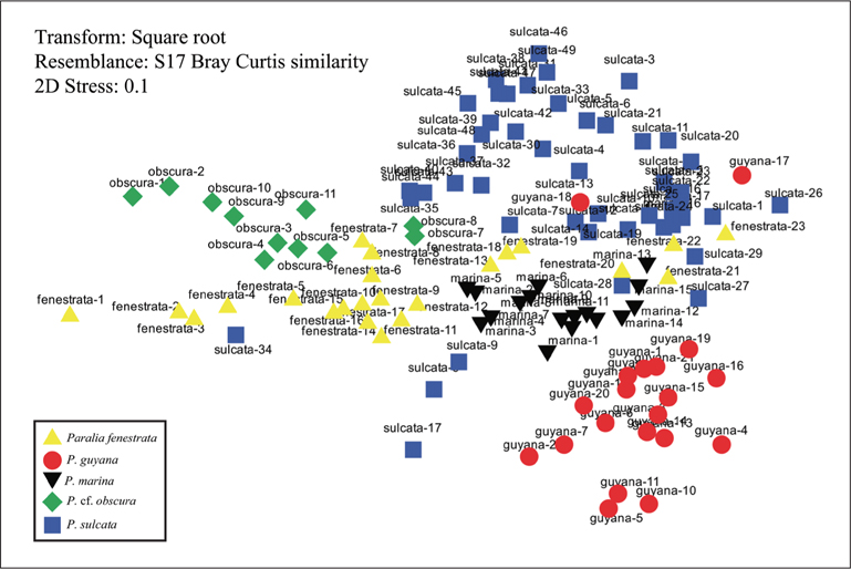
Data for morphological features were analysed using the statistical software Primer v6 (Primer-E Ltd., Lutton, UK) (Clarke 1993, Clarke and Ainsworth 1993, Clarke and Warwick 2001). Non-metric multidimensional scaling (MDS) was applied to complement the characteristics of
Five species belonging to the genus
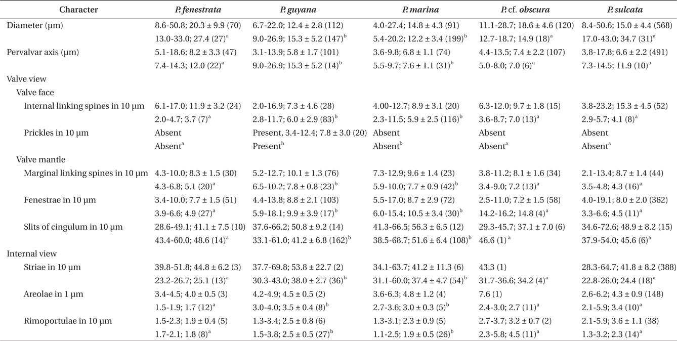
Comparison of some morphological features and biometric data of Paralia species from Korean coastal waters
>
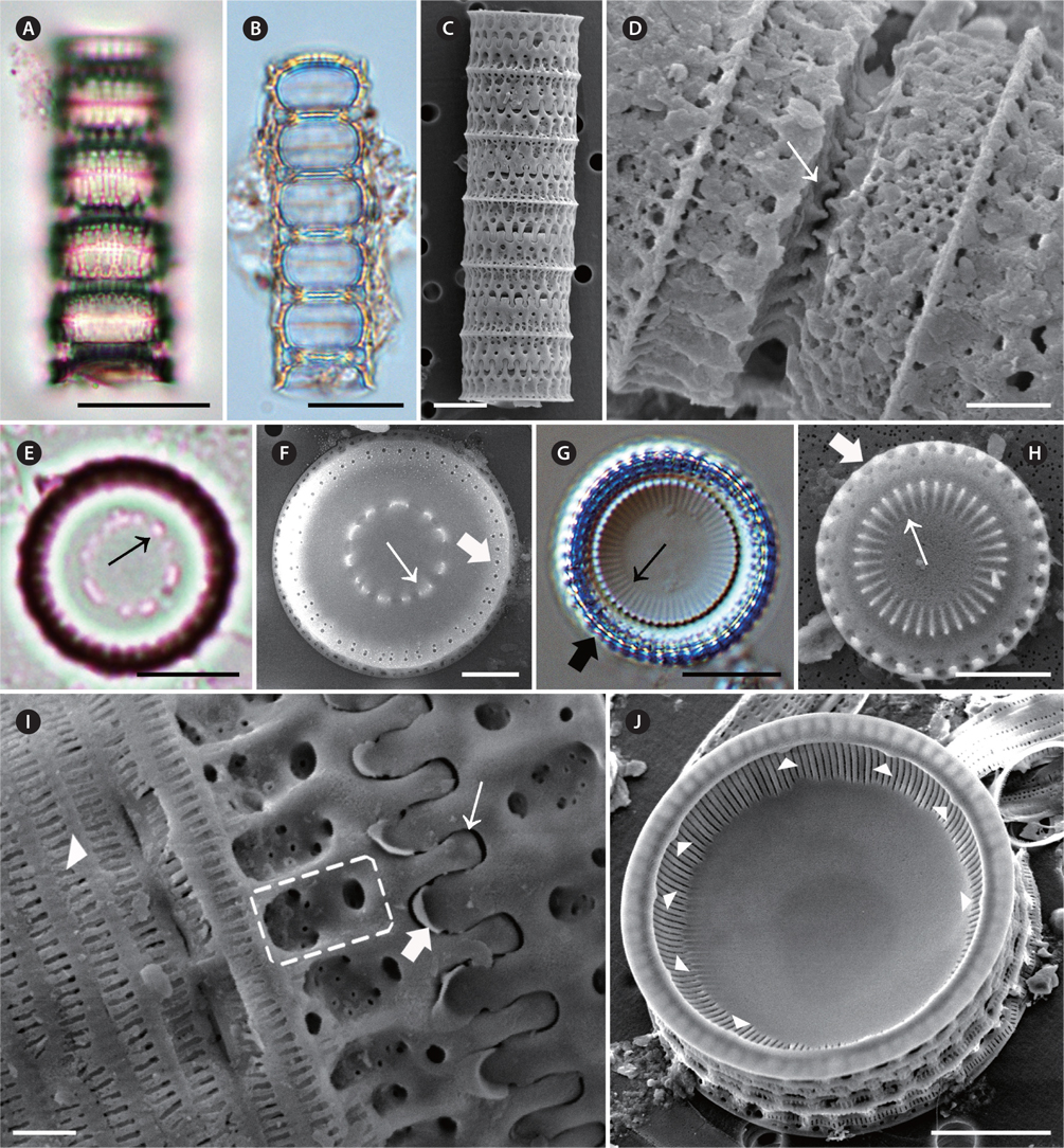
Paralia fenestrata Sawai & Nagumo (
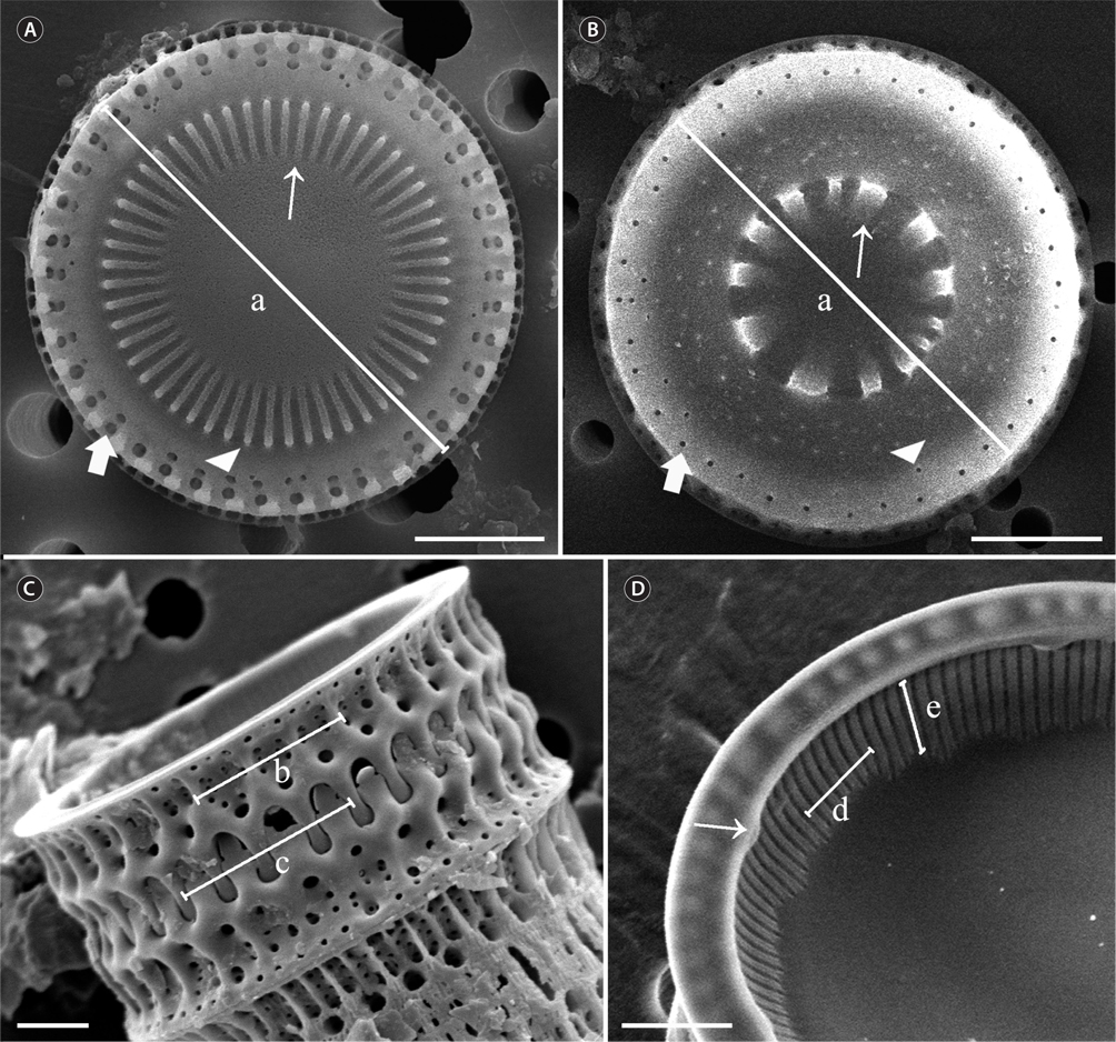
Description of specimen morphology based on LM. Straight chains were formed of interlocking cells (Fig. 2A). Cell chains were mostly <20 cells. Frustules were cylindrical and strongly silicified, 8.6-50.8 μm in diameter and 5.1-18.6 μm in pervalvar axis. Sibling valve chains linked with well-developed interlocking (Fig. 2A). Valves almost circular (Fig. 2C & E). Separation valves had lined up along the central region of internal spines (Fig. 2C, thin arrow) and fenestrae (Fig. 2C, thick arrow). Internal valves had lined up along the central region of internal spines (Fig. 2E, thin arrow) and fenestrae (Fig. 2E, thick arrow).
Description of specimen morphology based on SEM. Straight chains were formed of interlocking cells (Fig. 2B). Two types of valves were observed, separation (Fig. 2D) and intercalary valves (Fig. 2F), each type with two very similar forms, relief and intaglio. Spatulate marginal linking spines (Fig. 2G, thick arrow), 4.3-10.0 in 10 μm, occurred only along the face margin of intercalary valves and fit into notches (Fig. 2H) between the marginal region. Internal linking spines of varied size, 6.1-17.0 in 10 μm, tapered in height towards the smooth valve central area (Fig. 2F & G, thin arrow). The marginal and internal linking spines of sibling valves interlocked to united cells in colonies (Fig. 2B & H, thick and thin arrow). Separation valves were without marginal linking spines (Fig. 2B & D). Separation valves had smooth and convex valves, lined up along the central region of internal spines (Fig. 2B & D, thin arrow). Commonly, double rings of larger and smaller pores were irregularly present at the valve face margin of marginal and intercalary valves (Fig. 2D & F, thick arrow). The most exterior pores on the valve face contained larger pores than the interior pores (Fig. 2D & F, thick arrow). The arrangement of the interior ring of pores was variable (Fig. 2D & F). In some valves, pores were irregularly spaced (Fig. 2D). The valve faces were slightly convex, flat, and with a cylindrical girdle outline of the frustule (e.g., Fig. 2A & B). All valves were perforated by small circular areolae, in parallel crossing rows forming a decussate pattern (Fig. 2G, dashed square), 3.4-4.5 in 1 μm in internal valve view. The fenestrae were obvious at the base of the valve face and the mantle; 3.4-10.0 in 10 μm in girdle view. Above each fenestra was a large circular pore in the valve face (Fig. 2G & H), which corresponded to the presence of a marginal linking spine in intercalary valves. Thus, fenestrae and marginal linking spines nearly occurred at a ratio of 1 : 1 (Fig. 2G & H). Cingula were composed of a number of regularly spaced copulae. Cingula were plain except for regularly spaced slits, 28.6-49.1 in 10 μm (Fig. 2H, arrowhead). The mantle rim was flat and smooth in internal valve view (Fig. 2I). Small, simple slit rimoportulae were located near the mantle edge and were irregularly spaced; 1.5-2.3 in 10 μm (Fig. 2I, arrowheads). The external openings of the rimoportulae were clear (Fig. 2G, arrowhead). Striae ran below the mantle edge in internal view. Striae were perpendicular to the mantle edge and were parallel to each other when they were present (39.8- 51.8 in 10 μm).
Remark. Sawai et al. (2005) recently recorded
>
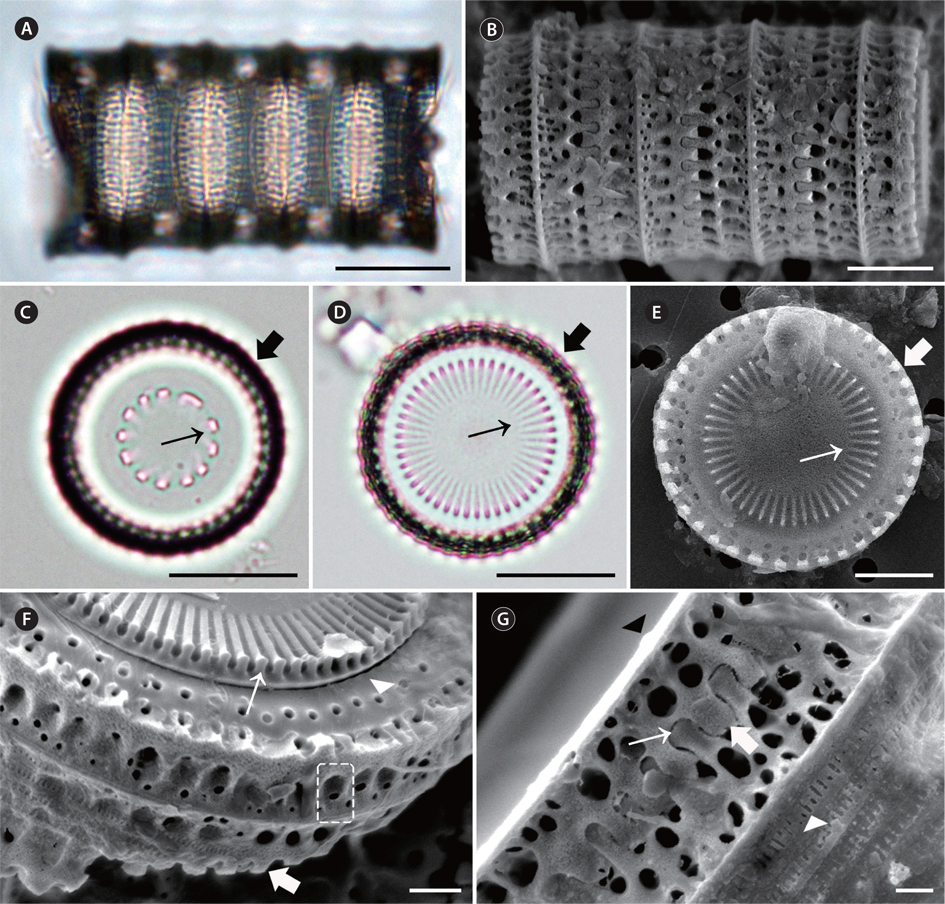
Paralia guyana MacGillivary (
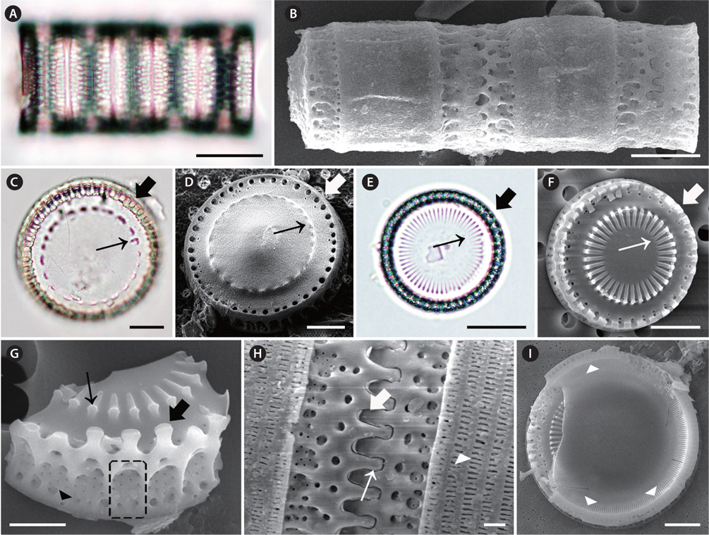
Description of specimen morphology based on LM. Straight chains were formed of interlocking cells (Fig. 3A & B). Sibling valve chains linked with well-developed interlocking. Cell chains were mostly <20 cells. Frustules were cylindrical and heavily silicified, 6.7-22.0 μm in diameter and 9.0-26.9 μm in pervalvar axis. Valves robust, circular (Fig. 3F). Internal valves had lined up along the central region of internal spines (Fig. 3F, thin arrow) and marginal pore (Fig. 3F, thick arrow).
Description of specimen morphology based on SEM. Straight chains were formed of interlocking cells (Fig. 3C & D). Two types of valves were observed, separation (Fig. 3E) and intercalary (Fig. 3G), each with two forms: relief (long, capitate marginal spines) (Fig. 3H, thin arrow) and intaglio (short, blunt marginal spines, Fig. 3H, thick arrow). Marginal linking spines occurred only along the face margin of intercalary valves and fit into notches (Fig. 3H) between the marginal, short, blunt, square-shouldered spines of a sibling valve. Internal linking spines were of varied size, 2.0-16.9 in 10 μm, tapered in height towards the central valve area (Fig. 3D, E & G). The marginal and internal linking spines of sibling valves interlocked to maintain cells in colonies (4.3-10.0 in 10 μm) (Fig. 3H). Commonly, only one ring of regular, mediumor small-sized pores was present on valves (Fig. 3E & G, thick arrow). Separation valve faces were free of marginal spines, but may have had prickles (Fig. 3D, thin arrow). Separation valves had convex valve faces (Fig. 3D) and were lined up along the central region of internal spines (Fig. 3E, thin arrow) connecting sibling separation valves (Fig. 3D, thick arrow). Valves ware perforated by small or large, poroid areolae in a cross pattern (Fig. 3G), 4.2-4.9 in 1 μm in internal valve view. The fenestrae were clearly at the base of the valve face and the mantle part; 4.4-13.8 in 10 μm in girdle view. Closer to the valve face, above each fenestra, was a large circular pore (Fig. 3H, dashed square), which corresponded to the presence of a marginal linking spine in intercalary valves. Thus, fenestrae and marginal linking spines occurred in an almost 1 : 1 ratio. (Fig. 3H). The cingula were composed of a number of copulae with regularly spaced, centrally located slits (Fig. 3D, arrowhead). The mantle rim was smooth in internal valve view (Fig. 3I). Small, slit rimoportulae were located just below the overhanging edge of the mantle, and each rimoportula had irregular space; 1.3-3.4 in 10 μm (Fig. 3I, arrowheads). Striae ran below the mantle edge in internal view. Striae were perpendicular to the mantle edge and were parallel to each other when they were present (37.7-69.8 in 10 μm). Striae varied in length and did not extend to the valve centre.
>
Paralia marina (W. Smith) Heiberg (
Basionym:
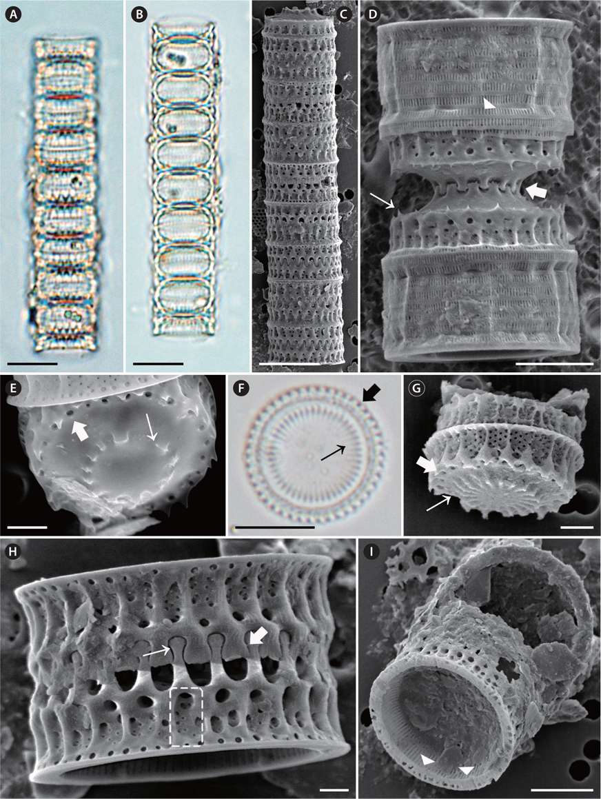
Description of specimen morphology based on LM. Cells commonly shortly cylindrical, linked to form straight chains (Fig. 4A). Cell chains were mostly <30 cells. Frustules were cylindrical and strongly silicified, 4.0-27.4 μm in diameter and 3.6-9.8 μm in pervalvar axis. Valves almost circular (Fig. 4D & F). Separation valves had lined up along the central region of internal spines (Fig. 4D, thin arrow) and marginal pore (Fig. 4D, thick arrow). Internal valves had lined up along the central region of internal spines (Fig. 4F, thin arrow) and marginal pore (Fig. 4F, thick arrow).
Description of specimen morphology based on SEM. Straight chains were formed of interlocking cells (Fig. 4B & C). Two types of valves were observed, separation (Fig. 4C & E) and intercalary (Fig. 4G & H), each with two forms, relief (long, capitate marginal spines) (Fig. 4B & H) and intaglio (short, blunt marginal spines) (Fig. 4B & H). Capitate marginal linking spines (Fig. 4H, thin arrow) occurred only along the face margin of intercalary valves and fit into notches (Fig. 4H, thick arrow) between the marginal, short, blunt, square-shouldered spines of a sibling valve. Internal linking spines of varied size, 4.0-12.7 in 10 μm, tapered in height towards the smooth central valve area (Fig. 4C, E & G, thin arrow). The marginal and internal linking spines of sibling valves interlocked to maintain cells in colonies (Fig. 4C, thin arrow). Generally, two rings of larger and smaller pores were irregularly present at the valve face margin of intercalary valves (Fig. 4G, thick arrow). Separation valves did not have marginal linking spines (Fig. 4C, E & G). Separation valves had smooth valve faces and were lined up along the central region of internal spines (Fig. 4E, thin arrow). Normally, one ring of regular, large- to medium-size pores was present on the valve face (Fig. 4E, thick arrow). Valve faces were flat and with a cylindrical girdle outline of the frustule (e.g., Fig. 4B & C). All valves were perforated by small or large, poroid areolae in a decussate pattern (Fig. 4C, dashed square), 3.6-6.3 in 1 μm in internal valve view. The fenestrae were mostly obvious at the base of the valve face and the mantle met part; 5.5-17.0 in 10 μm in girdle view (Fig. 4H, dashed square). Closer to the valve face, above each fenestra was a large circular pore (Fig. 4B & H), which corresponded to the presence of a marginal linking spine in intercalary valves. Thus, fenestrae and marginal linking spines occurred in a 1 : 1 ratio (Fig. 4B & H). Cingula were composed of a number of copulae, and each copula carried regularly spaced, centrally located slits (Fig. 4C, arrowhead). The mantle rim was smooth in internal valve view (Fig. 4I). Small, slit rimoportulae were located just below the mantle overhanging edge, and each rimoportula had irregular space; 1.3-3.1 in 10 μm (Fig. 4I, arrowheads). Minute external openings of the rimoportulae were observed in many valves (Fig. 4H, arrowhead). Striae began slightly below the mantle edge in internal view. Striae were perpendicular to the mantle edge and were parallel to each other when they were present (34.1-63.7 in 10 μm). Striae varied in length and did not extend to the valve centre.
>
Paralia cf. obscura MacGillivary (
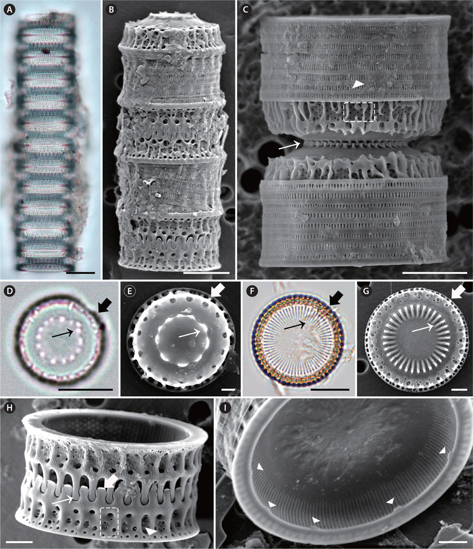
Description of specimen morphology based on LM. Straight chains were formed of interlocking cells (Fig. 5A). Cell chains were mostly <20 cells. Frustules were cylindrical and circular, strongly silicified, 11.1-28.7 μm in diameter and 4.4-13.5 μm in pervalvar axis. Valves almost circular (Fig. 5C & D). Separation valves had lined up along the central region of internal spines (Fig. 5C, thin arrow) and marginal pore (Fig. 5C, thick arrow). Internal valves had lined up along the central region of internal spines (Fig. 5D, thin arrow) and marginal pore (Fig. 5D, thick arrow).
Description of specimen morphology based on SEM. Straight chains were formed of interlocking cells (Fig. 5B). Two types of valves were observed, separation (Fig. 5C) and intercalary (Fig. 5D & E). Separation and intercalary valves were also observed in this obscured fenestra morphotype; intaglio and relief valves were observed in intercalary valves; relief (rounded tips of spatulate spines) (Fig. 5G, thin arrow) and intaglio (short, flat tips of marginal spines) (Fig. 5F & G, thick arrow). Separation valves were not found, Marginal linking spines, 3.8-11.2 in 10 μm, were present on intercalary valves (Fig. 5G, thin arrow). Internal linking spines varied in size, 6.3-12.0 in 10 μm, tapered in height towards the smooth valve central area (Fig. 5E & F, thin arrow). The marginal and internal linking spines of sibling valves interlocked to maintain cells in colonies (Fig. 5B & G). The exterior ring of pores on the valve face contained large (outside) and small (inside) pores (Fig. 5E, thick arrow). In most specimens, each pore in the exterior ring was bordered on each side by a marginal linking spine (Fig. 5E, thick arrow & F, arrowhead). The arrangement of the interior ring of pores was variable (Fig. 5E, thick arrow). In some valves, pores were irregularly spaced (Fig. 5E). The fenestrae were obscure at the base of the valve face and the mantle met part; 2.5-11.0 in 10 μm in girdle view (Fig. 5F, dashed square). Mantle fenestrae were incompletely silicified valves when observed in SEM. A siliceous cover normally obscured fenestrae and each section of this siliceous cover. Each fenestra was a large circular pore (Fig. 5B, F & G), which corresponded to the presence of a marginal linking spine in intercalary valves. The fenestrae and marginal linking spines mostly occurred in a ratio of 2 : 1 (Fig. 5G), but sometimes occurred in a ratio of 1 : 1 (Fig. 5B). Cingula were composed of a number of copulae and were plain except for regularly spaced slits (Fig. 5G, arrowhead). Internally, the mantle edge was flat and smooth. Small, simple slit rimoportulae were located just below the mantle edge and each rimoportula had irregular space; 2.7-3.7 in 10 μm (Fig. 5G, black arrowhead). Striae began slightly below the mantle edge in internal view. Striae were perpendicular to the mantle edge and were parallel to each other when they were present (46.6 in 10 μm). Striae varied in length and did not extend to the valve centre.
Remark. MacGillivary and Kaczmarska (2013) recently recorded
>
Paralia sulcata (Ehrenberg) Cleve (
Basionym.
Synonym.
Description of specimen morphology based on LM. Cells cylindrical, united in filamentous colonies (Fig. 6A & B). Sibling valve chains linked with well-developed interlocking (Fig. 6A). Chains could reach >50 cells, but were mostly <20 cells. Frustules were circular, cylindrical, and strongly silicified, 8.4-50.6 μm in diameter and 3.8- 17.8 μm in pervalvar axis. Separation valves had lined up along the central region of internal spines (Fig. 6E, thin arrow). Internal valves had lined up along the central region of internal spines (Fig. 6G, thin arrow) and marginal pore (Fig. 6G, thick arrow).
Description of specimen morphology based on SEM. Cells cylindrical, united in filamentous colonies (Fig. 6C). Two types of valves, intercalary and separation, were found. Intercalary valves were observed in intaglio and relief forms (sensu Crawford 1979). Separation valves were also found in intaglio forms and relief forms, but the two forms of valves were nearly identical. Internal linking spines of varied size, 3.8-23.2 in 10 μm, tapered in height towards the valve centre and were almost flat (Fig. 6F, thin arrow). The marginal and internal linking spines of sibling valves interlocked to form colonies (Fig. 6C & D). Intercalary valves had slender, spatulate marginal linking spines with round tips (Fig. 6I, thin and thick arrow): 2.1-13.4 in 10 μm. The tips of the spines were sometimes broken off in specimens. Generally, two rings of pores were found at the marginal area of the valve face; the external ring was made of larger pores, while the inner ring was made of small pores. The inner ring pores were sometimes irregular (Fig. 6F & H, thick arrow). Separation valves lacked external linking spines at the edge of the valve face and had less pronounced knob-like internal linking spines toward the central area (Fig. 6F, thin arrow). The junction of the valve face (Fig. 6D, thin arrow) and mantle produced a square shape in both the intercalary and separation valves. The basal silica layer was composed of coarse pores arranged in striae. Each stria consisted of pores which stretched parallel to the pervalvar axis and formed a decussate pattern, 2.6-6.2 in 1 μm in internal valve view. The U-shaped fenestrae were mostly obvious at the base of the valve face and the mantle met part; 4.0-19.1 in 10 μm in girdle view (Fig. 6I, dashed square). Above each fenestra was a large circular pore in the valve face (Fig. 6I, dashed square), and a marginal spine was located the apex of each fenestra (Fig. 6I). Thus, fenestrae and marginal linking spines mostly occurred in a 1 : 1 ratio (Fig. 6C & I). Cingula were composed of a number of copulae, and each copula had regularly spaced, centrally located slits (Fig. 6I, arrowhead). The mantle rim was smooth in internal valve view (Fig. 6J). Small, lip-like rimoportulae were located on the projection of the mantle edge and were irregularly spaced; 2.1-5.9 in 10 μm (Fig. 6J). Striae began slightly below and ran perpendicular to the projection of the mantle edge and were parallel to each other (Fig. 6J); 28.3-64.7 in 10 μm. Within each stria, regularly spaced areolae particularly appeared in valves (Fig. 6J).
>
Comparison of Paralia morphology
We found that the following characteristics are important for separating the genus
Most morphological differences of the genus
>
Comparison of morphometric criteria for identification
Although we tried to confirm the morphological differences among species, most quantitative and qualitative characteristics of the five species were similar and overlapping in ranges of length (Table 1). It is difficult to verify a clear difference between previously studied data (Sims and Crawford 2002, Sawai et al. 2005, Konno and Jordan 2008) and this study. Thus, distinguishing characteristics must be modified for precise identification of
[Table 2.] Comparison of ratio of Paralia species from Korean coastal waters
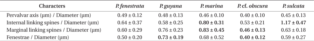
Comparison of ratio of Paralia species from Korean coastal waters