검색 전체 메뉴
PDF
맨 위로
OA 학술지
멍게(Halocynthia roretzi) 비타민 D 수용체 상동체 동정 및 전사활성 Characterization and Transcriptional Activity of a Vitamin D Receptor Ortholog in the Ascidian Halocynthia roretzi
  • 비영리 CC BY-NC
  • 비영리 CC BY-NC
ABSTRACT
멍게(Halocynthia roretzi) 비타민 D 수용체 상동체 동정 및 전사활성
KEYWORD
cDNA, Tunicate , Vitamin D receptor , Vitamin D responsive reporter gene , Halocynthia roretzi
  • 서 론

    비타민 D 수용체(vitamin D receptor, VDR, NR1I1)는 리간드에 의해 활성화되는 전사인자인 핵수용체 superfamily에 속한다. 핵수용체는 공활성인자(coactivator) 및 공억제인자(corepressor)와 협동적으로 유전자의 발현을 조절한다. 핵수용체 단백질들은 특징적인 domain 구조를 공유하고 있는데 N-말단부터 부분적인 전사활성을 가진 A/B domain, DNA-binding domain (DBD, C domain), hinge D domain, ligand-binding domain (LBD, E domain) 및 공활성인자/공억제인자 결합 부위이며 전사활성을 나타내는 F domain으로 구성된다. VDR은 다른 핵수용체와 유사하게 레티노이드 X 수용체(retinoid X receptor, RXR)과 이형이량체(heterodimer)를 형성하고 각각의 리간드와 결합한 후 비타민 D 반응 영역(vitamin D responsive element, VDRE, PuGGT/GTCANNNPuGGT/GTCA)에 결합하여 최대한의 전사활성을 나타낸다(Sánchez-Martínez et al., 2006; Koszewski et al., 2010; Landry et al., 2011).

    척추동물의 VDR은 콜레스테롤을 전구체로 하며 스테로이드 구조를 공유하는 1α,25-(OH)2-vitamin D3 (calcitriol; 1,25 D3)와 높은 친화력으로 결합하며 칼슘 및 인산염 항상성 조절과 같은 전형적인 calcitriol 효과를 나타낸다(Norman et al., 2002). VDR 유전자들은 포유동물, 조류, 양서류, 파충류, 경골어류 이외에도 무악동물 먹장어(Petromyzon marinus)에서도 동정되었다(Bouillon and Suda, 2014). 현재까지 분석된 포유류 게놈은 하나의 VDR유전자를 가지고 있으며, 뇌 조직을 포함하는 광범위한 조직(심장, 간, 췌장, 근육 등)에서 유전자발현이 관찰되었다. 일부 경골 어류, 복어(Takifugu rubripes), 넙치 (Paralichthys olivaceus)는 두 종류의 VDR 유전자(VDRα, VDRβ)를 가진 것으로 보고되어 있다(Suzuki et al., 2000; Maglich et al., 2003). 다른 척추동물의 VDR과 유사하게 복어 VDRα, 넙치 VDRβ는 광범위한 조직 분포를 보이지만 대조적으로 복어 VDRβ는 장에서만 발현되고 간, 아가미, 근육에서는 거의 발현되지 않는 차이점이 있다(Suzuki et al., 2000; Maglich et al., 2003).

    기능적인 비타민D (1,25 D3)는 자외선B (UVB)에 의해 7-dehydrocholesterol의 광이성질체에 기원을 둔 척추동물 진화 초기에 생체내에서 합성된 호르몬으로 생각되고 있다(Bouillon and Suda, 2014). 또한 1,25 D3는 UVB에 의한 DNA 손상을 예방해 줄 수 있는 해양동물의 효과적인 보호물질로 추정된다(Holick, 2011). 한편, 해양무척추동물 피낭류 유령멍게 Ciona intestinali로부터 척추동물의 VDR과 유사한 다수의 핵수용체 유전자들이 밝혀지고 이 유전자를 포함하는 동물군의 계통분류학적 기원에 대한 관심이 높아지게 되었다(Yagi et al., 2003). 유사한 기능을 가질 것으로 추정된 Ciona VDR은 척추동물의 VDR과 달리 1,25 D3에 의한 전사활성은 나타나지 않았으며 그 기능은 불확실한 채로 남아 있다(Reschly et al., 2007).

    본 연구에서는 해양무척추동물 VDR 유전자와 단백질 특성을 규명하기 위하여, 멍게(우렁쉥이, Halocynthia roretzi) VDR의 full-length cDNA (HrVDR)를 cloning하고 유령멍게 Ciona VDR과 함께, 해양 무척추동물 VDR의 아미노산 서열과 핵수용체의 특징적인 domain을 척추동물 ortholog들과 비교 분석하였다. 또한, VDRE-luciferase reporter assay를 실시하여 척추동물의 VDR 및 Ciona VDR과 결합한다고 알려진 리간드에 의한 HrVDR 전사활성도를 조사하였다.

    재료 및 방법

      >  RNA 추출 및 cDNA 합성

    멍게의 VDR cDNA HrVDR를 cloning하기 위해, 경남 통영에서 구입한 멍게의 근육 조직을 적출하여 total RNA를 추출하기 전까지 −80℃에 저장하였으며, Qiagen RNeasy Mini kit(Qiagen, Valencia, CA, USA)를 사용하여 total RNA를 추출하였다. SMART RACE (Rapid Amplification of the cDNA Ends) cDNA 합성 kit (Clontech Lab, Mountain View, CA, USA)를 이용하여 추출된 근육 total RNA (0.5 μg)로 부터 cDNA를 합성하였다.

      >  Polymerase Chain Reaction (PCR)

    Full-length HrVDR를 cloning하기 위해, Magest (http://magest.hgc.jp/) database에 등록되어 있는 VDR partial sequence를 바탕으로 HrVDR의 oligo primer들을 설계하였다(Table 1). 멍게 근육의 total RNA로부터 합성한 cDNA를 template로 하여 10X nTaq buffer 2 μL, dNTP mix, primer 각각 10 μM, nTaq DNA polymerase 0.2 μL (Enzynomics, Seoul, Korea)를 사용하여 총량 20 μL로 PCR을 수행하였다. PCR 조건은 Pre-denaturing step을 94℃ 5분, denaturing step을 94℃ 30초, annealing step을 55℃ 30초, extension step을 72℃ 1분, post-extension step은 72℃ 7분으로 총 30 cycle을 실행한 후 증폭된 DNA 산물을 1% agarose gel전기영동을 실시하여 확인하였다.

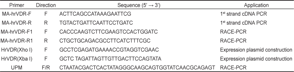
    [Table 1.] Oligo primers for polymerase chain reaction

    label

    Oligo primers for polymerase chain reaction

      >  RACE-PCR 분석

    Total cDNA를 template로 하여 VDR 5′말단과 3′말단 방향으로 각각, 10X BD advantage 2 PCR buffer 2.5 μL, dNTP mix (10 μM) 0.5 μL, 50X BD advantage 2 polymerase mix 0.5 μL, Universal Primer A Mix (UPM) (10X) 2.5 μL, forward primer (MAhrVDR-F), reverse primer (MAhrVDR-R) (10 μM 각각) 0.5 μL, 5′–RACE CDS Primer A, 3′–RACE CDS Primer A (Clontech Lab) (Table 1)를 사용하여 총 25 μL로 5′ RACE-PCR과 3′ RACE-PCR을 수행하였다. Pre-denaturing step 94℃ 3분, denaturing step 94℃ 30초, annealing step 72℃ 2분 5 cycle 실행한 뒤, 다시 denaturing step 94℃ 30초, annealing step 70℃ 30초, annealing step 68℃ 30초, extension step 72℃ 2분 25 cycle 실시한 다음, post-extension step 72℃ 7분 반응하였다. 총 35 cycle을 각각 실행하고 증폭된 DNA 산물은 1% agarose gel로 전기영동 분석하였다.

      >  Sequence 분석

    증폭된 PCR산물을 pGEM-T easy vector (Promega, Madison, WI, USA)에 삽입하고, Escherichia coli XL1-blue competent cell에 transformation 시켰다. Alkaline lysis miniprep 방법을 사용하여 각각의 단편이 삽입된 plasmid DNA 추출 후 5′ 및 3′ 방향으로 Sanger 염기서열 분석 방법으로 분석을 의뢰하였다(Solgent, Daejeon, Korea). 분석된 5′ RACE 및 3′ RACE clone들의 염기서열은 NCBI BLAST search program, NCBI ORF (Open Reading Frame) Finder와 NCBI Conserved Domain Search (CD Search) program으로 확인하였다. 아미노산 서열 비교는 CLUSTAL W 및 MultAlin multiple sequence alignment program을 사용하여 분석하였다. 분자 계통수는 MrBayes (Huelsenbeck et al., 2001)의 Bayesian 방법을 사용하여 제작하였다.

      >  Plasmid 제작

    HrVDR cDNA의 단백질 번역 부위를 증폭하기 위한 primer set (HrVDR-F, HrVDR-R; Table 1)를 설계하고 conventional PCR 방법으로 증폭하였으며, pcDNA3-HA-NLS expression vector (Maeng et al., 2012a)에 삽입하고 상법에 따라 E.coli XL1-blue competent cell에 transformation하고 plasmid DNA를 추출하였다. HrVDR cDNA가 삽입된 plasmid DNA를 Xho I, Xba I 제한효소 처리 하여 삽입된 insert를 확인한 후, T7 및 SP6 primer를 이용하여 5′ 및 3′ 방향으로 Sanger 염기서열 분석 방법으로 분석하였다(Solgent, Daejeon, Korea).

      >  Luciferase reporter assay 분석

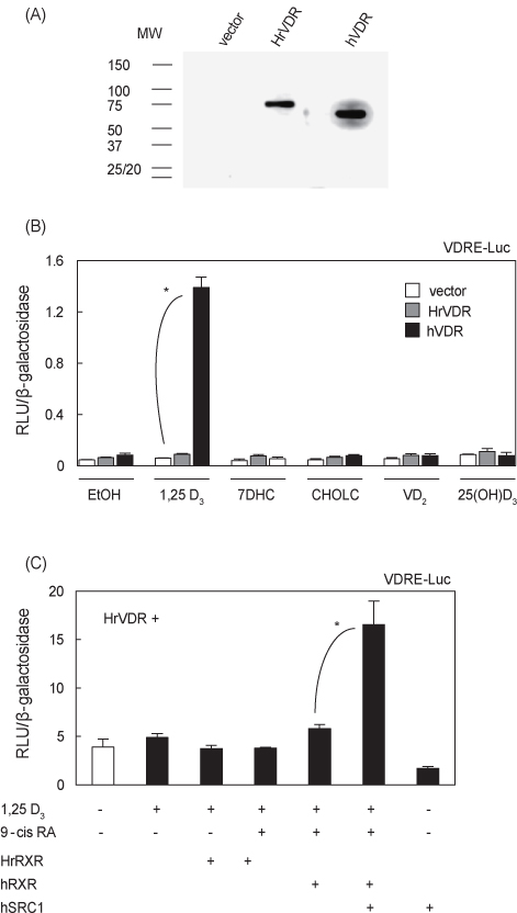
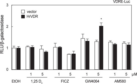
    실험 24시간 전에 분주한 human embryonic kidney 유래의 HEK293 세포(5×10⁴)에 HrVDR, HrRXR, human RXR (hRXR), human steroid receptor coactivator (hSRC1) (Maeng et al., 2012b) 및 전남대학교 이영철 교수님으로부터 제공받 은 human VDR 발현용 vector (hVDR), osteocalcin promoter 유래의 VDRE로 구성된 VDRE-luciferase reporter (VDRE-Luc) (Kim et al., 2009), β-galactosidase 발현용 pRSV-β-gal vector (Maeng et al., 2012a)를 LipofectamineTM 2000 (Invitrogen, Grand Island, NY, USA)으로 transfection하였다. 3시간 후에 10% FBS와 1% 항생제를 포함하는 배지로 교환하였다. 1α,25-(OH)2-vitamin D3 (1,25 D3), 7-dehydrocholesterol (7DHC), Cholecalciferol (CHOLC), Ergocalciferol (vitamin D2), 25-hydroxyvitamin D3 [25(OH)D3], 6-formylindolo [3,2-b]carbazole (FICZ), fanesoid X receptor agonist (GW4064), benzoic derivative retinoic acid (AM580), 9-cis retinoic acid (9-cis RA) (Sigma-Aldrich)를 EtOH에 희석하여 처리하고 24시간 동안 37℃, CO2 5% incubator에 배양하였다. 세포배양액 중의 EtOH 농도는 0.1%이하로 유지하였다. 24시간후, 1X cell lysis reagent (Promega)을 사용하여 세포를 용해 시키고, 실험군 간의 형질 도입률을 보정하기 위하여 β-galactosidase assay buffer (200 mM sodium phosphate buffer pH 7.3, 2 mM MgCl2, 100 mM β-galactosidase (98%), 1.33 mg/mL O-nitrophenyl-β-D-galactopyranoside (OPNG)를 처리하여 흡광도 405 nm에서 β-galactosidase의 활성을 조사하였으며, luciferase assay buffer (1M KH2PO4 pH 7.8, 0.5 M MgCl2, 0.1 M ATP, 10 mM luciferin)와 luminometer (Berthold Co., Wildbad, Germany)를 사용하여 luciferase reporter의 활성도를 측정하였다. HrVDR 벡터의 단백질 발현을 확인하기 위하여 HA-epitope를 인식하는 monoclonal 1차항체(mouse anti-HA, H9658, Sigma-Aldrich, St. Louis, MO, USA), goat 2차항체(anti-mouse IgG-HRP, sc2005, Santa Cruz Biotech., Dallas, TX, USA)를 각각 5,000배 희석하여 상법에 따라 western immunoblot을 실시하였다.

    결 과

    HrVDR을 암호화하고 있는 cDNA 전체 염기서열은 5′ 및 3′ RACE 방법으로 얻은 cDNA 산물을 NCBI ORF Finder와 Conserved Domain Search (CD Search) program, CLUSTAL W 및 MultAlin multiple sequence alignment program을 사용하여 분석하였다. HrVDR full-length cDNA는 총 559 amino acids (aa)를 암호화하고 있으며 1680 nucleotides (nt)의 번역서열과 149 nt의 5′ 비번역 부위, 1633 nt의 3′ 비번역 부위로 구성된 총 3462 nt로 밝혀졌다(GenBank Accession no. KT988062). N-말단부터 A/B domain 26 aa, DBD 77 aa, hinge domain 161 aa, LBD 161 aa로 구성되어 있으며 다른 척추동물의 hinge domain에 비하여 긴 특징을 보인다(Fig. 1). HrVDR을 다른 동물의 VDR과 상동성을 비교하였을 때, 예상한 바와 같이 유령멍게 C. intestinalis의 VDR-b 타입과 가장 높은 상동성을 나타내었다(56%) (Table 2). 비록 다른 척추동물의 VDR과 전체적인 상동성, LBD영역의 상동성은 비교적 낮았지만(31–35%), DBD영역의 상동성은 비교적 높았으며 (62–63%) (Table 2) 시스테인 잔기가 동일한 부위에 잘 보존되어 있다(Fig. 1). Bayesian software를 사용하여 분자계통수를 작성한 결과, HrVDR은 고등한 척추동물의 VDR과는 다른 분지에 위치하며 Ciona VDR-b 타입과 함께 피낭류 VDR 그룹을 형성하고 있음을 알 수 있었다(Fig. 2).

    [Table 2.] Amino acid sequence identities with Halocynthia roretzi VDR, HrVDR

    label

    Amino acid sequence identities with Halocynthia roretzi VDR, HrVDR

    Luciferase reporter assay를 실시하기에 앞서 HEK293세포에 transfection된 HA-HrVDR 발현벡터가 발현되는지 HA epitope 항체를 이용한 western immunoblot 방법으로 조사하였다. 예상한 바와 같이 약 70 kDa 부근에 HrVDR 단일 밴드로 검출되었고, 동일하게 vehicle vector를 transfection한 세포에서는 밴드가 관찰되지 않았으며 hVDR 도입한 세포에서는 약 60 kDa 부근에 나타났다(Fig. 3A).

    VDRE-luc reporter를 이용한 luciferase assay에서는 positive control hVDR을 도입한 HEK293 세포에서만 활성형 비타민 D3 (1,25 D3)에 의해 전사활성이 보였고, 다른 비타민D3 전구체 7DHC, CHOLC, 25(OH)D3 및 식물성 비타민 D2에 의한 활성은 보이지 않았고, HrVDR은 실험에 이용된 모든 리간드에 반응을 나타내지 않았다(Fig. 3B). 척추동물의 VDR은 RXR과 heterodimer를 형성하여 최대한의 전사활성을 나타내며 공활성인자가 관여하기 때문에(Sánchez-Martínez et al., 2006), HrRXR, hRXR 및 hSRC1을 transfection하고 전사활성을 추가적으로 조사하였다. HrVDR/HrRXR, HrVDR/hRXR transfection 세포에 1,25 D3 및 9-cis RA 처리에 의해 변화가 없었지만, 공활성인자 hSRC1의 cotransfection은 전사활성을 유의적으로 증가시켰다(Fig. 3C; P<0.05). 한편, 고농도의 FICZ 및 AM580 처리에 의해 Ciona VDR 전사활성이 증가된 선행연구가 있었으므로(Reschly et al., 2007), 비교적 고농도(1–5 μM)로 관련 리간드를 처리하여 동일하게 VDRE reporter assay를 실시하였다. 그 결과, HrVDR은 1,25 D3, FICZ, AM580에 의한 전사활성은 나타나지 않았지만, 고농도의 GW4064 (5 μM) 처리에 의해 전사활성이 증가하였다(Fig. 4; P < 0.05).

    고 찰

    본 연구에서는 멍게 VDR을 암호화하는 full-length cDNA (HrVDR)를 cloning하고 유령멍게 Ciona VDR 및 척추동물의 해당 ortholog들과 비교 분석하였다. 또한 HrVDR과 결합하는 리간드를 조사하기 위하여 비타민 D3 반응형 VDRE reporter 유전자 및 다양한 비타민 D3 관련 화합물을 이용하여 HrVDR 전사활성도 변화를 측정하였다. 본 연구는 무척추동물 VDR의 리간드 탐색 및 기능을 연구할 때 필요한 중요한 기초자료로서 활용될 것으로 생각된다.

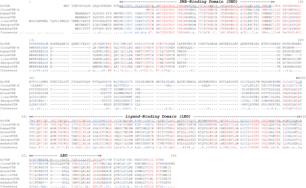
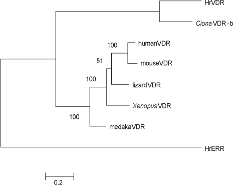
    VDR 유전자는 인간, 마우스, 닭, 도마뱀, Xenopus 등 대표적인 척추동물에서는 한 종류만 존재하지만, 송사리(Oryzias latipes), 넙치, 복어 등 경골어류는 유전자 중복에 의해 VDRα, VDRβ 두 종류가 존재한다(Suzuki et al., 2000; Maglich et al., 2003; Howarth et al., 2008). 본 연구에서 멍게 HrVDR 전체 아미노산 서열을 분석한 결과, 계통분류학상 근연종인 C. intestinalis의 VDR-b subtype과 가장 유사한 것으로 밝혀졌으며 이는 두 종이 동일한 피낭류 해초강에 속한 것을 반영하고 있다. 한편, 멍게의 다른 핵수용체 superfamily 유전자를 조사한 바에 의하면, estrogen receptor-related receptor (ERR), farnesoid X receptor (FXR), liver X receptor (LXR) 및 RXR은 splicing variant가 존재할 경우도 있으나, 각각 한 종류의 유전자만 존재한다(Park et al., 2009; Maeng et al., 2012a, 2012b; Raslan et al., 2013). 따라서, HrVDR을 포함하여 멍게의 핵수용체들은 각각의 단일 유전자들에 의해 암호화되어 있을 가능성이 높다.

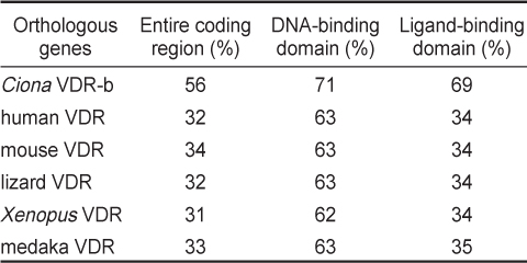
    다른 척추동물의 VDR 단백질의 핵심적인 기능 domain들로 판단되고 있는 DBD 및 LBD 영역에 대하여 HrVDR의 대응 영역과 비교하였을 때, 다소간 아미노산서열의 변이가 높은 수준임을 알 수 있었다. 비록 NCBI Blast 분석 결과 척추동물의 가장 유사한 분자로서 VDR 단백질들이 검색되었지만, 호르몬 결합 부위인 LBD의 변이가 많았고 유사 종 C. intestinalis의 VDR LBD 영역과도 69%의 상동성에 불과하였다. 따라서, 비타민 D가 리간드인 척추동물의 VDR과 같은 양식으로 피낭류 VDR LBD에도 용이하게 비타민 D가 결합할 것으로는 추정되지 않는다. 멍게와 C. intestinalis 핵수용체 LBD 영역은 인간의 해당 ortholog들과 비교한 상동성은 38–78%인 점을 고려하면 HrVDR의 아미노산 상동성(34%)은 조사된 핵수용체들 중에서 가장 낮음을 알 수 있다(Park et al., 2009; Maeng et al., 2012a, 2012b; Raslan et al., 2013). 한편, DBD 영역에서 DNA 결합에 핵심적인 구조로 알려진 zinc finger구조를 형성하는 4개의 시스테인 site는 모든 동물들에서 매우 잘 보존되고 있어서 VDRE를 포함하는 유전자 상류 영역에 결합하는 능력은 있을 것으로 판단된다. 향후, 해양무척추동물 멍게류 VDR이 더 밝혀져서 VDR아미노산 변이와 전사인자로서의 기능에 관련된 분자진화학적인 연구가 기대된다.

    척추동물의 VDR과 현저한 아미노산 변이를 보이는 HrVDR 및 C. intestinalis의 LBD 영역, 그리고 1,25 D3Ciona VDR의 리간드로서 작용하지 않았다는 연구 결과(Reschly et al., 2007)는 멍게 HrVDR의 비타민 D3 비의존성을 예상할 수 있었다. 실제로 1,25 D3는 물론 그 전구체인 7DHC, CHOLC, 25(OH)D3 및 식물성 비타민 D2에 의한 전사활성 역시 관찰되지 않았고 현재로서는 HrVDR의 내재적 리간드 및 agonist는 불확실한 실정이다. 한편, FXR 리간드인 GW4064를 비교적 고농도인 5 μM로 처리한 결과 HrVDR의 약한 전사활성이 나타난 점은 해양 무척추동물 피낭류 핵수용체의 리간드 선택성은 잘 알려진 척추동물 핵수용체의 리간드 선택성과는 많은 차이점을 시사하고 있다. 실례로 멍게 FXR 및 LXR은 각각의 척추동물 agonist로 알려진 GW4064 및 GW3965에 의한 전사활성이 보이지 않았다(Maeng et al., 2012a, Raslan et al., 2013). 전술한 바와 같이, 고농도로 FICZ 및 AM580를 처리한 경우 Ciona VDR의 전사활성이 약하게 관찰된 결과(Reschly et al., 2007)와 대조적으로 HrVDR은 전사활성이 보이지 않았다. 이는 Ciona VDR의 LBD와도 아미노산서열 차이가 현저한 점을 반영하고 있으며 피낭류 VDR 리간드 및 agonist의 다양성을 시사하고 있다. 향후, 1,25 D3와 유사한 척추동물형 비티민D계열의 리간드 존재 및 HrVDR결합affinity를 조사하는 추가적인 연구가 필요할 것으로 사료된다.

참고문헌
  • 1. Bouillon R, Suda T 2014 Vitamin D: calcium and bone homeostasis during evolution [Bonekey Rep] Vol.3 P.480 google cross ref
  • 2. Holick MF 2011 Vitamin D: evolutionary, physiological and health perspectives [Curr Drug Targets] Vol.12 P.4-18 google cross ref
  • 3. Howarth DL, Law SH, Barnes B, Hall JM, Hinton DE, Moore L, Maglich JM, Moore JT, Kullman SW 2008 Paralogous vitamin D receptors in teleosts: transition of nuclear receptor function [Endocrinology] Vol.149 P.2411-2422 google cross ref
  • 4. Huelsenbeck JP, Ronquist F, Nielsen R, Bollback JP 2001 Bayesian inference of phylogeny and its impact on evolutionary biology [Science] Vol.294 P.2310-2314 google cross ref
  • 5. Kim JY, Son YL, Lee YC 2009 Involvement of SMRT corepressor in transcriptional repression by the vitamin D receptor [Mol Endocrinol] Vol.23 P.251-264 google cross ref
  • 6. Koszewski NJ, Herberth J, Malluche HH 2010 Retinoic acid receptor gamma 2 interactions with vitamin D response elements [J Steroid Biochem Mol Biol] Vol.120 P.200-207 google cross ref
  • 7. Landry CS, Ruppe MD, Grubbs EG 2011 Vitamin D receptors and parathyroid glands [Endocr Pract] Vol.17 P.63-68 google cross ref
  • 8. Maeng S, Lee JH, Kim GJ, Kim SH, Kwon HC, Shin YK, Sohn YC 2012a olecular and expression analysis of the farnesoid X receptor in the urochordate Halocynthia roretzi [Comp Biochem Physiol B Biochem Mol Biol] Vol.161 P.189-196 google cross ref
  • 9. Maeng S, Lee JH, Choi SC, Kim MA, Shin YK, Sohn YC 2012b The retinoid X receptor in a marine invertebrate chordate: evolutionary insights from urochordates [Gen Comp Endocrinol] Vol.178 P.380-390 google cross ref
  • 10. Maglich JM, Caravella JA, Lambert MH, Willson TM, Moore JT, Ramamurthy L 2003 The first completed genome sequence from a teleost fish (Fugu rubripes) adds significant diversity to the nuclear receptor superfamily [Nucleic Acids Res] Vol.31 P.4051-4058 google cross ref
  • 11. Norman AW, Okamura WH, Bishop JE, Henry HL 2002 Update on biological actions of 1α,25(OH)2-vitamin D3(rapid effects) and 24R,25(OH)2-vitamin D3 [Mol Cell Endocrinol] Vol.197 P.1-13 google cross ref
  • 12. Park W, Kim GJ, Choi HS, Vanacker JM, Sohn YC 2009 Conserved properties of a urochordate estrogen receptor-related receptor (ERR) with mammalian ERRalpha [Biochim Biophys Acta] Vol.1789 P.125-134 google cross ref
  • 13. Raslan AA, Lee JH, Shin J, Shin YK, Sohn YC 2013 Transcriptional activity and expression of liver X receptor in the ascidian Halocynthia roretzi [Zoolog Sci] Vol.30 P.731-741 google cross ref
  • 14. Reschly EJ, Bainy AC, Mattos JJ, Hagey LR, Bahary N, Mada SR, Ou J, Venkataramanan R, Krasowski MD 2007 Functional evolution of the vitamin D and pregnane x receptors [BMC Evol Biol] Vol.12 P.7-222 google
  • 15. Sanchez-Martinez R, Castillo AI, Steinmeyer A, Aranda A 2006 The retinoid X receptor ligand restores defective signalling by the vitamin D receptor [EMBO Rep] Vol.7 P.1030-1034 google cross ref
  • 16. Suzuki T, Suzuki N, Srivastava AS, Kurokawa T 2000 dentification of cDNAs encoding two subtypes of vitamin D receptor in flounder, Paralichthys olivaceus [Biochem Biophys Res Commun] Vol.270 P.40-45 google cross ref
  • 17. Yagi K, Satou Y, Mazet F, Shimeld SM, Degnan B, Rokhsar D, Levine M, Kohar Y, Satoh N 2003 A genomewide survey of developmentally relevant genes in Ciona intestinalis. III. Genes for Fox, ETS, nuclear receptors and NFkappaB [Dev Genes Evol] Vol.213 P.235-244 google cross ref
OAK XML 통계
이미지 / 테이블
  • [ Table 1. ]  Oligo primers for polymerase chain reaction
    Oligo primers for polymerase chain reaction
  • [ Table 2. ]  Amino acid sequence identities with Halocynthia roretzi VDR, HrVDR
    Amino acid sequence identities with Halocynthia roretzi VDR, HrVDR
  • [ Fig. 1. ]  Amino acid sequence alignment of VDR proteins. Multalin program (Corpet 1988) was used to generate a multiple alignment with VDRs: Halocynthia roretzi HrVDR, (GenBank Accession no. KT988062), Ciona intestinalis CionaVDR-b (NP_001037831.1), human VDR (J03258.1), mouse VDR (BAA06737.1), Anolis carolinensis lizard VDR (XP_008101719), Xenopus laevis XenopusVDR (NP_001079288.1), and Oryzias latipes medakaVDR (NP_001121988.1). The DNA-binding domain (DBD) and ligand-binding domain (LBD) were predicted by NCBI domain architecture analysis program. Highly conserved amino acid sequences are shown in the consensus sequence.
    Amino acid sequence alignment of VDR proteins. Multalin program (Corpet 1988) was used to generate a multiple alignment with VDRs: Halocynthia roretzi HrVDR, (GenBank Accession no. KT988062), Ciona intestinalis CionaVDR-b (NP_001037831.1), human VDR (J03258.1), mouse VDR (BAA06737.1), Anolis carolinensis lizard VDR (XP_008101719), Xenopus laevis XenopusVDR (NP_001079288.1), and Oryzias latipes medakaVDR (NP_001121988.1). The DNA-binding domain (DBD) and ligand-binding domain (LBD) were predicted by NCBI domain architecture analysis program. Highly conserved amino acid sequences are shown in the consensus sequence.
  • [ Fig. 2. ]  A phylogenic tree for VDR proteins. Phylogenetic tree construction was generated with MrBayes software and the reliability of the tree was assessed by bootstrapping using 10,000 bootstrap replications. The abbreviated amino acid sequence names for VDRs are noted in Fig. 1. Estrogen receptor-related receptor of Halocynthia roretzi, HrERR, was used as outgroup.
    A phylogenic tree for VDR proteins. Phylogenetic tree construction was generated with MrBayes software and the reliability of the tree was assessed by bootstrapping using 10,000 bootstrap replications. The abbreviated amino acid sequence names for VDRs are noted in Fig. 1. Estrogen receptor-related receptor of Halocynthia roretzi, HrERR, was used as outgroup.
  • [ Fig. 3. ]  Transcriptional activity of HrVDR. (A) HEK293 cells in a 24-well plate were transfected with HA-HrVDR expression vector (100 ng) and cultured for 48 h. Total cell lysates were resolved by 12% SDS-PAGE and blotted with the HA primary antibody and HRP-conjugated secondary antibody. The maternal vehicle vector and HA-hVDR expression vector were used for controls. (B-C) The vehicle vector, HrVDR, hVDR, hRXR and hSRC1 expression vectors (100 ng each) were transfected into HEK293 cells in 24-well plates, along with the VDRE-Luc reporter (100 ng) and β-galactosidase expression vector (pRSV-β-gal; 100 ng). After 24 h of transfection, the cells were incubated in the presence of the indicated ligands (10?7 M each) or the same amount of EtOH for 18 h. All cells were lysed, and the luciferase activities were measured and normalized against the β-galactosidase expression as an internal control. The relative luciferase activity represent the mean±SE of four separate transfections expressed as a relative light unit (RLU). Statistical changes were determined by the Student’s t test (two-tailed). *, P<0.05.
    Transcriptional activity of HrVDR. (A) HEK293 cells in a 24-well plate were transfected with HA-HrVDR expression vector (100 ng) and cultured for 48 h. Total cell lysates were resolved by 12% SDS-PAGE and blotted with the HA primary antibody and HRP-conjugated secondary antibody. The maternal vehicle vector and HA-hVDR expression vector were used for controls. (B-C) The vehicle vector, HrVDR, hVDR, hRXR and hSRC1 expression vectors (100 ng each) were transfected into HEK293 cells in 24-well plates, along with the VDRE-Luc reporter (100 ng) and β-galactosidase expression vector (pRSV-β-gal; 100 ng). After 24 h of transfection, the cells were incubated in the presence of the indicated ligands (10?7 M each) or the same amount of EtOH for 18 h. All cells were lysed, and the luciferase activities were measured and normalized against the β-galactosidase expression as an internal control. The relative luciferase activity represent the mean±SE of four separate transfections expressed as a relative light unit (RLU). Statistical changes were determined by the Student’s t test (two-tailed). *, P<0.05.
  • [ Fig. 4. ]  Transcriptional activation of HrVDR by FXR agonist GW4064. Vehicle and HrVDR expression vectors were transfected into HEK-293 cells as indicated. Treatment of ligands, luciferase reporter assays and statistical analysis are same with Fig. 3.
    Transcriptional activation of HrVDR by FXR agonist GW4064. Vehicle and HrVDR expression vectors were transfected into HEK-293 cells as indicated. Treatment of ligands, luciferase reporter assays and statistical analysis are same with Fig. 3.
(우)06579 서울시 서초구 반포대로 201(반포동)
Tel. 02-537-6389 | Fax. 02-590-0571 | 문의 : oak2014@korea.kr
Copyright(c) National Library of Korea. All rights reserved.