Carbon nanotubes (CNTs), as a form of carbon in which the carbon atoms are arranged in hollow cylindrical structures with diameters of the order of a nanometer, have attracted great attention from chemists and scientists due to their exceptional properties, such as their electronic, mechanical, optical, and chemical characteristics [1]. These properties of CNTs make them potentially suitable for applications in many fields, such as adsorbents [2,3], corrosion protection [4], cancer therapy [5], and drug delivery [6]. Also, on account of the size and physicochemical properties of CNTs their biomedical applications are being intensively investigated. However, because of the high hydrophobicity, low functionality, and large size of pure CNTs, their medical and biological applications are limited. Thus, the surface functionalization of CNTs with polar substituents is essential. To this end, oxygenated CNTs possessing carboxyl moieties have been used as starting materials for sidewall functionalization in several studies, which can be linked to a wide variety of anticancer drugs or active molecules, which is a key step for biomedical applications [7,8]. For example, Pastorin et al. [9] have developed CNT-methotrexate conjugates via covalent linkage for the use of CNTs as multimodal drug delivery systems. Extensive research has been carried out on the functionalization of CNTs based on cycloaddition [10], carbanion addition [11], radical addition [12] and amidation [13-18]. In this current study, carboxylated multi-wall carbon nanotubes (MWCNT)-COOH were first modified by 2-aminobenzamide to form an MWCNT-amide and then by phosphoryl chloride (POCl3) to produce MWCNT-quin. The percentage of functionalization of MWCNTs with these species was estimated by thermal analysis.
Because of the many biological applications of CNTs mentioned above and their annual global production on a massive scale, considerable concerns have been raised about the risks related to CNTs because these materials are easily transported to the environment, and they interact with cell materials, thereby developing their eventual toxic effects [19]. Therefore, it is essential to thoroughly investigate the toxicity and biocompatibility of CNTs in relation to human health and the environment.
Reports made so far on the bio-toxicity of CNTs have been controversial with some of them emphasizing the toxic effects of CNTs, while others have demonstrated their biocompatibility with live cells [20-24]. Taking this background into account and considering the biologic and antitumor activities of quinazoline derivatives [25-29], the purpose of the present work was to study the cytotoxicity of modified MWCNTs on a human noncancerous cell line (HEK293) and a cancerous one (SKBR3). Our study indicates that MWCNT-quin is highly toxic to cancer cells, and this effect is highly significant compared to its effect on noncancerous cell samples.
2.1. Materials and characterizations
All reagents and solvents, including 2-aminobenzamide, POCl3, tetrahydrofuran (THF), and dimethylformamide (DMF) were purchased from Merck Chemical Inc. and MWCNTCOOH (95% purity, O.D.: 10-20 nm, Length: 0.5-2 µm, content of COOH groups: 2 wt%, Neutrino Co., New York, NY, USA) was used as received. Fourier transform infrared spectroscopy (FT-IR) spectrum was recorded using KBr tablets on a Thermo Nicolet Nexus 870 FTIR spectrometer (Thermo Nicolet, USA). The Raman spectra were recorded on an Almega Thermo Nicolet Dispersive Raman Spectrometer (532 nm of an Nd:YLF laser, Thermo Nicolet). Field emission scanning electron microscopy (FE-SEM) was used to study the morphology of the MWCNTs. FE-SEM measurement was carried out on a Hitachi S4160 (cold field emission) electron microscope (Hitachi, Tokyo, Japan). Elemental analyses of C, H, and N were performed with a SERIES (ІІ) 2400 from Perkin Elmer, Waltham, MA, USA. The samples were subjected to thermogravimetric analysis (TGA) (NETZSCH TG 209 F1 Iris, NETZSCH Co., Selb, Germany) in the N2 (10℃/min). Fluorescence spectra were measured using a JASCO SP-6200 spectrofluorometer (JASCO, Tokyo, Japan). Samples were prepared by ultrasonic dispersion in DMF for 30 min. To record a fluorescence spectrum, 5 mL of the obtained suspension was placed in the cell.
2.2. Preparation of MWCNT-amide
An amount of 150 mg MWCNT-COOH was suspended in 30 mL SOCl2 and 1 mL DMF. The mixture was stirred at 70℃ for 48 h in a 100 mL round-bottom flask with a heater and a condenser. Subsequently, the residual SOCl2 was removed by reduced pressure distillation to yield acyl chloride-functionalized MWCNTs. This material was mixed with 250 mg 2-aminobenzamide in 25 mL DMF, and the reaction mixture was stirred at 100℃ for 96 h. The mixture was cooled to room temperature, filtered, and washed thoroughly with DMF, ethyl alcohol, and THF. Subsequently, the black solid was dried at room temperature for 8 h under vacuum.
2.3. Preparation of MWCNT-quin
An amount of 100 mg MWCNT-amide was sonicated in 20 mL of POCl3 in a 50 mL round-bottom flask for 15 min to give a homogeneous suspension. The suspension was stirred at 80℃ for 96 h with a heater and a condenser. After it was cooled to room temperature, the reaction mixture was separated by centrifugation. Then, the sediment of the centrifuged mixture was washed thoroughly with THF. Finally, the obtained solid was dried under vacuum for 8 h.
SKBR3 and HEK293 cells were maintained and expanded in Dulbecco modified Eagle medium (DMEM, Gibco/Invitrogen, San Diego, CA) supplemented with 10% fetal bovine serum and 1% L-glutamine, penicillin, and streptomycin and incubated at 37℃ in a 5% CO2 atmosphere. The cells were split by trypsinization when a confluence of 85%-90% was reached. All cell culture supplies were purchased from Gibco/Invitrogen. To assess the effects of MWCNT exposure on cell survival, 70%-80% confluent cultures were washed twice with ice-cold phosphate buffered saline (PBS) solution and then cultured in 24 wells for 24 hr. The modified MWCNTs were dissolved in sterile water to make serial concentrations. Then, the cells were treated with these serial concentrations of MWCNTs. After 24 h, the cells were analyzed by a 3-(4,5-dimethylthiazol-2-yl)-2,5-diphenyl tetrazolium bromide (MTT) viability assay.
2.4.1. MTT viability assay
A standard procedure was followed to carry out the MTT assay to evaluate the proliferative activity of the cells. A 0.5-mg/mL stock of MTT (Sigma-Aldrich, USA) was prepared in PBS (pH 7.2) at a concentration of 5 mg/mL and filter-sterilized before storage at 4℃ until use. From this stock, we added 20-180 µL serum-free DMEM to each well and incubated the cells at 37℃ with 5% CO2 for 3 h. Finally, we replaced this medium with 200 µL dimethyl sulfoxide, incubated the cells for 5 min, and read the absorbance at 580 nm.
2.4.2. Statistical analyses
Data in the figures are represented as the mean ± standard error of the mean of 3 or more separate experiments carried out in triplicate. The relative growth rates (RGR) of the cell samples were calculated using the formula of RGR = OD of each group / OD of natural group × 100%. To determine the cytotoxic response of the MWCNTs to cells in culture, 6-graded toxicity and other analytical methods were applied. All statistical analyses were performed using SPSS version 17.0 (SPSS Inc., Chicago, IL, USA). We used the independent Student
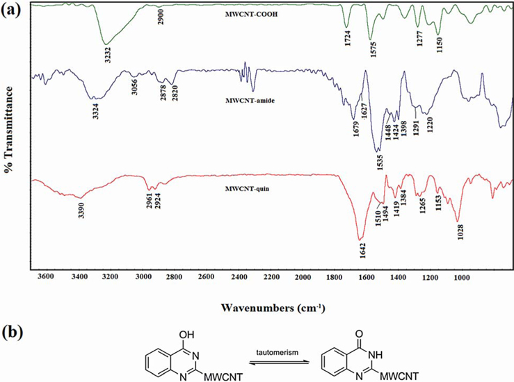
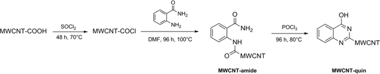
Fig. 1 illustrates the procedure for the functionalization of MWCNT-COOH by 2-aminobenzamide and 4-hydroxyquinazoline. The products were characterized by elemental analysis, FT-IR, Raman, scanning electron microscopy (SEM), TGA, derivative thermogravimetric (DTG) analysis, steady-state fluorescence spectroscopy, and solubility testing.
The elemental analysis results of the modified MWCNTs are shown in Table 1. Apart from the carbon values, the atomic percentages of: H (2.06%) and N (2.22%) in MWCNT-amide (as compared to MWCNT-COOH) indicated that MWCNT-COOH is functionalized with 2-aminobenzamide. On the other hand, the decreasing of percentage of H from 2.06% to 1.84% for MWCNT-quin confirms the 4-hydroxyquinazoline formation as compared to MWCNT-amide. Based on these data coupled with the assumption that the atomic percentages of nitrogen and hydrogen originated from the employed 2-aminobenzamide and 4-hydroxyquinazoline without elimination, we confirmed the functionalization of MWCNT-COOH with these compounds.
[Table 1.] Elemental analysis results of modified MWCNTs
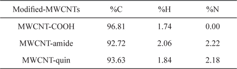
Elemental analysis results of modified MWCNTs
Fig. 2a shows the FT-IR spectra of the modified MWCNTs. For MWCNT-COOH, the peak at 1575 cm–1 is due to the C=C stretch mode in the MWCNTs, and the peaks at 1724 and 1150 cm–1 apparently correspond to the stretching modes of the carboxylic groups [17,18]. The two bonds at 2800-2970 cm–1, which are seen in all spectra, are assigned to the CH stretching of MWCNT-COOH defects. In the spectra of MWCNT-amide, the two prominent peaks at 1679 [-C(=O)NH linkage] and 1627 cm–1 [NH2-(C=O) of benzamide] can be assigned to the amide carbonyl groups (as compared to 1724 cm–1 for MWCNTCOOH), which confirms the formation of MWCNT-amide. In the spectrum of MWCNT-quin, the peak of -C(=O)NH linkage has disappeared, and the remarkable peaks at around 3390 and 1642 cm–1 appear, which can be attributed to NH and C=O stretching mode, respectively, which is due to the tautomerism process of enol to the amide group (Fig. 2b). Also, the other peaks in the spectra of MWCNT-amide and MWCNT-quin at around 3200-3400, 3056, 1520-1600, 1400-1520, 1200-1400, and 1000-1200 cm–1 could corresponded to NH, CH aromatic ring, C=C, C=N, C-N, and C-O stretching modes, respectively [15-18]. Thus, the FT-IR spectra confirm that MWCNT-COOH has been successfully modified by 4-hydroxyquinozaline.
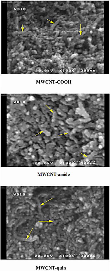
More evidence on the grafting has been obtained from FE-SEM imaging, which is a powerful tool to study the surface morphological structure of modified MWCNTs, as shown in Fig. 3. These images show that the resulting samples have the morphology of nanotubes, in which they are highly tangled and agglomerated. Also, the images suggest that the organic moieties are glued on the MWCNTs by strong interaction, and that these compounds did not alter the morphological structure of the MWCNT surfaces significantly, as observed in the SEM images of MWCNT-amide and MWCNT-quin.
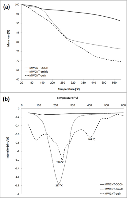
Thermal defunctionalization as observed by TGA, is the best evidence for the functionalization of MWCNTs. This is based on the observation that the temperature of MWCNT decomposition is considerably higher than the necessary temperature for the decomposition of functional groups bound to nanotubes. Thus, the TGA results can provide useful information concerning the functionalization of MWCNTs. According to Fig. 4a, since the TGA curve of MWCNT-COOH is almost thermally stable, the weight loss before decomposition of MWCNTs can be used to estimate the quantity of various groups attached to nanotubes. As seen in Fig. 4a, the MWCNT-amide sample exhibits one major decomposition at around 120℃-320℃ with a weight loss of about 16.5% which can be attributed to decomposition of the 2-aminobenzamide attached to MWCNTs. The TGA curve of MWCNT-quin shows two decomposition regions. The first region (180℃-300℃) corresponds to the decomposition of 2-aminobenzamide groups (as compared with the TGA curve of MWCNT-amide) with a weight loss of about 10.44%; these show some residual 2-aminobenzamide groups on the MWCNT surface. The second region is seen at around 300℃-500℃ with a weight loss of about 9.45%, which arises from the decomposition of the 4-hydroxyquinazoline on the MWCNT surface. Also, in the TGA curve of MWCNT-quin, a weight loss below 120℃ is observable, which can probably be attributed to residual humidity on the CNTs. These results indicate that there is one 2-aminobenzamide group for MWCNT-amide per 58.4 and one 4-hydroxyquinazoline for MWCNT-quin per 102.2 carbon atoms of MWCNTs, respectively, at 500℃. In other words, the proportions of carbon atoms that were functionalized with 2-aminobenzamide and 4-hydroxyquinazoline groups at 500℃ were calculated to be about 1.71 and 0.98 wt%, respectively.
The DTG curve provides further evidence for the covalent modification of MWCNTs (Fig. 4b). This curve can be used to determine the decomposition temperature of CNTs, which is the temperature at the highest peak for the CNTs on the DTG curve, and to define a mass-loss event as a single decomposing species (e.g., single peak) or as multiple decomposition events (e.g., double peaks, shouldered peaks). According to Fig. 4b, the one major peak at 217℃ could be attributed to the loss of 2-aminobenzamide groups bonded to MWCNTs. On the other hand, the DTG curve of MWCNT-quin shows two peaks at 240℃ and 405℃, which can be attributed to the loss of the residual 2-aminobenzamide on the MWCNTs and 4-hydroxyquinazoline groups, respectively. Overall, these results successfully confirm the functionalization of MWCNT-COOH with these compounds.
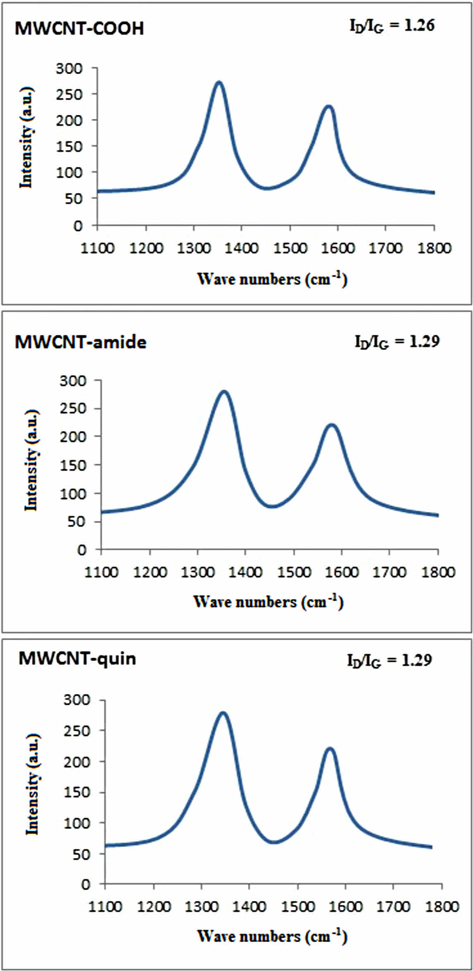
The Raman spectra of the modified MWCNTs are shown in Fig. 5. The modified MWCNTs exhibit a disorder mode at around 1330-1360 cm–1 (D band) and a tangential mode at around 1560-1590 cm–1 (G band), which originates from the in-plane tangential stretching mode of carbon-carbon bonds [30,31]. Furthermore, a comparison of the D and G band intensity ratios (ID/IG) of the three samples (Fig. 5) shows that the graphite structure of the MWCNTs is not changed by these reactions [11,18].
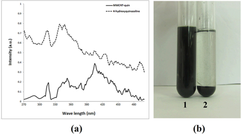
Direct evidence for functionalization and interaction between 4-hydroxyquinazoline and MWCNTs in the excited state is given by the fluorescence spectroscopy results (Fig. 6a). The emission spectrum of 4-hydroxyquinazoline with MWCNT-quin are compared by adjusting the absorbance of the organic moieties to be identical at the excitation wavelength (λex) of 320 nm. These two species show substantial quenching of fluorescence, again suggesting an energy exchange or interaction between the excited 4-hydroxyquinazoline and MWCNTs [32,33]. This demonstrates the existence of strong fluorescence quenching for organic moieties bonded to the MWCNT backbone [34]. In other words, comparing the two spectra confirms the functionalization of MWCNTs with 4-hydroxyquinazoline. Also, dispersion testing is a good means of assessment of the functionalization of MWCNTs because it shows whether modification of MWCNTs has been achieved. Fig. 6b presents the dispersed photographs of MWCNT-quin (1) and MWCNT-COOH (2) in DMF in which the samples had been sonicated and stored, respectively. As seen in Fig. 6b, MWCNT-COOH was insoluble in DMF, while MWCNT-quin can be dispersed in DMF homogeneously, and no precipitation was found even after it was sealed for 6 months at room temperature. These results indicate that MWCNT-COOH was functionalized by 4-hydroxyquinazoline.
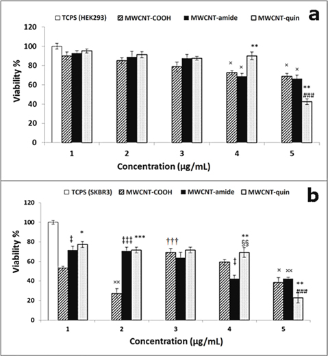
Fig. 7 show the results of the MTT assay carried out on the human HEK293 and SKBR3 cell lines each treated with TCPS (control), MWCNT-COOH, MWCNT-amide, and MWCNT-quin. Both cell lines were incubated for 24 h with varying concentrations of MWCNTs and the control materials. In Fig. 7a, a dose-dependent decline in cell viability can be observed. The percentage of viable cells did not significantly change within the 1-3 µg/mL concentration range regardless of which compound was applied to the HEK293 cells. However, when the concentration was increased to 4 µg/mL, the percentage of viable cells treated with either MWCNT-COOH or MWCNT-amide significantly declined compared to their counterparts treated with 3 µg/mL of either compound (
Similar to the HEK293 cells, increasing concentrations of each compound induced cell death among the SKBR3 cancer cells except for MWCNT-COOH, which caused a variable cell response (Fig. 7b). Also, these cells showed greater sensitivity to higher concentrations of MWCNT-quin than to the other two compounds. As for MWCNT-COOH, when the concentration was increased from 1 to 2 µg/mL a dramatic decline in the percentage of viable cells was observed (
We have introduced 4-hydroxyquinazoline groups on the surface of CNTs, and the results demonstrated the functionalization of MWCNT-COOH by this compound. Also, the modified MWCNTs can potentially be used as cellular killers, especially of cancerous cells. Our data allowed us to monitor the range of concentrations at which these derivatives can be applied safely for human health and to determine which compound might be more relevant to use for a particular cell type.
No potential conflict of interest relevant to this article was reported.


![(a) Emission spectra (in dimethylformamide [DMF]) of multiwalled carbon nanotubes (MWCNT)-quin in comparison with 4-hydroxyquinazoline at a matching absorbance (320 nm). (b) Photographs of dispersions of (1) MWCNT-quin and (2) MWCNT-COOH in DMF (1 mg/7 mL) after standing for 6 months. The samples were sonicated for 10 min, and photographs were taken 6 month after putting bottles.](http://oak.go.kr/repository/journal/20826/HGTSB6_2016_v17n1_45_f006.jpg)