Worldwide, there are around 278 million people with hearing impairments, and this number continues to rise with increasing environmental pollution and longer life expectancies [1]. Cochlear implants electrically stimulate nerve tissue, and can improve speech recognition in quiet environments for people with moderate to severe hearing impairments. However, cochlear implants are less effective in noisy environments. This is due to inherent disadvantages of electrical stimulation of nerve tissue, including the current-spreading effect, small number of electrodes, low spatial selectivity, stimulus artifacts, and direct contact of electrodes with tissues [2].
Laser stimulation has many advantages compared to electrical stimulation. First, due to improved directionality, laser stimulation has a decreased spreading effect, which can reduce signal disturbance among different channels. Second, lasers scatter very little and therefore have good spatial selectivity. Third, the laser source does not directly contact tissue, leading to improved safety. Finally, laser stimulation creates no stimulus artifacts [3-9].
Currently, the infrared laser stimulation technique has been widely used in many fields of nerve stimulation. In the auditory field, Izzo
In previous studies of laser stimulation of the auditory nerve, visible light (532 nm) [14], near-infrared laser light (813 and 808 nm) [15, 16], and midinfrared long-wavelength laser light (1.84-2.12 μm) [4, 10-13] were used to stimulate the cochlea.
Wells
However, experiments using the above two lasers adopted relatively high radiant exposure, which required high output power of the laser. For example, the radiant exposure for the 808-nm laser was about 52-1015 mJ/cm2 [16]. Actually, in experiments with a midinfrared long-wavelength laser, the radiant exposure was about 10-100 mJ/cm2 [4, 10-13], sometimes even higher [12]. In experiments with a 532-nm laser, the radiant exposure was about 0-1171.4 mJ/cm2 according to calculations [14]. To study the feasibility of a near-infrared short-wavelength laser with low energy to evoke auditory neural response, this paper adopts low laser radiant exposure of only 9.6-32.79 mJ/cm2. ,
In addition, although there have been several experiments focusing on optically evoked auditory response with different laser wavelengths, it is still necessary to find the most effective and safe wavelength to stimulate spiral ganglion cells in the cochlea.
Finally, in cochlear implants, electric currents of different frequencies excite different places to the maximum extent. It is reasonable to deduce that lasers with different simulation parameters (e.g. wavelength, radiant exposure, repetition rate, and pulse duration) will result in different auditory responses. Thus, it is necessary to study the effects of laser stimulation parameters for different wavelengths.
The current paper used a 980-nm pulsed near-infrared laser to stimulate the cochlea in guinea pigs, to investigate the feasibility of evoking auditory nerve impulses, and to provide reference laser stimulation parameters for optical cochlear implants.
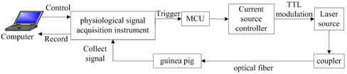
Figure 1 shows the schematic diagram of the current experiment. The computer controlled a physiological signal acquisition instrument (RM6240C, Chengdu Instrument Factory, Chengdu, China) to trigger a microcontroller unit (MCU, MC9S12XS128, Freescale Semiconductor, Inc.). The laser source (LSR980H-4W, Ningbo Yuan Ming Laser Technology Co., Ltd., Ningbo, China) generated continuous laser light with high power, which was modulated by a current source controller (LSR-PS-FA, Ningbo Yuan Ming Laser Technology Co., Ltd., Ningbo, China) driven by the MCU. Transistor-transistor logic (TTL) modulation was used. The output laser light with a certain repetition rate and pulse duration was coupled into an optical fiber 105 μm in diameter by an optical coupler (APFC-3AT, Zolix, Beijing, China). The laser light at the distal end of the fiber was measured and used to stimulate spiral ganglion cells of guinea pigs. The physiological signal acquisition instrument was used to collect signals, which were transmitted to a computer for further analysis.
The numerical value of the fiber’s aperture was about 0.2. In the experiment, the distal end of the fiber was placed about 1 mm away from the target modiolus using a micro-manipulator (MP-225, American Sutter Instrument Company). According to calculations, the area of the radiated modiolus was 0.0873 mm2, which approximately equaled the area of the distal end of the fiber (0.0866 mm2). Thus the radiant exposure was approximately the same between the end of the fiber and the target modiolus.
Ten adult Hartley guinea pigs were used for the experiments. Each animal was anesthetized with urethane (1.2 ml per 100 g body weight, 10% concentration, i.p.). Maintenance doses of 0.5 ml of 10% urethane were given throughout the experiment whenever the animal showed signs of arousal, which were assessed every 30 minutes. The animal was considered fully anesthetized when corneal reflection disappeared. Once anesthetized, the animal was fixed in a supine position on a heating pad, which maintained body temperature at 38℃. The skin was incised along the rim of the auricle root. The skin and connective tissue were removed from the upper part of the ear canal and the back of the skull. The tympanic cavity was then opened to expose the cochlea. A silver recording electrode was hooked onto the bony rim of the round window of the cochlea, a ground electrode was inserted into the dorsal skin, and a reference electrode was placed at the skin wound of the pinna. The surgical platform containing the animal was then moved onto a vibration-isolation table in a soundproof booth.
To create the animal model of deafness, the tympanic membrane was damaged with a scalpel. ACAPs were measured before and after damage to verify the success of the process.
2.4. Acoustical Stimulation and Measurements
The acoustical stimuli consisted of clicks (0.1 ms duration, 80 dB SPL intensity), generated by the physiological signal acquisition instrument and played through a speaker (SPA 2380/93). The acquisition instrument was also used to measure ACAPs at a sampling rate of 100 kHz with bandpass filtering range of 50-3000 Hz. For each animal, 30 waveforms were averaged to obtain an ACAP.
2.5. Optical Stimulation and Measurements
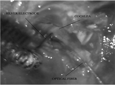
The laser light at the distal end of the optical fiber had a wavelength of 980 nm and repetition rate of 0.5 Hz, which was generated by the system shown in Fig. 1. Once ACAP measurements were completed at baseline and after the deafening procedure, a triangular needle was used to drill a hole 1 mm in diameter in the basal turn of the cochlea. The optical fiber was inserted through this hole and oriented toward the modiolus using a micromanipulator. The fiber was fixed in place, and was not in direct contact with cochlear structures or tissues. Figure 2 shows the placement of optical fiber and silver electrode. A total of 30 OCAPs were measured using the previously mentioned electrodes and acquisition instrument, and then averaged to obtain a final OCAP for each animal.
Four experiments were conducted in this paper. Experiment 1 validated the animal model of deafness, by comparing ACAPs before and after damage to the tympanic membrane. Experiment 2 tested the ability to evoke OCAPs in deaf animals using 980-nm pulsed near-infrared laser stimulation with a radiant exposure of 24.29 mJ/cm2, pulse duration of 100 μs, and repetition rate of 0.5 Hz. Experiment 3 investigated the effect of varying radiant exposure on OCAPs: The pulse duration of the laser was held at 100 μs, while the intensities of the laser were 9.6, 18.27, 24.29, and 32.79 mJ/cm2. Experiment 4 investigated the effect of varying pulse duration on OCAPs: The intensity of the laser was held at 32.79 mJ/cm2, and the pulse duration varied between 100 and 2000 μs.
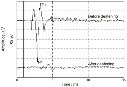
Experiment 1 verified that damaging the tympanic membrane caused deafness. Figure 3 shows ACAPs measured in response to stimulation (heavy vertical line at 1 ms), before and after damage to the membrane. The ACAP measured from the intact animal showed salient peaks and valleys. The most salient valley was marked as N1, followed by a salient peak marked as P1. Several small but still salient peaks and valleys appeared before N1, which represented cochlear microphonics (CMs). Note that CM, N1, and the following P1 are signs of ACAPs. Before tympanic membrane damage, the N1/P1 peak-to-peak amplitude was 310.7 μV; each grid represented 50 μV. In the same animal after tympanic membrane damage, no ACAP, CM, N1, or P1 was detected. These data support previous observations of absence of ACAPs after deafening animals by injecting kanamycin or neomycin [10, 12], or by destroying the basilar membrane with a dissecting probe [16]. Thus, this method of damaging the tympanic membrane was demonstrated to effectively cause deafness.
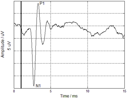
Experiment 2 tested the feasibility of 980-nm pulsed near-infrared laser stimulation to evoke OCAPs. Figure 4 shows an OCAP measured in a deaf animal in response to stimulation with a radiant exposure of 24.29 mJ/cm2, pulse duration of 100 μs, and repetition rate of 0.5 Hz. Similar to the ACAP observed in the intact animal, this OCAP had salient valleys and peaks, marked as N1 and P1, respectively, which were signs of compound action potentials (CAPs). However, there were no CMs in the OCAP waveform, similar to previous findings [13]. The mean N1/P1 peak-to-peak amplitude was 33.22 μV. Thus, we demonstrated that the 980-nm near-infrared laser could evoke auditory nerve impulses.
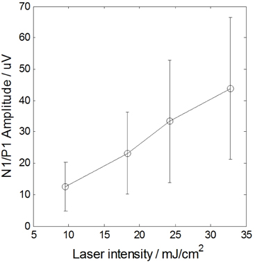
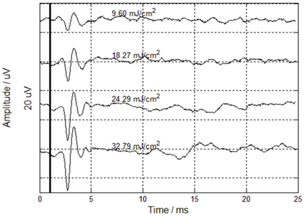
Experiment 3 investigated the effects of varying laser radiant exposure on OCAPs, as shown in Fig. 5. The mean N1/P1 peak-to-peak amplitudes were 12.46, 23.2, 33.4, and 43.89 μV for laser intensities of 9.6, 18.27, 24.29, and 32.79 mJ/cm2 respectively; grids represented 20 μV (ordinate) and 5 ms (abscissa). As shown in Fig. 6, OCAP amplitude increased with increasing infrared radiation energy, which agreed with previous findings [10, 13, 16]. A one-way analysis of variance (ANOVA) showed significant effects of laser radiant exposure on OCAPs (
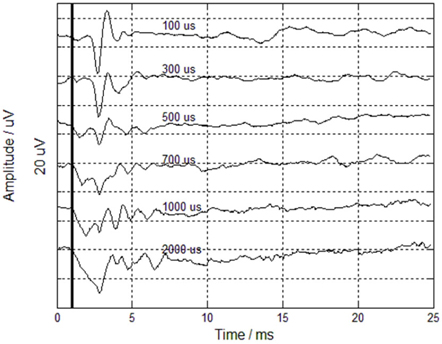
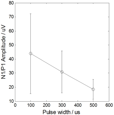
Experiment 4 investigated the effect of varying pulse duration between 100 and 2000 μs on OCAPs, while holding the intensity constant at 32.79 mJ/cm2. Figure 7 shows that as pulse duration increased, the mean N1/P1 peak-to-peak amplitude of the OCAP decreased. Specifically, the peak-to-peak amplitudes for pulse durations of 100, 300, and 500 μs were 43.89, 30.86, and 18.47 μV respectively, as quantitatively shown in Fig. 8. ANOVA also showed significant effects of pulse duration on OCAPs (
When the pulse duration was longer than 500 μs, no salient OCAPs were observed. Thus, near-infrared laser stimulation with a pulse duration below 500 μs activated auditory neurons and produced OCAPs. In previous research using pulsed infrared laser stimulation, radiant exposures measured at a fixed OCAP threshold were similar for pulse durations of 5, 10, 30, and 100 μs. However, for longer pulse durations (>100 μs), to elicit the same OCAP amplitude, higher radiant exposures were required as pulse duration increased [10, 11, 13, 19]. In other words, when the cochlea was stimulated at a fixed radiant exposure, OCAP amplitude decreased as pulse duration increased. This was also observed in the current study.
The current experiments demonstrated that a 980-nm pulsed near-infrared laser with relatively low energy can be used to activate the auditory nerve. The OCAPs evoked were similar to ACAPs. This indicates that, similar to acoustical stimulation, optical stimulation has high spatial selectivity, superior to that of electrical stimulation. Furthermore, the current data demonstrated that OCAP amplitude increased with increasing radiant exposure, and decreased with increasing pulse duration.
4.1. Mechanism of Low-Energy Laser Stimulation of Auditory Nerve
The underlying mechanism for low-energy laser stimulation of the auditory nerve has not been thoroughly described to date. Possible mechanisms may include photochemical, photomechanical, or photothermal effects.
According to Wells
Photomechanical effects might play a role in optical stimulation, when tissue is heated rapidly by a short laser pulse (pulse duration < 1 μs). Subsequently, laser-induced pressure waves are generated, which disrupt spiral ganglion cells and tissues [21]. For example, Wenzel
However, a photomechanical mechanism is not sufficient to explain the results obtained in the current paper, for two reasons. First, the laser pulse durations used here (100-2000 μs) were much larger than 1 μs, which violated the foundation of the photomechanical effect. Second, unlike ACAPs, OCAP waveforms showed no CMs, indicating no deflection of cochlear hair-cell cilia in response to mechanical perturbation of cochlear fluids. Thus, generation of OCAP in the current experiment does not involve electromechanical transformation, ruling out a photomechanical process.
For the present laser parameters, the most likely mechanism underlying optical stimulation of auditory nerves is a photothermal effect. Wells
4.2. Effect of Radiant Exposure on OCAP
In the current experiment, OCAP amplitude increased as laser radiant exposure increased. This strongly supports the photothermal effect of laser stimulation, because stronger laser intensity would deliver more laser energy to the cochlea per unit time. More auditory neurons in the laser beam path would then be activated, resulting in a higher transient temperature of the tissue and larger OCAP amplitudes.
In fact, previous studies have reported similar findings [10-12, 14]: (1) when radiant exposure was below a threshold, the laser could not activate auditory nerves; (2) when radiant exposure was above the threshold, the amplitude of auditory neural impulses increased with radiant exposure, as shown in the current paper; and (3) as the radiant exposure continued to increase, the amplitude of the auditory response reached a plateau, or even decreased. Under such conditions, lasers have very high energy levels, which damage neurons and tissues. Thus the radiant exposure of the infrared laser should be between the threshold and the plateau value. If an infrared laser is used to stimulate spiral ganglion cells in the cochlea, the highest radiant exposure to obtain suitable OCAP is recommended to be 3 J/cm2 [10]. On the other hand, if an infrared laser is used to stimulate the osseous spiral lamina, the required radiant exposure for suitable optical auditory brainstem evoked response (OABR) is much lower, with a maximum of 13-15 μJ/pulse [14].
The nonlinearity of auditory neural impulses in response to radiant exposure of laser stimulation reflects the nonlinearity of the auditory system. When the signal is too strong, the auditory neural system can adjust to protect itself.
4.3. Effect of Pulse Duration on OCAP
Previous studies found that to obtain a fixed OCAP threshold, incident laser exposure must increase as pulse duration increases [11, 13]. In other words, when the incident laser intensity is fixed, the amplitude of OCAP should decrease with increasing pulse duration, due to a higher activation threshold, as shown in the current paper. Typically, Izzo
These data indicate that optical stimulation is not governed by the total amount of energy that is delivered, but rather the duration over which the energy is delivered. When the duration is longer, more energy from each individual pulse will diffuse away from the absorption site in the tissue, and less energy will be absorbed to evoke auditory responses. Thus, for a fixed incident radiant exposure, OCAP amplitude decreases with increasing pulse duration.
It is worthwhile to note that the laser energy diffusing away over time will accumulate in tissue and eventually cause damage. Thus, shorter pulses are safer for optical stimulation. However, shorter pulses require the stimulating device to generate greater peak power, which is difficult to achieve. To balance these two factors, a pulse duration less than 100 μs seems to be optimal for optical stimulation of the auditory nerve [13].
This paper has demonstrated the feasibility of a pulsed near-infrared laser with low energy to effectively evoke OCAPs with a wavelength of 980 nm, pulse duration of 100-2000 μs, repetition rate of 0.5 Hz, and radiant exposure of 9.6-32.79 mJ/cm2 . The data provide theoretical and experimental support for studying the mechanism of optical stimulation of the auditory nerve, and provide effective ranges of stimulation parameters (e.g. radiant exposure, pulse duration) for the realization of single-channel optical cochlear implants. In addition, the experimental data for guinea pigs are a reference for future experiments with laser stimulation in human beings. Moreover, in a subsequent study the authors plan to increase the diversity of data measurement methods, and to increase data samples. For example, ABR and CAP can be measured sequentially for the same animals, and statistical analysis can compare the effects of different measurement methods on the results.