Sesamin is a lignan from sesame seed, and many bioactivities of sesamin for human health were reported.1-3 Their analytical methods using mass spectrometry (MS) are useful, but G. Yan reported that sesamin decomposed in electrospray ionization (ESI) ion-trap MS and that the protonated molecule of sesamin [M + H]+ was not detected but the dehydrated molecule [M − H2O + H]+ or dehydrogenated molecule [M − H2 + H]+ were clearly detected,4,5 although the fragmentation from those in-source ion were useful.
Matrices in matrix-assisted laser desorption/ionization (MALDI) are labile and decompose under the laser irradiation. Yamaguchi reported that the matrix-cyclodextrin complex used as a matrix and the cyclodextrin complex suppress the in-source decomposition of the matrix under the laser irradiation.6
Here, we thought that the labile compound of sesamin in the ionization is protected by the sesamin-cyclodextrin complex. A conventional cyclodextrin consists of α-Dglucopyranoses, and its ionization efficiency as the protonated molecule [M + H]+ was expected to be lower than that of the compound having amino group (-NH2).7 So, we used amino-β-cyclodextrin instead of conventional cyclodextrin, which has the amino group and high ionization efficiency.
Recently, ion-mobility MS was applied to the separation of organic compounds, lipids, carbohydrates, peptides, and proteins. We reported that ion-mobility MS was a powerful tool for distinguishing among the sugar chain isomers such as the branching isomers and glycosidic linkage isomers.8,9 The self-association property of the molecules was clearly revealed by ion-mobility MS technique for the different linkage sugar chains such as cello-oligosaccharides (β1-4 linkage), laminarioligosaccharides (β1-3), malto-oligosaccharides (α1-4 linkage), isomalto-oligosaccharides (α1-6 linkage). We are interested in the intermolecular binding complexes with ion-mobility MS, and in this study, we analyzed the complex between cyclodextrin and lignans such as sesamin, episesamin, and sesamolin.
Positive ion electrospray time-of-flight mass spectrometry was carried out by using a Waters Synapt G2-S quadrupole time-of-flight mass spectrometer (Waters Inc., Milford, USA) equipped with travelling-wave ion mobility spectrometry. For the detection of amino-β-cyclodexitrin (NβCyD) complexes with lignans, each sample (1 mM) was prepared in acetonitrile/0.1% aqueous formic acid (1:1, v/v) and mixed at the molar ratio of NβCyD/lignans (2/1), which was directly infused into the ion source at 180 µL/h of flow rate. Optimum positive-ion electrospray conditions included the following: capillary voltage, 1.5 kV; sampling cone voltage, 20 V; ion source temperature, 80 ℃; desolvation temperature, 280 ℃; cone gas flow rate, 0 L/h; desolvation gas flow rate, 500 L/h. The optimized ion mobility separation parameters at an IMS gas flow rate of 90 mL/min (N2 ) included a wave velocity of 1000 m/s and a ramping wave height from 10 V to 40 V. Data were acquired by using MassLynx software Ver. 4.1 (Waters Inc., Milford, USA) and processed by using Driftscope Ver. 2.1 software (Waters Inc, Milford, USA).
Solvents used in mass spectrometry were HPLC grade (NACALAI TESQUE, INC. Kyoto, Japan) and H2O used was purified by using ultrapure water preparing systems (Adventure A and Elix-UV3, Millipore, Darmstadt, Germany). 3A-Amino-3A-deoxy-(2A
Episesamin was chemically prepared from sesamin. To a solution of HCl (5-10%, v/v) in 50 mL methanol, 50 mg of sesamin was added and heated at 90 ℃ for 19 h. Obtained reaction mixture was subjected twice to a preparative thin layer chromatography (PLC silica gel 60 F254, 2 mm, Merck, Germany) with the eluent of hexane-ethyl acetate (7/3, v/v) at Rf 0.46. Finally episesamin was purified with a chiral column CHIRALPAK IB (id 20 × 250 mm) (Dicel Chemical Industries, LTD., Osaka, Japan), and it was identified by 1H and 13C NMR spectroscopy.
>
Detection of NβCyD-sesamin-NβCyD complex
NβCyD and sesamin were dissolved and mixed in the water/acetonitrile as described in Experimental section. The mixture was analyzed with (+) ESI Q-TOF MS. The mono isotope peak at
>
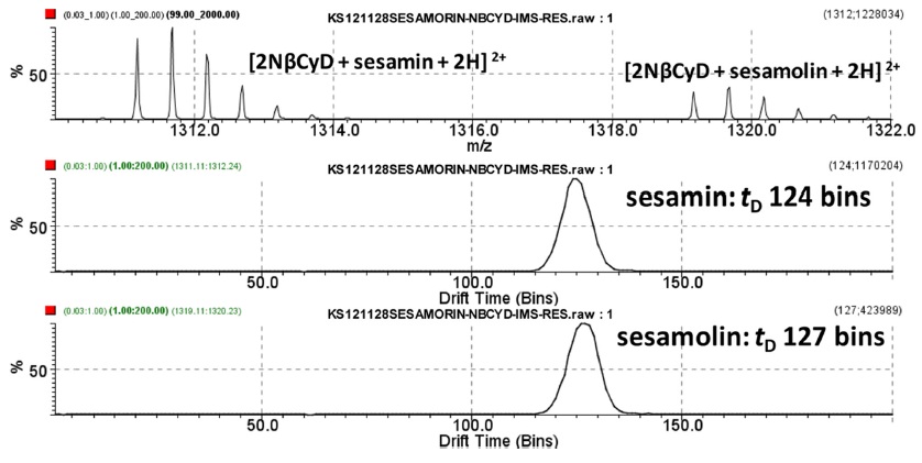
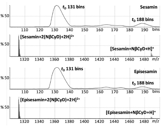
Ion-mobility MS of sesamin/episesamin-2NβCyD complexes
The sesamin-2NβCyD and episesamin-2NβCyD complexes were analyzed by ion-mobility MS. The ionmobility peak of the sesamin-2NβCyD complex was detected predominantly at 131 bins as shown in Figure 3. The low abundance and broad peak was observed at 188 bins, which suggested that the peak from the sesaminNβCyD (1:1) complex. Unfortunately, the NβCyD complexes of sesamin and episesamin were not separated by their ionmobility MS.
>
Ion-mobility MS of NβCyD-sesamolin-NβCyD complex
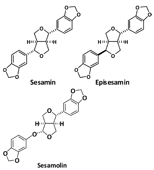
Sesamolin has one more oxygen atom between methylenedioxyphenyl group and furan ring at the ether part from sesamin (Figure 1). The complex of sesamolin and aminoCyD was analyzed by ion-mobility MS (Figure 4). The complex consisted one sesamolin and two amino-CyD molecules with the doubly charge from the
A labile compound of sesamin was ionized without decomposing of the molecule as the amino-βCyD complex because the ionization efficiency of amino-CyD itself is very high. The results suggested that the amino-CyDs assist the ionization of the labile molecules and low polar compounds without their decomposition by the formation of the aminoCyD complexes with these hydrophobic ligands, those are like amino-CyD complex-assisted ionization. The sesamin-2NβCyD and episesamin-2NβCyD complexes showed almost the same separation in the ion-mobility. The ion-mobility of the sesamolin-2NβCyD complexes was longer than those of the complexes of sesamin-2NβCyD and episesamin-2NβCyD.