Gami-jakyak gamcho buja decoction (Mecasin) was developed for treating amyotrophic lateral sclerosis patients with pain, joint contracture and muscular weakness. gami-jakyak gamcho buja decoction, which is the major component of Mecasin, has been used in traditional medicine to relieve pain, muscle spasms and cold syndrome due to blood deficiency [1]. A recent study showed that jakyak gamcho decoction (JGT) and its constituents had a protective effect against tert-butyl hydroperoxide (t-BHP)-induced cytotoxicity in the hippocampal (HT22) cell line [2]. In addition, jkyak gamcho buja decoction, which consists of JGT and
The constituents of Mecasin are
Mecasin was provided by the Nervous & Muscular System Disease Clinical Research Center of Wonkwang University Gwang-ju Korean Medical Hospital. It was mixed with pure water, without change of acidity, and the concentrations were 50 mg/mL, 100 mg/mL, and 200 mg/mL. The completed extracts were stored at room temperature (15 — 25°C).
The animals used in this study were 6-week-old SD rats. The reason SD rats were chosen is that they have been widely used in safety tests in the field of medicine, so the results can be easily compared with many other databases. The mean weights of the rats were 167.6 — 185.7 g and 146.2 — 159.8 g, respectively, for the male and the female rats at the time of Mecasin administration. For all animals, a visual inspection was conducted; all animals were weighed at the beginning. During 7 days of acclimatization, the general symptoms of the rats were observed once a day. The weights of the rats were recorded on the last day of acclimatization. No abnormalities were found.
The temperature of the lab was 20.1 — 21.5°C, and the humidity was 44.3% — 56.8%. Enough food (Rodent Diet 205053) and reverse osmosis water were provided.
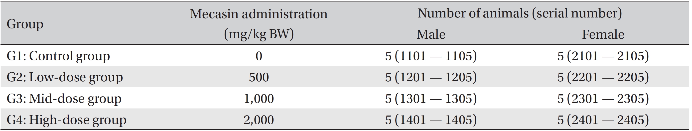
Groupings were done after 7 days of acclimatization. Animals were selected if their weights were close to the mean weight. In total, 20 male rats and 20 female rats were selected. The animals were randomly distributed into 4 groups (5 male and 5 female rats per group) as shown in (Table 1).
In this study 2,000 mg/kg was set as a high dose, and 1,000 mg/kg and 500 mg/kg were set as mid and low doses, respectively. In the control group, 10 mL/kg body weight (BW) of normal saline solution was administered. Mecasin and normal saline were administered into the mouths of the rats in all groups by using syringes once a day for 4 weeks. This study was conducted under the approval of the Institutional Animal Ethics Committee of KTR Co., Ltd.
Throughout the treatment, the general symptoms (side effects, revealing time, recovery time, etc.) and mortality were examined once a day. The weights were measured immediately before treatment, at grouping, once a week during treatment, and at the time of necropsy. The amounts of food and water were measured once a week before they were supplied to each cage, and the leftover food and water were measured on the next day. The difference was calculated and regarded as the daily food and water consumption.
The animals were fasted for 18 hours prior to necropsy and blood collection. Blood samples were drawn from the abdominal aorta by using a syringe needle under isoflurane anesthesia. Blood samples were collected into tubes containing ethylene diamine tetraacetic acid (EDTA) and were analyzed by using a blood counting analyzer (AD-VIA 120, Siemens, U.S.A.) to determine the red blood cell count (RBC), hemoglobin concentration (HGB), hematocrit (HCT), mean corpuscular cell volume (MCV), mean corpuscular hemoglobin (MCH), mean corpuscular cell hemoglobin concentration (MCHC), platelet count (PLT), white blood cell count (WBC), reticulocytes (Reti), prothrombin time (PT), and activated partial thromboplastin time (APTT).
The serum biochemistry analysis was performed using an auto-analyzer (TBA-120FR, TOSHIBA, Japan). Blood samples were centrifuged at 3,000 rpm for 10 minutes. Serum biochemistry parameters, including alanine aminotransferase (ALT), aspartate aminotransferase (AST), alkaline phosphatase (ALP), total bilirubin (T-BIL), alanine aminotransferase (ALT), gamma-glutamyl-transferase (GGT), blood urea nitrogen (BUN), creatinine (Crea), total protein (TP), albumin (Alb), albumin/globulin ratio (A/G ratio), total cholesterol (T-Chol), triglycerides (TGs), glucose (Glu), calcium (Ca), inorganic phosphorus (IP), creatine kinase (CK), sodium (Na+), potassium (K+), and chloride (Cl-) were examined.
The organs and tissues of all the animals were visually examined. Net weights were measured for the following organs: the brain, heart, liver, spleen, adrenal gland, testes, ovaries, uterus, thymus, lungs, prostate, pituitary gland, and kidneys. A relative weight ratio was calculated based on the fasting weight. The tissues from the following organs were obtained from all the animals and were fixed with 10% neutral buffered formalin solution: the brain, thymus, thyroid and parathyroid, heart, tongue, rahea, esophagus, lungs with bronchus, heart, liver, spleen, kidneys, adrenal gland, pituitary gland, stomach, small and large intestines, urinary bladder, pancreas, skin and mammary glands, salivary gland, seminal vesicle, prostate gland, ovaries, uterus, vagina, sternum, femur, spinal cord, mesenteric lymph node, skeletal muscles, and peripheral nerves. Testis and epididymis were fixed with Bouin solution. The eyeball was fixed with Davidson solution. Especially, liver, spleen, kidney, heart, and lung tissues were routinely processed, embedded in paraffin, and sectioned. The sections were stained with hematoxylin & eosin (H&E) dye for microscopic examination. All organs and tissues taken from all animals were examined microscopically.
Data on the weights, food consumption, hematology, serum biochemistry, and organ weights were tested using the SPSS program (SPSS 19.0). A Levene test was conducted to evaluate the homogeneity of the variance and the significance. The one-way analysis of variance (ANOVA) test was conducted when the homogeneity of the variance was recognized.
No mortality was observed in any of the groups of SD rat during the 4-week observation period. No clinical signs occurred in the male and the female SD rats. However, one animal in the mid dosage (1,000 mg/kg) male group experienced slight loss of fur, but this symptom was assumed to be the result of an accidental change. No significant differences in weights were observed between the experimental groups and the control group during the 4 weeks (Table 2).
No significant differences in hematology and organ weights were observed between the experimental groups and the control group. Serum biochemistry revealed that the low (500 mg/kg) and the high (2,000 mg/kg) dosage male groups showed significant decreases in IP (
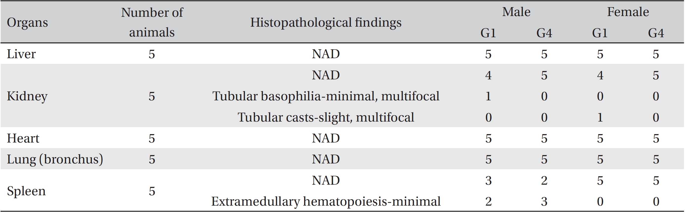
In the histopathological findings, a tubular basophilia of the kidney was found in one male rat in the control group, and a tubular cast of the kidney was found in one female rat in the control group. In addition, the two male rats in the control group showed extramedullary hematopoiesis of the spleen. In high dosage male group, an extramedullary hematopoiesis of the spleen was observed. However, those changes were minimal and had occurred naturally or sporadically. No other changes were observed in the livers, lungs, hearts of the control and the experimental groups (Table 4).
Mecasin has been utilized in clinics to relieve pain, joint contracture and muscular weakness, especially for patients whith amyotrophic lateral sclerosis [4]. A recent study showed that JGT and its constituents had a protective effect against t-BHP-induced cytotoxicity in the hippocampal HT22 cell line [2]. In addition, Jakyak Gamcho buja decoction, which consists of JGT and
Although Mecasin has been used in clinics, safety studies on Mecasin are insufficient. Toxicity tests provide important data and are essential for evaluating the safety of test substances in medications [24]. The current research trend for oral toxicity testing of extracts is to study subacute toxicity through GLP regulations. All the experiments for this research were conducted at the KTRI, an institution authorized to perform non-clinical studies, under the GLP regulations. Animal testing is the most fundamental and basic way to perform safety assessments [25]. The Korea Food & Drug Administration has testing protocol guidelines for the study of toxicity, and all experiments should be conducted following GLP regulations [26]. The present study was designed to evaluate a 4-week, repeated, oral dose, toxicity test of Mecasin in SD rats. Our study showed no significant changes in the weights, hematology, serum biochemistry, organ weights, necropsy findings, and histopathology of the rats. Also, no mortalities were observed. Although some changes were observed in several organs, those changes seemed to have occurred naturally or sporadically. During this 4-week, repeated, oral dose, toxicity test of Mecasin in SD rats, no toxicity changes due to Mecasin were observed in any of the male or female rats in the high dosage group. Thus, we suggest that the doses in a 13-week, repeated oral dose, toxicity test should be 0, 500, 1,000, and 2,000 mg/kg, respectively.
The results showed that administration of 500 — 2,000 mg/ kg BW of Mecasin to SD male and female rats did not cause any changes in weight or in the results of necropsy examinations. It also did not result in any mortalities, which indicated that the lethal dose of Mecasin was higher than 2,000 mg/kg. During this 4-week, repeated, oral toxicity test of Mecasin in SD rats, no toxicity changes due to Mecasin were observed in any of the male or the female rats in the high dosage group. Thus, we suggest that the doses in a 13- week, repeated test should be 0, 500, 1,000, and 2,000 mg/ kg, respectively.
Groups of animals
[Table. 2] Body weights in grams
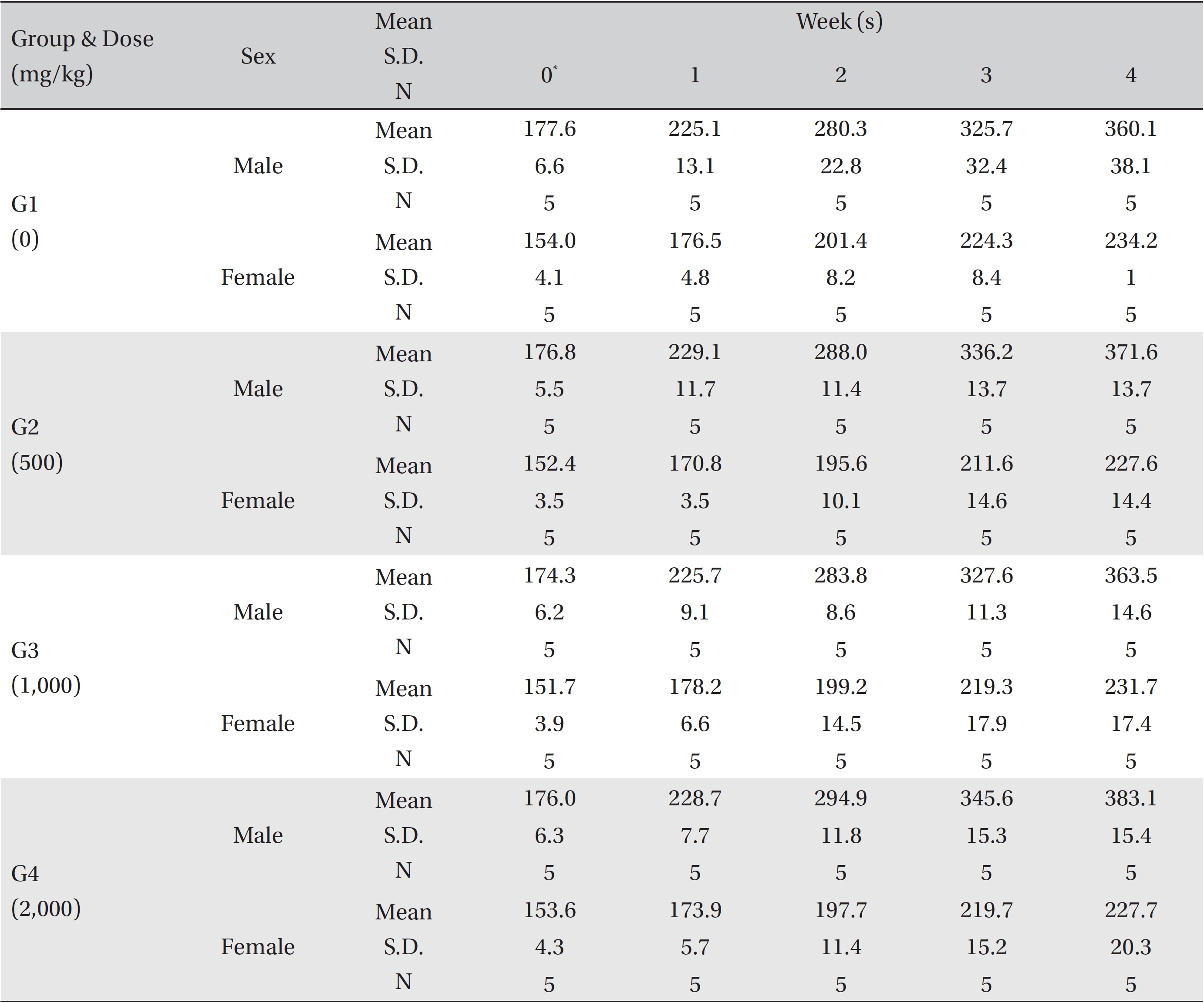
Body weights in grams
[Table. 3] Necropsy findings of male and female rats
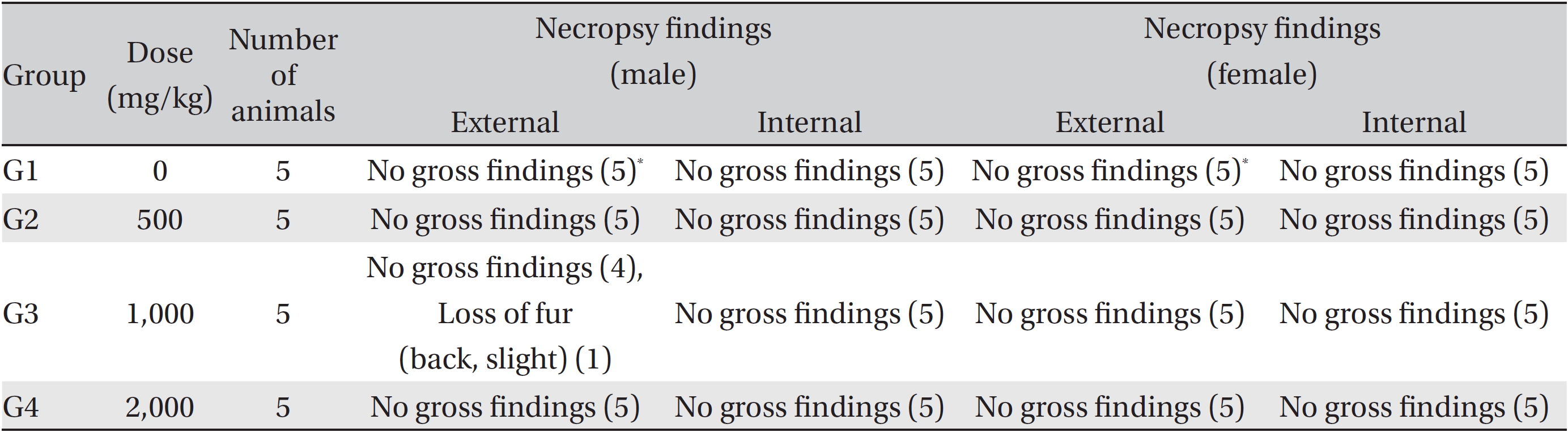
Necropsy findings of male and female rats
[Table. 4] Histopathological findings of rats

Histopathological findings of rats