The family Radiococcaceae is distributed in freshwater as well as terrestrial habitats worldwide. This family is defined broadly as coccoid green algae with mucilaginous cover reproducing only by autospores, and it is one of the most taxonomically difficult groups in green algae (Pazoutova 2008). The name Radiococcaceae was first reported by Fott (1959) in German but was not valid according to International Botanical Nomenclature. This family name has been previously used by some researchers (Bourrelly 1966, Philipose 1967, Hindak 1977), but since then the family Radiococcaceae have been determined to be valid by the description of Komarek (1979) in Latin. As stressed by Hindak (1977) the present state of the taxonomy of the family Radiococcaceae is not satisfactory (Hindak 1980). Hindak (1980) explained that the acceptance of traditionally acknowledged genera based on characters of a lower taxonomically value in other groups of green algae produced a subdivision within the system and many other genera. The classification principles of Chlorococcales were adopted based on those for other groups of algae. For example, the presence or absence of mucilage used to be deemed taxonomically significant, but Hindak (1980) pointed out that mucilage sheath was not a meaningful taxonomic characteristics. In the Radiococcaceae, however, several genera were classified according to spherical or oval cells (Korshikov 1953, Hindak 1977, 1980, Komarek and Fott 1983). In addition, many taxa in the genera are classified based on if the mother cell wall gelatinizes relatively quickly after protoplast division and in the process of autospore growth such that its remnants remain only temporarily in the gelatinous envelope of a colony. Some taxa have the characteristics that the remnants of cell wall exist for a long time in the colonial gelatinous envelope.
The family Radiococcaceae is composed of 4 subfamilies that are Radiococcoideae, Disporoideae, Dictyochlorelloideae and Palmodictyoideae. Of these, Radiococcoideae includes the genera
The purpose of this study is to review and add newly recorded species of 3 genera and 5 taxa in which the remnants of the mother cell wall exist for a long time in the colonial gelatinous envelope.
The samples of coccoid green algae were collected 290 sites including ponds, swamp, reservoirs, lakes and rivers from May 2011 to January 2014. Thirteen species in these 4 genera were identified and classified from 24 sites (Table 1). Sampling sites were located throughout Korea. All samples were collected by 10- or 20-μm mesh-sized plankton nets with vertical and/or horizontal towing, or were submerged benthic or soil algae collected with spoid or brush. Coccoid green algae samples were immediately fixed with Lugol’s iodine solution (0.5%) for immobilizing the cells to facilitate microscopic examination. To examine the fine structures and cellular shapes, and to identify and classify species of the coccoid green algae, temporary slides were made by the following steps: 1) the phytoplankton samples (coccoid green algae) were mixed with glycerin in micro tubes. 2) The mixed sample was placed, drop-wise on a slide glass, and was fixed in position with cover slide. Permanent slides were made using the following steps. 1) The phytoplankton samples (coccoid green algae) were mixed with liquid glycerol gelatin for mounting histochemical slides (Sigma-Aldrich, St. Louis, MO, USA); 2) the mixed sample was placed drop-wise on a slide glass and was fixed in position with a cover slide; 3) The margin of the cover glass was cemented with manicure (Thecashop, Seoul, Korea). The temporary and permanent slides were observed at ×200 to ×1,000 magnification using light microscopy (LM) (Axioskop 20 and Axio Imager A2; Carl Zeiss, Jena, Germany) with an attached digital camera (Axiocam HRc; Carl Zeiss) used to capture images. Specimens numbers are listed on the National Institute of Biological Resources (NIBR). The asterisk (*) mark indicates a newly recorded species and two asterisk (**) mark indicates a newly reported genus in Korea.

Sampling sites of genera Coenochloris, Radiococcus, Schizochlamydella and Thorakochloris from 2011 to 2014
At each station, physical and chemical factors of the water were recorded during the sampling periods. Water temp. (Water temperature, ℃) and EC (electric conductivity) was measured in situ using a portable thermometer and EC meter (Orion 5-star; Thermo Scientific, Waltham, MA, USA) and pH was measured in situ using a pH meter (Ultrabasic-5; Denver Instrument, Bohemia, NY, USA). This study obtained data of total nitrogen (TN) and total phosphate (TP) concentrations at each sampling site from the water information system of the Ministry of Environment (NIER 2014).
The identification of coccoid green algae is mainly based on guidelines from Komarek and Fott (1983), John and Tsarenko (2002), Hindak (1977, 1980, 1984, 1988), Prescott (1962), and Yamagishi and Akiyama (1984-1997).
The family Radiococcaceae is composed of 6 genera, 24 species, 1 variety based on Komarek and Fott (1983), Hindak (1977, 1980, 1984, 1988), John and Tsarenko (2002) and Komarek and Jankovska (2001).
This study details the taxonomic information, illustrations, classifications, references, basionyms, synonyms and distributions of the newly recorded species of these families.
Class Chlorophyceae
Order Chlorococcales
Family Radiococcaceae
Subfamily Radiococcoideae
>
Genus Schizochlamydella Korshikov 1953: 331
*
Basionym:
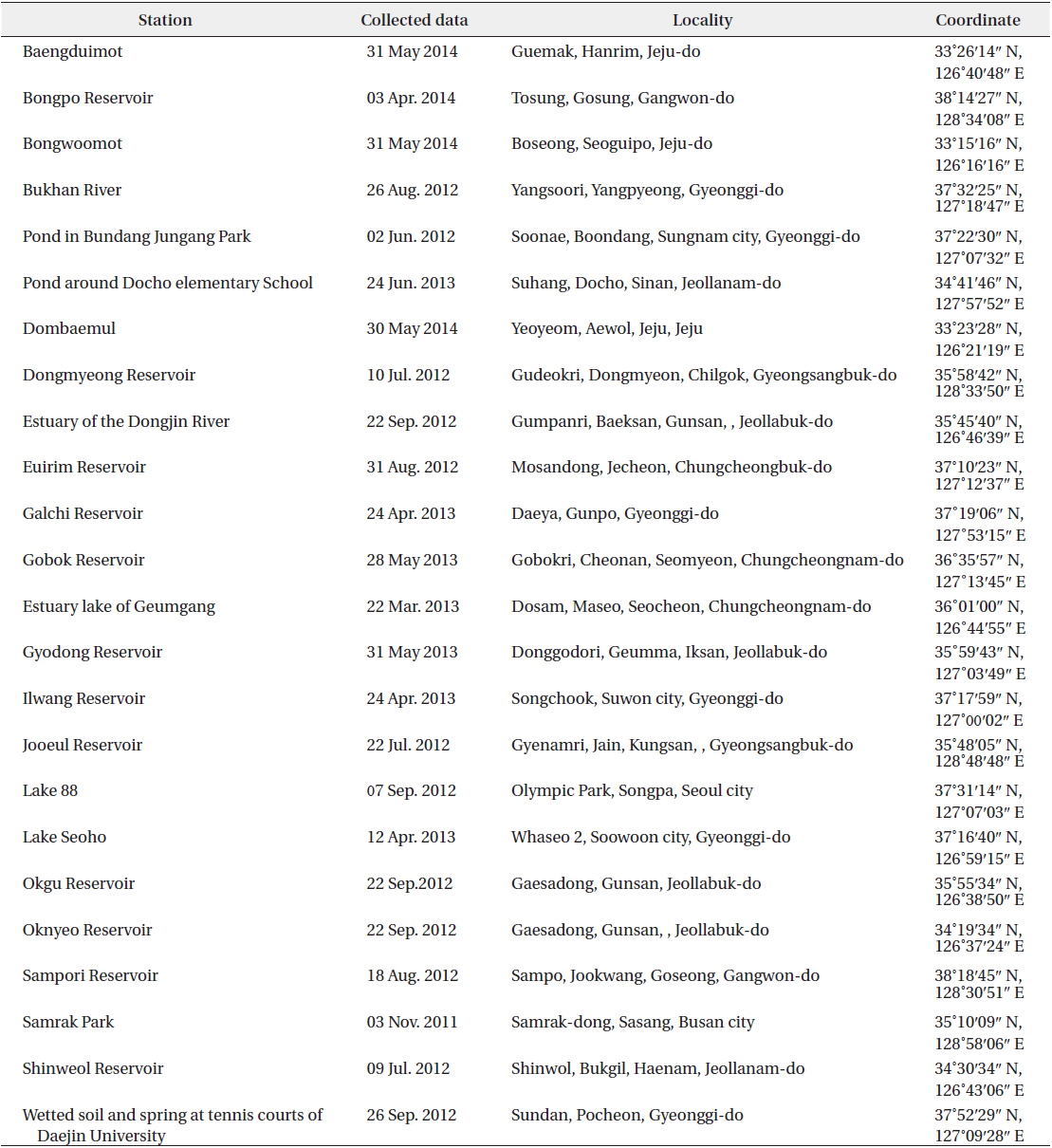
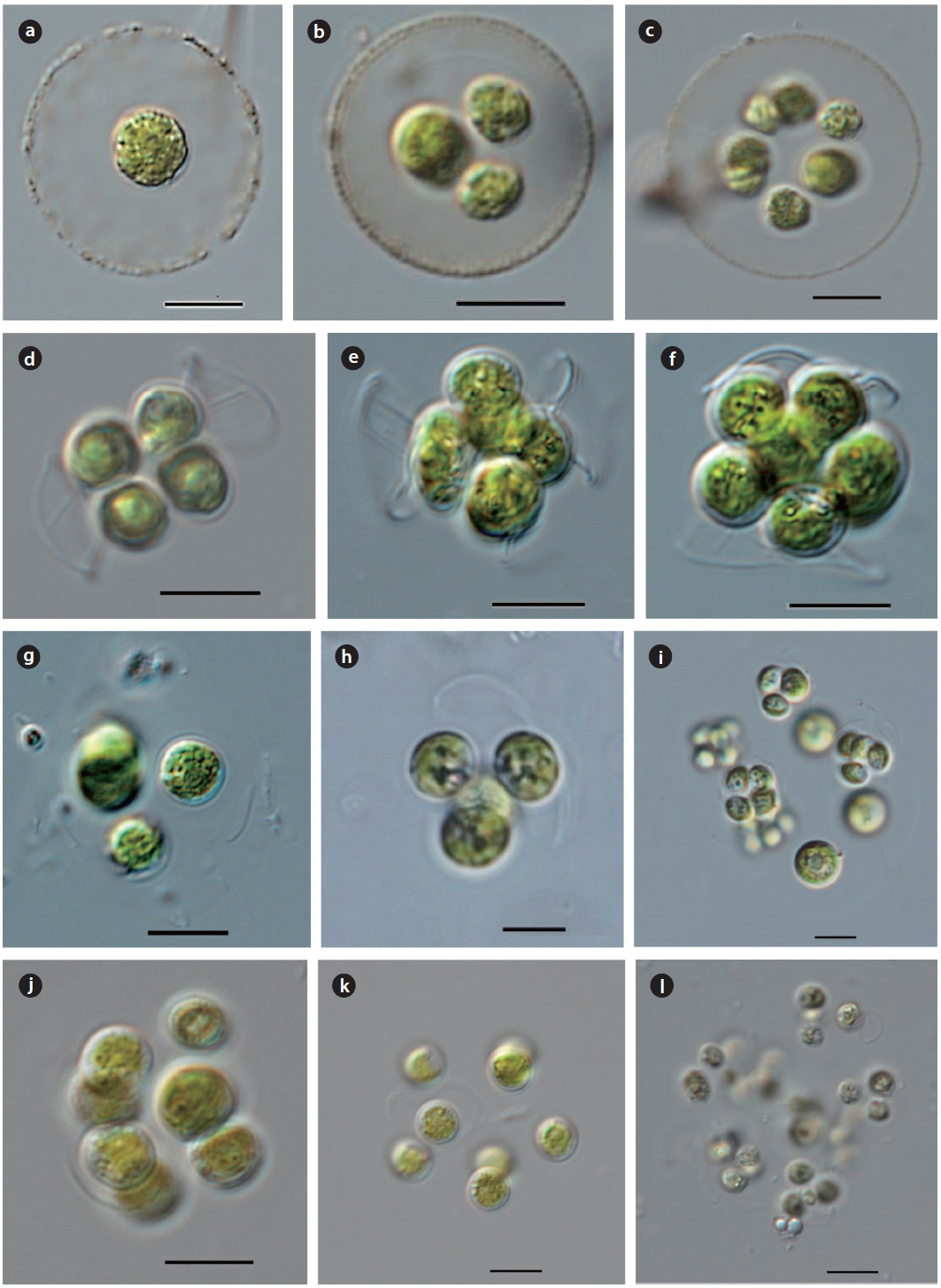
Illustration: Cells spherical, enclosed by colorless, non-stratified mucilaginous envelope. Cell wall thin, smooth or covered with a few warts. Chloroplast parietal, with a pyrenoid. 4 autospores in the mother cell wall (Fig. 1b, and c), released by the splitting of the mother cell wall. Cells 7−12 µm in diameter, colonies 30−50 µm in diameter of gelatinous envelope.
Key Reference: Fott (1974).
Specimens: DJGC20130424, DJGD20130531
Information of sampling sites: This species inhabits lakes and reservoirs as plankton and on wetted soil collected from Galchi Reservoir (24 April 2013; water temp. 23.4℃, pH 6.9, EC 143 µS cm-1), and Gyodong Reservoir (31 May 2013; water temp. 25.3℃, pH 8.5, EC 204 µS cm-1).
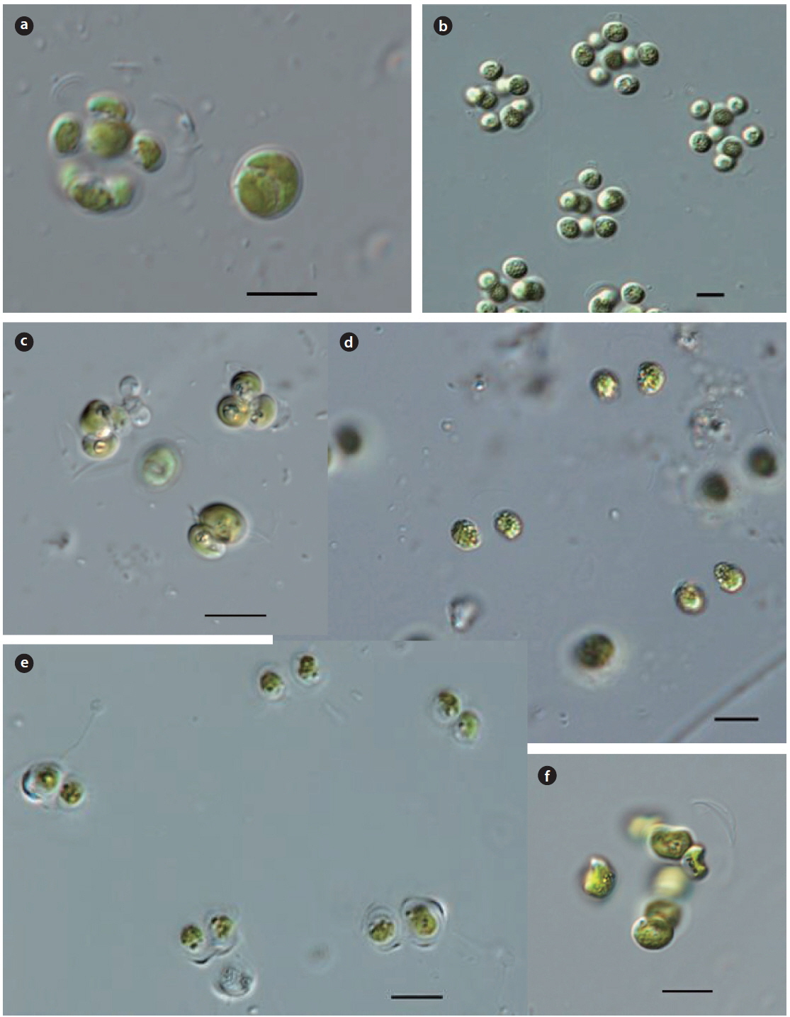
Illustration: Cells spherical, enclosed by colorless, nonstratified mucilaginous envelope. Cell wall thin, smooth or covered with a few warts. Chloroplast parietal, with a pyrenoid. 4−8 autospores in the mother cell wall (Fig. 2b and 2c), released by the splitting of the mother cell wall. Cells 7−10 µm in diameter, colonies 30−50 µm in diameter of mucilaginous envelope.
Key Reference : Watanabe (1977).
Specimens: KOSPCL0000108451, KOSPCL0000108706.
Information of sampling sites: This species inhabits lakes and reservoirs as plankton and on wetted soil collected from Lake 88 in Olympic Park (19 June 2013; water temp. 29.8℃, EC 193 µS cm-1), an estuary of Dongjin River (02. August 2012; water temp. 31.6℃, pH 8.5, EC 1,000 µS cm-1, TN 1.189 mg L-1, TP 0.100 mg L-1), wetted soil at tennis courts of Daejin University (26 September 2012), Baengduimot (30 May 2014; water temp. 31.7℃, pH 6.7, EC 160.4 µS cm-1), and Bongwoomot (31 May 2014; water temp. 30.4℃, pH 7.3, EC 251 µS cm-1).
>
**Genus Thorakochloris
*
Illustration: Colonies spherical, consisting of 4 cells, enclosed by colorless, non-stratified gelatinous envelope. Cell wall thin, smooth or covered with a few warts. Chloroplast parietal, with a pyrenoid. 4 autospores arranged in tetrahedral to quadrilateral shapes in the mother cell wall (Fig. 1d−f), released by the splitting of the mother cell wall. cells 5−8 × 4−6 µm, colonies 30−50 µm in diameter of mucilaginous envelope.
Key Reference: Fott (1933).
Specimens: DJBD20140530, DJDB20140530.
Information of sampling sites: This species inhabits lakes and reservoirs as plankton and wetland from Baengduimot (30 May 2014; water temp. 31.7℃, pH 5.3, EC 91.3 µS cm-1), and Dombaemul (30 May 2014; water temp. 28.7℃, pH 6.2, EC 417.1 µS cm-1).
Remarks:
>
Genus Coenochloris
Illustration: Colonies spherical or ovoid, consisting of 4-8 cells in a gelatinous envelope. Cells spherical or ovoid, which develop 4 autospores in the mother cell wall. 2 fragments of remnants persist after by the splitting of the mother cell wall. Chloroplast single parietal, with a pyrenoid. Cells 5−10 µm in diameter, colonies 20−40 µm in diameter
Key Reference: Fott (1974).
Specimens: KOSPCL0000108394, DJDJ20120926
Information of sampling sites: Wetted soil and spring at tennis courts in Daejin University (26 September 2012), Okgu Reservoir (22 September 2012; water temp. 25.2℃, pH 8.5, EC 231 µS cm-1, TN 1.315 mg L-1, TP 0.322 mg L-1), Oknyeo Reservoir (22 September 2012; water temp. 25.2℃, pH 9.0, EC 572 µS cm-1, TN 0.855 mg L-1, TP 0.194 mg L-1), and Bongpo Reservoir (03 May 2014; water temp. 22.3℃, pH 8.1, EC 125.7 µS cm-1, TN 0.536 mg L-1, TP0.036 mg L-1).
Remarks: This species was first reported from Prague in Czech Republic by Fott (1974). This species has also been reported in several other regions – in fish pond at Zelezna Studienka, Slovakia (Hindak 1977, 1980). In a small pool at the Little Carpathians Mountains near Bratislava, Slovakia (Hindak1988), and in a small pond at Prague, Czech Republic (Komarek and Fott 1983). Fott (1974) reported cells be enclosed by a gelatinous envelope, but their gelatinous envelope has not been investigated from this study
Synonym:
Illustration: Colonies spherical or irregular shaped, consisting of 4−8 cells in a gelatinous envelope. Cells spherical. 4 autospores enclosed with the mother cell wall, released by the splitting the mother cell wall, with wall remnants persisting thereafter. Cells 7−10 µm in diameter, colonies 40−70 µm in diameter.
Key Reference: Komarek (1979).
Specimens: DJSW20120709, DJOP20130619.
Information of sampling sites: Shinweol Reservoir (09 July 2012; water temp. 25.2℃, pH 9.0, EC 572 µS cm-1, TN 0.855 mg L-1, TP0.194mg L-1), Lake 88 in Olympic park (19 June 2013; water temp. 29.8℃, EC 572 µS cm-1), and Galchi Reservoir (24 April 2013; water temp. 23.4℃, pH 6.9, EC 143.2 µS cm-1).
Remarks: This species is widespread throughout the world as plankton in eutrophic or sometimes oligotrophic lakes, ponds, slow flowing river and swamps. This species was first reported from Lake Cicovske Jazero, near the Danube river, Slovakia (Komarek 1979), and was reported from Lake Cicovaske Jazero near the Danube, Slovakia (Hindak 1988 ) and rivers in Bratislava, Czech Republic (Komarek and Fott 1983).
Basionym:
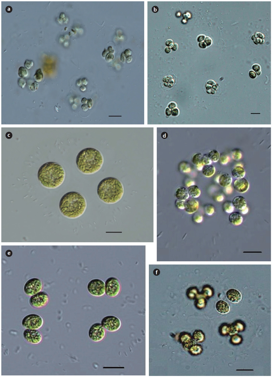
Illustration: Colonies spherical, consisting of 4−8 or- 16cells in a gelatinous envelope. Cells spherical, 4−8 autospores in the mother cell wall, released by the splitting of the mother cell wall. Chloroplast single parietal, invisible pyrenoid. Cells 5−10 µm in diameter, colonies 40−70 µm in diameter.
Key Reference: Hindak (1977).
Specimens: DJDM20120610, DJON20120922.
Information of sampling sites: Dongmyeong Reservoir (10 July 2012; water temp. 28.7℃, pH 7.3, EC 240.3 µS cm-1, TN 2.321 mg L-1, TP0.046mg L-1), Oknyeo Reservoir (22 September 2012; water temp. 25.2℃, pH 9.0, EC 572 µS cm-1, TN 0.855 mg L-1, TP0.194mg L-1), and Gobok Reservoir (28 May 2012; water temp. 27.2℃, pH 7.6, EC 209 µS cm-1).
Remarks: This species is widespread throughout the world as plankton in eutrophic or sometimes oligotrophic lakes, ponds, slow flowing rivers and swamps. This species (strain hindak 1975/158) was first reported from Lake Strokovec, Bratislava in south and west Slovakia by Hindak (1977). This species was also reported from Lake Sempachersee, Switzerland (Hindak 1980), and lakes and ponds from Czech Republic and Ukraina (Komarek and Fott 1983).
Cells length 5−10 µm, width 4-8 µm, colonies 40−70µm in diameter.
Illustration: Colonies spherical or irregular shape, 4−8 or16 cells in a gelatinous envelope. Cells ellipsoid or wide ovoid. Eight spherical autospores in the mother cell wall, released by the splitting of the mother cell wall, with 1−2 fragments of wall remnants persisting thereafter. Chloroplast parietal, with a pyrenoid. Cells 8−6 µm in diameter, colonies more than 70 µm in diameter.
Key Reference: Korshikov (1953).
Specimens: KOSPCL0000107362, DJDM20120610.
Information of sampling sites: Dongmyeong Reservoir (10 July 2012; water temp. 28.7℃, pH 7.3, EC 240.3 µS cm-1, TN 2.321 mg L-1, TP0.046mg L-1), the Bukhan River at Yangsoori (26 August 2012; water temp. 30.2℃, EC 238 µS cm-1), Ilwang Reservoir (24 April 2013; water temp. 20.2℃, pH 6.8, EC 253.2 µS cm-1), and Pond around Docho elementary school (24 June 2013; water temp. 30.2℃, EC 347 µS cm-1).
Remarks: This species is widespread throughout the world as plankton in eutrophic or sometimes oligotrophic lakes, ponds, slow flowing rivers and swamps. This species was first reported from the Kharkov Area, Lake Liman, Ukraine by Korshikov (1953). It has also been recorded from several other regions: in a fish pond of Central Bohemia (Fott 1974), in stagnant and flowing waters in Bratislava, in the river Danube, in a fish pond at Zelezna Studienka, Slovakia (Hindak 1977), and in an inundation lake at the Morava River at Bratislava-Devin, Slovakia (Hindak 1980).
Synonym:
Illustration: Colonies spherical or irregular shaped, consisted of 4−8 or 16 cells in a mucilaginous envelope. Cells ellipsoid or slightly asymmetrically shaped. 8 autospores in the mother cell wall, released by the splitting of the walls, with wall remnants persisting thereafter. Chloroplast single parietal, with a pyrenoid. Cells 5−12 µm in length, 4−8 µm in width. Colonies 40−80 µm in diameter.
Key Reference: Hindak (1980).
Specimens: KOSPCL0000107417, KOSPCL0000108372.
Information of sampling sites: Wet soil and spring at tennis courts at Daejin University (26 September 2012; no data), the Bukhan River at Yangsoori (26 August 2012; water temp. 30.2℃, EC 238 µS cm-1), Lake 88 in Olympic Park (19 June 2013; water temp. 29.8℃, EC 572 µS cm-1), and Jooeul Reservoir (22 September 2012; water temp. 22.7℃, EC 267 µS cm-1).
Remarks: This species is widespread throughout the world as plankton in eutrophic or sometimes oligotrophic lakes, ponds, slow flowing rivers and swamps. This species was first reported from the small dystrophic lake Gerzensee, Kastanienbaum, Switzerland by Hindak (1978, 1980) and littoral of a fish pond in Thienemann-Weiher, near Seeon, Bavaria, Germany (Hindak 1980).
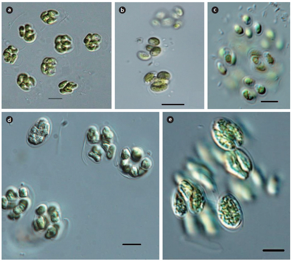
Illustration: Colonies spherical or irregular ovoid, consisted of 4−8 or 16 cells or temporarily 64 cells in a gelatinous envelope. Cells elongated asymmetric reniform or ovoid, cell wall smooth. Usually, 4 autospores in the mother cell wall, released by the splitting of the mother cell wall, with wall remnants persisting thereafter for a long time. Chloroplast single parietal, with a pyrenoid. Cells 8−12 µm in length, and 4−8 µm in width. Colonies 40−100 µm in diameter.
Key Reference: Hindak (1977).
Specimens: KOSPCL0000108410, DJBR20120826.
Information of sampling sites: the Bukhan River at Yangsoori (26 August 2012; water temp. 30.2℃, EC 238 µS cm-1), Sampori Reservoir (18 August 2012; water temp. 30.1℃, EC 208 µS cm-1), Okgu Reservoir (22 September 2012; water temp. 25.2℃, pH 8.5, EC 231 µS cm-1, TN 1.315 mg L-1, TP0.322mg L-1), Euirim Reservoir (31 August 2012; water temp. 28.2℃, pH 7.6, EC 138 µS cm-1), and Bongpo Reservoir (03 May, 2014; water temp. 20.8℃, pH 7.6, EC 125.7 µS cm-1).
Remarks: This species is widespread throughout the world as plankton in eutrophic or sometimes oligotrophic lakes, ponds, slow flowing rivers and swamps. This species was reported from plancto fluminum (flumen Danubius in Bratislava, locus classicus), lacus (Bratislava – Trávniky) piscinarumque (elezná Studienka, Bratislava), Slovacia by Hindak (1977). It has also been reported from several oher regions: in Lake Velky Les near the Danube in Gabeikovo, Slovakia (Hindak 1977), and in the inundation lake at the Morava River at Bratislava-Devin, Slovakia (Hindak 1980).
Basionym:
Illustration: Colonies spherical or irregular shaped, consisted of 4−8 or16 cells in a mucilaginous envelope. Cells wide ovoid, ellipsoid or reniform, cell wall smooth, usually 4 autospores in the mother cell wall, released by the splitting of the mother cell wall, with remnants persisting for a long time thereafter. Chloroplast single parietal, with a pyrenoid. Cells 10−15 µm in length and 5−10 µm in in width. Colonies 40−80µm in diameter.
Key Reference: Hindak (1980).
Specimens: DJOP20120907, DJJE20120922.
Information of sampling sites: Lake 88 in Olympic Park (07 September 2012; no data), and Jooeul Reservoir (22 September 2012; water temp. 22.7℃, EC 267 µS cm-1).
Remarks: This species is widespread throughout the world as plankton in eutrophic or sometimes oligotrophic lakes, ponds, slow flowing rivers and swamps. This species was first reported from Lake Nove Mestonad Vahom, Slovakia by Hindak (1980). It has also been reported from several other regions: in a gravel pit lake at Nove Mestonad Vahom, Slovakia (Hindak 1977) and eutrophic lakes throughout south−western Slovakia (Komarek and Fott 1983).
>
**Genus Radiococcus
*
Synonym:
Illustration: Colonies spherical, irregularly shaped, consisting of 4−8 or16 cells randomly arranged, enclosed by a mucilaginous envelope arranged with radial striation. Cells spherical. usually 4 autospores in the mother cell wall, released by the splitting of the mother cell wall, with cap-like remnants persisting thereafter. Chloroplast parietal, with a pyrenoid. Cells 5−10 µm in diameter, colonies 40−120µm in diameter.
Key Reference: Lund (1956).
Specimens: DJSH20120527, DJOP20130619.
Information of sampling sites: Lake Seoho (27 May 2012; water temp. 30.4℃, pH 7.3, EC 569 µS cm-1), and Lake 88 in Olympic Park (19 June 2013; water temp. 29.8℃, EC 572 µS cm-1).
Key Reference: Lund (1956).
Remarks: This species is widespread throughout the world as plankton in eutrophic or sometimes oligotrophic lakes, ponds, slow flowing rivers and swamps. This species was first reported from Lake District, Great Britain by Lund (1956). It has also reported from many other regions: in the Lake District and Windermere in the British Isles (John and Tsarenko 2002), a fishpond at Zelezna Studienka, at Bratislava and a small pond in the Little Carpathian Mountains. near Bratislava from Slovakia (Hindak 1988).
*
Basionym:
Synonym:
Illustration : Colonies spherical, consisting of 4−8 or 16cells, enclosed by a mucilaginous envelope arranged with radial striation. Cells spherical, 4 autospores in the mother cell wall, released by the splitting of the cell wall, with wall remnants persisting thereafter. Chloroplast parietal, with a pyrenoid. cells 5−12 µm in diameter, colonies 40−70 µm in diameter.
Key Reference: Schmidle (1902).
Specimens: KOSPCL0000106117, DJOP20130619.
Information of sampling sites: Samrak Park in Busan (15 June 2011; no data), and Lake 88 in Olympic Park (19 June 2013; water temp. 29.8℃, EC 572 µS cm-1).
Remarks: This species is widespread throughout the world as plankton in eutrophic or sometimes oligotrophic lakes, ponds, and swamps or epiphyton to under the surface of still water. This species was first reported from the Sinai Peninsula and Syria, Egypt by Schmidle (1902). It has also been reported from many other regions: in eutrophic lakes, rivers, and still water throughout the British Isles (John and Tsarenko 2002), in South Carolina and Tennessee of the southeastern United States (Dillard 1989), in Shinobazu, Japan (Yamagishi and Akiyama 1984-1997), in bodies of the water of eastern and north-eastern states throughout India (Jena and Adhikary 2007), in artificial lakes of Goias Mnicipality, Brazil (Nogueira and Oliveira 2009).
*
Basionym:
Synonym:
Illustration: Colonies spherical, consisting of 4−8 or 16 cells, enclosed by a mucilaginous envelope, arranged with radial striation. Cells spherical, 4 autospores in the mother cell wall, released by the splitting of the mother cell wall, with the remnants of the cell wall remaining thereafter. Chloroplast parietal, with a pyrenoid. cell 5−8 µm in diameter, colonies 40−70 µm in diameter.
Key Reference: Komarek (1979).
Specimens: KOSPCL0000108512, KOSPCL0000106002.
Information of sampling sites: Bundang Jungang Park (02 June 2012; water temp. 26.3℃, pH 8.2, EC 407 µS cm-1), and Lake 88 in Olympic Park (19 June 2013; water temp. 29.8℃, EC 572 µS cm-1)
Remarks: This species occurred throughout the world as plankton in eutrophic or sometimes oligotrophic lakes, ponds, slow flowing rivers and swamps. This species was first reported from Lake Cicovske Jazero, near the Danube River, Hungary by Komarek (1979). It has also been reported from many other regions: in the Danube Rivers, Hungary (Komarek 1979), in Chiemgau, Oberbayern, Germany (Komarek and Fott 1983).
Taxa belonging to 4 genera (
A study on fresh-water green coccoid algae was carried out at ponds, swamps, reservoirs, lakes and rivers (290 sites) from May 2012 to January 2014. In this study, 4genera and 13 taxa of the family Radiococcaceae having the remnants of the cell wall existing for a long time in the colonial gelatinous envelope are identified and classified from 24 sites. 8 taxa (1 genus) of the family Radiococcaceae in coccoid green algae are reported here based on characteristics by light microscopy: