The order Chlorococcales belongs to the division Chlorophyta and class Chlorophyceae, which widely inhabit freshwater and soil. It is composed of useful microorganisms with a short life cycle that are easily cultured (Richmond 1986, Haag 2007). Komárek and Fott (1983) reported that this order included 15 families, 148 genera, and 1,025 species, whereas Hindák (1977, 1980, 1984, 1988) reported that it included only 168 species, excluding the genus
In this study, aerial algae were collected from tree barks, stones, soil, mosses, and lichen around the Hongcheon-River. We aimed to add genera and species of Korea flora to the order Chlorococcales and newly described these discoveries.
Aerial algae were sampled from 28 sites around the Hongcheon-river at Gangwon-do from December 2011 to September 2012 (Table 1 and Fig. 1). The samples were collected from tree barks, stones, soil, and mosses around the river by using a soft brush and a spatula. Each sample was sealed and refrigerated in a light-tight container with sterilized distilled water and transferred to the laboratory (Crispim et al. 2004). Some of the samples were fixed and stored in 1% formalin. Enriched cultures of aerial algae were made on Bold’s basal medium (Stein 1973) and maintained in the algal culture collection of Kyonggi University (ACKU).
List of 28 sampling sites of Hongcheon-river at Gangwon-do from December 2011 to September 2012
Analyses of physico-chemical environmental factors, such as the surface temperature of the substrates, light intensity, and humidity, were conducted on all the sampling dates (Song et al. 2012). The temperature was measured with a stem thermometer, and the surface temperature was measured with a Testo 830-T1 infrared thermometer (Testo, Lenzkirch, Germany). Humidity was determined with a Testo 625 hygrometer (Testo), and light intensity was determined by using a LX-1108 light meter (Lutron, Taipei, Taiwan).
The taxonomic classification system of the order Chlorococcales was based on that of Komárek and Fott (1983), and the taxa were identified according to the system used by Hindák (1977, 1980, 1984, 1988), Chung (1993), Prescott (1973), Prescott et al. (1972, 1977, 1981, 1982), Hirose et al. (1977), John et al. (2002), and Wehr and Sheath (2003). The cultured samples were examined at ×400–1,000 magnification under a light microscope (BX41; Olympus, Tokyo, Japan), equipped with Nomarski differential interference optics. Species were illustrated in the drawing attachment, together with light microscope photographs.
In this study, seven genera belonging to the order Chlorococcales and eight species belonging to these seven genera were newly recorded in Korea. The newly recorded genera are
Family Chlorococcaceae
Subfamily Spongiococcoideae
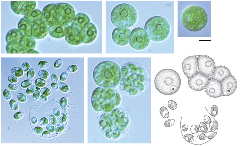
Genus
This genus was named by Deason in 1959, and newly recorded in this study in Korea. Deason (1959) isolated the genus
Distribution: Caribbean Islands, Alabama (Ettl and Gärtner 1995).
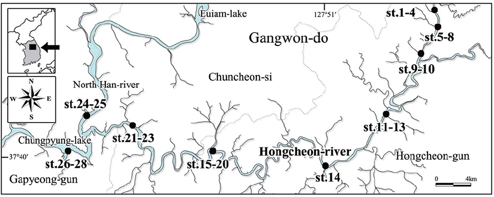
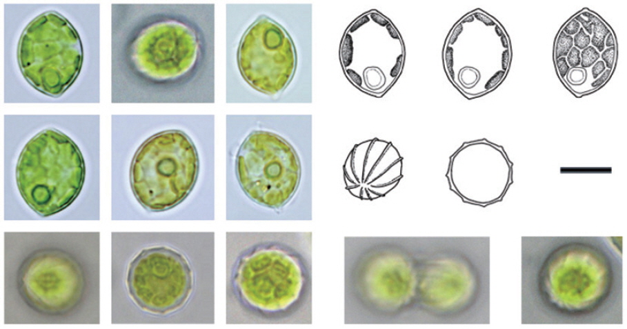
Spherical cells, 10–22 µm in diameter. A sponge-like chloroplast, with a pyrenoid usually situated in the center. Large and fragmented pyrenoid. Cells mostly solitary, with four young cells, which are sometimes enclosed in the wall of a mother cell. Cells up to 30 µm in diameter have been recorded (Deason 1971). Zoospores unobserved.
This species has been found among aerial algae (Deason 1959). In this study, this species was found in a wide range of habitats where the light intensity and humidity ranged from 3,915–48,000 lux and 26–65%, respectively (Table 2). However, it was found only in the moss samples, suggesting that the moss might protect it from environmental changes.
The environmental factors of 28 sites in Hongcheon-river from December 2011 to September 2012
Sites of collection: 1, 5, 19, 21, 27; hereafter, site numbers correspond to Fig. 1.
Specimen: NIBRCL0000104610, ACKU HC1-96.
Family Chlorococcaceae
Subfamily Spongiococcoideae
Genus
This genus was named by Brown et al. (1964) and recorded for the first time in Korea in this study. The genus
Distribution: in Europe, Italy (Ettl and Gärtner 1995); in North America, Mexico (Ettl and Gärtner 1995) and Texas (Ettl and Gärtner 1995); in Asia, Japan (Ettl and Gärtner 1995).
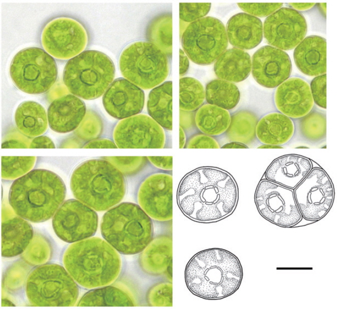
The cells are spherical, 15–30 µm in diameter. The species recorded in the present study was larger than 10 µm, which was larger than that observed by Nakano (1983). This species is distinguished from other species by a fragmented pyrenoid, a single nucleus, and an ellipsoidal zoospore. The cell wall is generally thin, but older cell walls may be thick. Sometimes, two or four young cells are enclosed in the mother cell wall. The zoospore is ellipsoidal, 9–12 µm long, and 3–6 µm wide. This species was found in soil samples and similar to that reported by Nakano (1983).
The genus
Sites of collection: 5, 6, 7, 9, 11, 15, 22, 24, 25.
Specimen: NIBRCL0000104612, ACKU HC4-128.
Family Palmellaceae
Subfamily Neochloridoideae
Genus
This genus was named by Printz (1921), and recorded for the first time in Korea in this study. The morphological characteristics of this genus are similar to those of the genus
Distribution: in Europe, Austria (Ettl and Gärtner 1995), Czech Republic and/or Slovakia (Ettl and Gärtner 1995), Romania (Caraus 2002), and Russia (Black Sea) (Ettl and Gärtner 1995); in Australia and New Zealand, Victoria (Day et al. 1995); in Antarctic and the subantarctic islands, Signy Island (Ettl and Gärtner 1995).
The cells are ellipsoidal or spherical, 8–15 µm wide, with a parietal cup-shaped chloroplast. This species is an aerial algae (Gomez et al. 2003). We isolated this species from the aerial environment of soils and rocks.
Sites of collection: 5, 8, 11, 19, 21, 22, 27.
Specimen : NIBRCL0000107672, ACKU HC1-65.
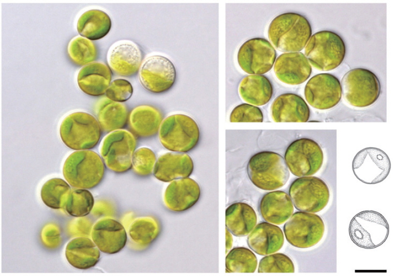
Family Radiococcaceae
Subfamily Radiococcoideae
Genus
This genus was named by Korshikov (1953). The genus
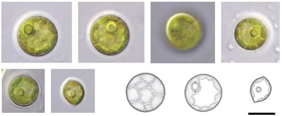
Cells 7–15 µm in diameter, surrounded by a mucilage layer. Parietal chloroplast, with a single pyrenoid. Four ellipsoidal cells surrounded by a mucilage layer when young, and a single spherical mature cell surrounded only by a mucilage layer.
We isolated this species from aerial environments of soils and mosses. In this study, this species was sampled at low light intensity (2,100–3,915 lux) and high humidity (60–61%) conditions (Table 2). We expected this species to inhabit areas with high temperatures but low surface temperatures.
Sites of collection : 1, 13.
Specimen : NIBRCL0000104596, ACKU HC1-74.
Family Chlorellaceae
Subfamily Chlorelloideae
Genus
This genus was named by Reisigl (1969), and first recorded in Korea. Komárek and Fott (1983) suggested that this could be transferred to
Distribution: in Europe, Austria (Ettl and Gärtner 1995) and Rumania (Caraus 2002).
Spherical cells, 5–13 µm in diameter. Parietal chloroplast parietal, 2 fragments, with a single fragmented pyrenoid.
kaloud (2009) recorded this species as aerial algae isolated from soil, but we sampled this species from tree bark lichens in this study.
Sites of collection : 3, 4, 14, 16, 26.
Specimen : NIBRCL0000104604, ACKU HC3-6.
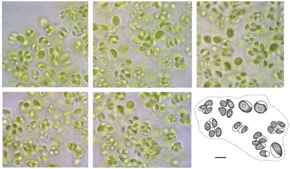
Family Chlorellaceae
Subfamily Ankistrodesmoideae
Genus
This genus was named by Korshikov (1953), and recorded for the first time in Korea in the present study. The genus
Synonym:
Distribution: in Europe, Britain (John et al. 2002), Czech Republic (Gross et al. 2002), Czech Republic and/ or Slovakia (Ettl and Gärtner 1995), Italy (Ettl and Gärtner 1995), Romania (Caraus 2002), and Russia (Black Sea) (Ettl and Gärtner 1995); in North America, Mac Robertson Land (Ettl and Gärtner 1995); in Asia, Japan (Ettl and Gärtner 1995); in Australia and New Zealand, New Zealand (Broady et al. 2012) and Victoria (Day et al. 1995).
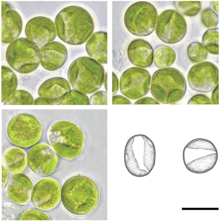
Ellipsoidal or oval cells, with the anterior end slightly pointed, 8–14 µm long, 3–5 µm wide. Parietal chloroplast, with a smooth margin, filling two-thirds of the cell, with a single nucleus, without a pyrenoid. The cells are attached at one or two ends by a cap (small mucilage pad).
This species is known to be aerial algae, which attaches to rocks. In this study, this species was found at low light intensities and in humid aerial environments of mosses.
Sites of collection: 2, 13, 22.
Specimen : NIBRCL0000104607, ACKU HC3-30.
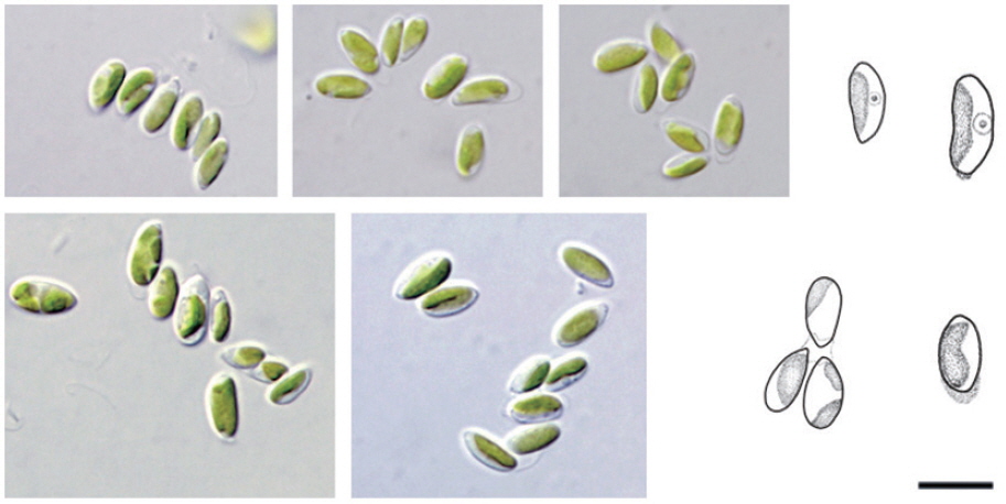
Family Scenedesmaceae
Subfamily Coelastroideae
Genus
This genus was named by Chodat (1922), and it belongs to the subfamily Scotiellocystoideae according to Kalina and Punčochářová (1987). Hanagata (1998) and Hegewald and Hanagata (2000, 2002) suggested that the subfamily Scotiellocystoideae should be removed and that
Synonym:
Distribution: in Europe, Britain (Ettl and Gärtner 1995, John et al. 2002), Czech Republic and/or Slovakia (Ettl and Gärtner 1995), and Spain (Rifón-Lastra and Noguerol-Seoane 1999).
Spindle-shaped, asymmetric cells, with both ends pointed. Parietal chloroplast, with several fragments and a single pyrenoid. The cell wall has 8–12 ribs.
According to John et al. (2011), this species mostly inhabits damp or wet terrestrial surfaces, including rocks, sand, and soil. In this study,
Sites of collection: 3, 12, 16, 17, 18.
Specimen: NIBRCL0000104595, ACKU HC3-59.
Family Scenedesmaceae
Subfamily Coelastroideae
Synonym:
Spherical or ellipsoidal cells, 5–14 µm in diameter. Young cells are spindle-shaped or ellipsoidal with both ends pointed. Mature cells are mostly spherical. Parietal chloroplast, with several fragments and a single distinct pyrenoid. The cell wall has several ribs.
The ribs of
Sites of collection: 10, 12, 20, 23, 28.
Specimen: NIBRCL0000107673, ACKU HC3-20.