검색 전체 메뉴
PDF
맨 위로
OA 학술지
Anti-inflammatory effect of polyphenol-rich extract from the red alga Callophyllis japonica in lipopolysaccharide-induced RAW 264.7 macrophages
  • 비영리 CC BY-NC
  • 비영리 CC BY-NC
ABSTRACT
Anti-inflammatory effect of polyphenol-rich extract from the red alga Callophyllis japonica in lipopolysaccharide-induced RAW 264.7 macrophages
KEYWORD
Callophyllis japonica , inflammation , macrophages , MAPKs
  • INTRODUCTION

    Inflammation is a defense response in a wide variety of physiological and pathological processes caused by stress, injury and infection (Kim et al. 2010). This process contributes to impairment on the immune system when persisted for a long period of time as activated macrophage produces toxic factors. Macrophages activated by lipopolysaccharide (LPS) or interferon gamma secrete nitric oxide (NO), cyclooxygenase-2 (COX-2) and other intermediates to destroy the remaining microorganisms in the inflammation response. In particular, exposure to LPS activates macrophage to secrete pro-inflammatory cytokines such as tumor necrosis factor-α (TNF-α), interleukin (IL)-1β and IL-6 through the expression of inducible nitric oxide synthase (iNOS) and NO whereas prosPhlorotaglandins production is mediated by COX-2 (Ulevitch and Tobias 1999, Akira et al. 2001). Moreover, this process activates a set of extracellular stimuli-dependent signal transduction cascades such as the mitogen-activated protein kinases (MAPKs) pathway (Akira et al. 2001). MAPK pathway, such as the extracellular signal-regulated kinase (ERK), p38 and c-Jun N-terminal kinase/stress-activated protein kinase (JNK) signaling pathways, regulate inflammatory and immune responses and are known to be involved in LPS-stimulated iNOS and COX-2 expression in macrophages (Kim et al. 2010).

    Marine algae or seaweeds are classified based on their colors as red algae (Rhodophyta), brown algae (Phaeophyta), and green algae (Chlorophyta). Red algae are more well known because 4,500 species have dominated along the coastal and continental shelf areas of tropical, temperate and cold-water regions for thousands of years, whereas the remaining algae include 1,000 species of brown and 900 species of green algae (Athanasiadis 2003). Red algae are ecologically significant as primary producers and providers of the structural habitats for other marine organisms, and their important role is in the primary establishment and maintenance of coral reefs. Moreover, the high level of minerals, vitamins, essential amino acids, indigestible carbohydrates and polyphenols, as well as their bioactive metabolites, in red and their relatively easy cultivation makes them attractive (Xu et al. 2003, Kelman et al. 2012) (Fig. 1). Therefore, many maritime countries have used red algae as a source of food, for industrial applications, a fertilizer and for medicinal purposes due to their biological activities.

    The red alga Callophyllis japonica, which is distributed in the coast of South Korea and Japan, has been eaten traditionally in Oriental food but was recently revealed by researchers to possess numerous biological activities such as antioxidant, hepatoprotective, anti-obesity and radioprotective properties (Kang et al. 2005, 2012, Park et al. 2005, Kim et al. 2008a, 2008c). These growing interests associated with the discovery of the biological activities of algae are based on the following two types of studies: pharmaceutical and neutraceutical field studies on the consumption of algae due to their bioactive substances that can reduce the progress of chronic diseases, and the study of algae as natural sources that are safer to intake than synthetic compounds (Yamamoto and Maruyama 1985, Okai et al. 1994). Additionally, the anti-inflammatory effects of C. japonica in RAW 264.7 macrophages and the molecular mechanisms regulating these effects remain largely unknown, while those effects of brown algae have recently been studied (Samarakoon et al. 2013). The present study aimed to assess the effects of the C. japonica ethanol extract (CJE) on LPS-induced inflammatory responses in RAW 264.7 macrophages and to explore the possible molecular mechanisms underlying these activities.

    MATERIALS AND METHODS

      >  Materials

    Dulbecco’s modified Eagle’s medium (DMEM), penicillin/ streptomycin, fetal bovine serum (FBS) and the other materials required for culturing cells were purchased from Gibco BRL Life Technologies (Grand Island, NY, USA). LPS of Escherichia coli 026:B6, Griess reagent and 3-(4,5-dimethyl-thiazol-2-yl)-2,5-diphenyltetrazolium bromide (MTT) were obtained from Sigma (St. Louis, MO, USA). The specific antibodies used for western blot analysis were purchased from Santa Cruz Biotechnology (Santa Cruz, CA, USA) and Amersham Pharmacia Biosciences (Piscataway, NJ, USA). All other reagents were of the highest grade available commercially.

      >  Sample preparation

    C. japonica was collected from the coast of South Korea in July 2012. The sample was washed three times with tap water to remove any salt, sand, and epiphytes attached to its surface. The sample was then rinsed carefully with fresh water and freeze-dried. The dried sample was homogenized with a grinder prior to extraction. For ethanol extraction, dried C. japonica powder (50 g) was extracted with methanol (500 mL) and filtered. The filtrate was evaporated to dryness under vacuum and dissolved in dimethylsulfoxide (DMSO), and then used for the experiments after adjusting the final concentration of DMSO in the culture medium to less than 0.1%.

      >  Cell culture and viability assay

    RAW 264.7 macrophages were purchased from the Korea Cell Line Bank (Seoul, Korea) and maintained in a 5% CO2 humidified atmosphere and at 37℃ in DMEM supplemented with 10% heat-inactivated FBS, 100 μg mL−1 streptomycin, and 100 U mL−1 penicillin. The cell viability was determined by the MTT reduction assay as described by Hansen (Hansen et al. 1989). RAW 264.7 macrophages plated on 96-well plates were pre-incubated and subsequently treated with LPS (1 μg mL−1) coupled with aliquots of the CJE at 37℃ for 24 h. The MTT stock solution (50 μL) was then added to each well to obtain a total reaction volume of 250 μL. After 4 h of incubation, the plates were centrifuged (800 ×g, 5 min), and the supernatants were aspirated. The formazan crystals in each well were dissolved in 150 μL of DMSO, and the absorbance was measured with an enzyme-linked immunosorbent assay plate reader (BioTek Instruments, Winooski, VT, USA) at 540 nm.

      >  NO assay

    The NO production in the culture supernatants was measured by the Griess reaction as described previously by Coker and Laurent (1998). After a 24 h pre-incubation of the RAW 264.7 macrophages with LPS (1 μg mL−1), the exquantity of nitrite accumulated in the culture medium was measured as an indicator of NO production. In brief, 100 μL of the cell culture medium was mixed with 100 μL of Griess reagent (1% sulfanilamide and 0.1% naphthylethylenediaminedihydrochloride in 2.5% phosphoric acid), the mixture was incubated at room temperature for 10 min, and the absorbance at 540 nm was measured using a micro-plate reader (BioTek Instruments). Fresh culture medium was employed as a blank in every experiment.

      >  Reverse transcription-polymerase chain reaction (RT-PCR) analysis

    The total RNA from the macrophages treated with LPS in the presence or absence of the CJE was extracted using the TRIzol reagent. Equal amounts of RNA were used for each cDNA synthesisreaction. Adjusted oligodT primers (10 μM) with DEPC were added, and the mixture was then incubated for 5 min at 70℃ and then snap-cooled on ice. The isolated mRNA was then used to synthesize cDNA according to the manufacter’s instruction (Promega, Madison, WI, USA). The PCR was performed in an automatic Whatman thermocycler (Biometra, Kent, UK). Single-stranded cDNA was amplified by PCR using specific primers. The primer sequences used to amplify the desired cDNA fragment were as follows: COX-2 forward and reverse primers: 5′-GGGGTACCTTCCAGCTGTCAAAA TCTC-3′ and 5′-GAAGATCTCGCCAGGTACTCACCTG-3′; iNOS forward and reverse primers: 5′-CCCTTCCGAAGTTTCTGGCAGCAGC-3′ and 5′-GGCTGTCAGAGCCTCGTGGCTTTGG-3′; IL-1β forward and reverse primers: 5′-ATGGCAACTGTTCCTGAACTCAACT-3′ and 5′-TTTCCTTTCTTAGATATGGACAGGAC-3′; IL-6 forward and reverse primers: 5′-AGTTGCCTTCTTGGGACTGA-3′ and 5′-CAGAATTGCCATTGCACAAC-3′; TNF-α forward and reverse primers: 5′-ATGAGCACAGAAAGCATGATC-3′ and 5′-TACAGGCTTGTCACTCGAATT-3′ and glyceraldehyde 3-phosphate dehydrogenase (GAPDH) forward and reverse primers: 5′-TGAAGGTCGGTGTGAACGGATTTGGC-3′ and 5′-CATGTAGGCCATGAGGTCCACCAC-3′. The following PCR conditions were applied for all amplifications by 30 cycles of 94℃ for 30 s (denaturing), 57℃ for 30 s (annealing), and 72℃ for 30 s (primer extension). The resulting cDNA was separated by electrophoresis on a 1% agarose gel for 15 min at 100 V and visualized under UV light after ethidium bromide staining. The bands of specific genes were normalized using GAPDH as s reference.

      >  Western blot analysis

    The cells were lysed in lysis buffer (20 mM Tris, 5 mM EDTA, 10 mM Na4P2O7, 100 mM NaF, 2 mM Na3VO4, 1% NP-40, 10 mg mL−1 aprotinin, 10 mg mL−1 leupeptin, and 1 mM PMSF) for 60 min and then centrifuged at 12,000 rpm and 4℃ for 15 min. The protein concentrations were determined using the BCA protein assay kit (Thermo scientific, Rockford, IL, USA). The lysate containing 40 μg of protein was subjected to electrophoresis on a sodium dodecyl sulfate-polyacrylamide gel, and the gel was transferred onto a nitrocellulose membrane. The membrane was blocked with 5% non fat dry milk in TBS-T (25 mM Tris-HCl, 137 mM NaCl, 2.65 mM KCl, and 0.05% Tween 20, pH 7.4) for 2 h. The primary antibodies were used at a 1:1,000 dilution. The membranes were incubated with the primary antibodies at 4℃ overnight, washed with TBS-T and then incubated with the secondary antibodies at 1:3,000 dilutions. The signals were developed using an ECL western blotting detection kit and quantified using the Multi Gauge V3.0 software (Fujifilm Life Science, Tokyo, Japan).

      >  Statistical analysis

    All of data are presented as the means ± standard deviation (SD). The mean values were calculated based on data from at least three independent experiments conducted on separate days using freshly prepared reagents. The data were analyzed using the analysis of variance (ANOVA) test of the statistical package for the social sciences (SPSS Inc., Chicago, IL, USA). Significance differences between treatment groups were determined using Duncan’s multiple range tests. The statistical significance of the differences was defined at the p < 0.05 and p < 0.01 levels.

    RESULTS

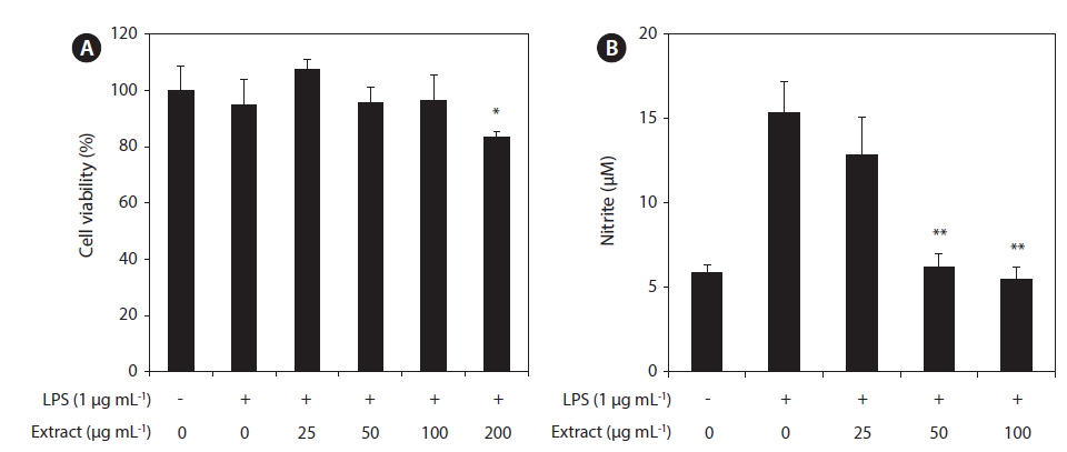
      >  Cytotoxicity of the CJE and its inhibitory effect on NO production

    In this study, the cytotoxicity of the CJE in RAW 264.7 macrophages was evaluated using the MTT assay at multiple CJE concentrations (25, 50, 100, and 200 μg mL−1). The CJE did not show cytotoxicity at concentrations up to 100 μg mL−1 compared with untreated cells (Fig. 2A). These lower concentrations were therefore used in the subsequent experiments. To evaluate whether the CJE exquantity erts a potential anti-inflammatory effect in LPS-stimulated RAW 264.7 macrophages, we investigated the inhibitory effect of the CJE on NO production. LPS alone was able to markedly induce higher NO production in these cells than that observed in the untreated cells. However, the CJE significantly inhibited the levels of NO production induced by LPS in a dose-dependent manner (Fig. 2B).

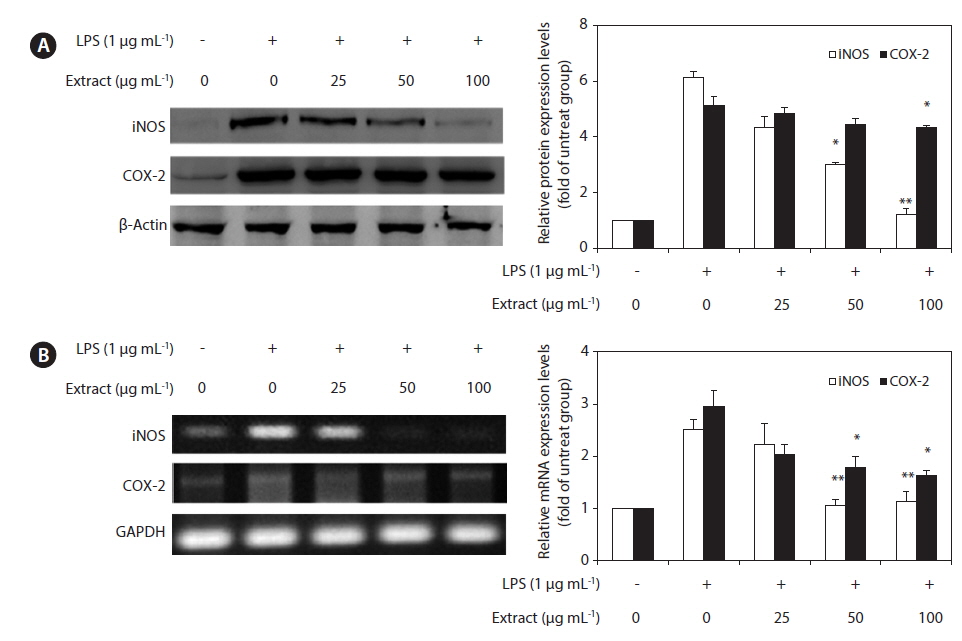
      >  Effects of the CJE on LPS-induced iNOS and COX-2 protein and mRNA expressions

    To elucidate the mechanism involved in the inhibition of NO generation by the CJE in LPS-induced RAW 264.7 cells, we further studied the effect of the CJE on iNOS and COX-2 protein and gene expression by western blot and RT-PCR analysis (Fig. 3). In response to LPS, the expression of iNOS was markedly increased, and the CJE significantly inhibited the iNOS and COX-2 protein expression levels in a dose-dependent manner (Fig. 3A). Under the same conditions, the levels of iNOS and COX-2 mRNA expression were correlated with their protein levels (Fig.3B). Similar patterns were observed in the analysis of the effect of the CJE on LPS-induced iNOS and COX-2 protein and mRNA expression.

      >  Effects of the CJE on LPS-induced transcription of pro-inflammatory cytokines

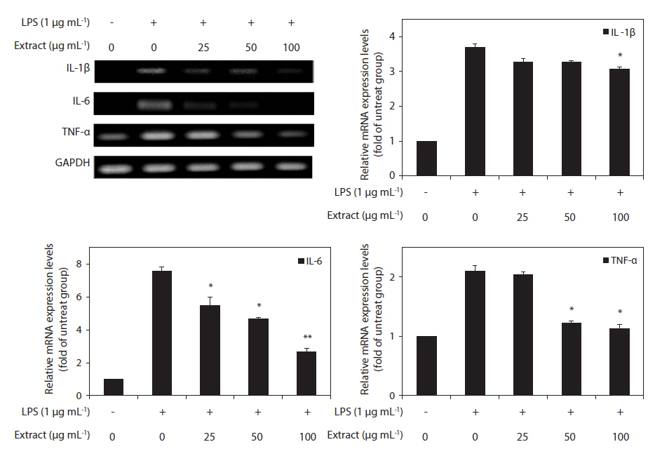
    Because IL-1β, IL-6, and TNF-α are pro-inflammatory cytokines that are secreted early and because their levels are elevated in various acute and chronic inflammatory diseases, we determined the effects of the CJE on the mRNA levels of IL-1β, IL-6, and TNF-α in macrophages after treatment with LPS. RT-PCR was performed to determine whether the CJE reduced the expression of those cytokines at the mRNA levels. All of the mRNA levels were increased by treatment with LPS, and these increases were significantly decreased in a concentration-dependent manner by treatment with the CJE (Fig. 4). This finding suggests that the CJE is capable of disrupting key signal transduction pathways activated by LPS in macrophages, subsequently preventing the transcription of pro-inflammatory mediators.

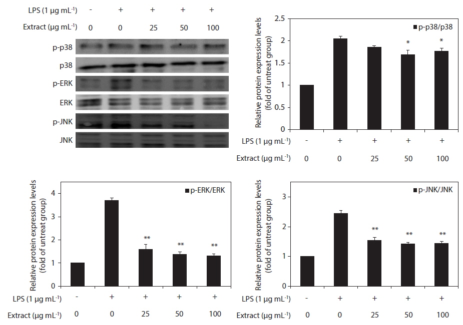
      >  Effect of CJE on LPS-induced MAPKs activation

    To elucidate the mechanism of the action of the CJE, we investigated the effect of the CJE on the activation of MAPKs in LPS-activated macrophages by western blot analysis. The levels of phosphorylated ERK, JNK, and p38 MAPK were elevated in the macrophages after treatment with LPS, whereas the levels of total ERK, JNK, and p38 MAPK did not show any significant changes (Fig. 5). However, compared with the cells treated with LPS, the macrophages pretreated with the CJE showed significantly lower LPS-induced activation levels of these proteins (Fig. 5). This result indicates that the phosphorylation of MAPKs was effectively blocked by the CJE in LPS-activated macrophages. Together with the present results, these findings support the conclusion that the CJE, which is obtained from red algae that contains polyphenols, inhibits the inflammation process through inhibiting the expression of pro-inflammatory cytokines in macrophages by down-regulating the LPS-induced activation of MAPK signaling.

    DISCUSSION

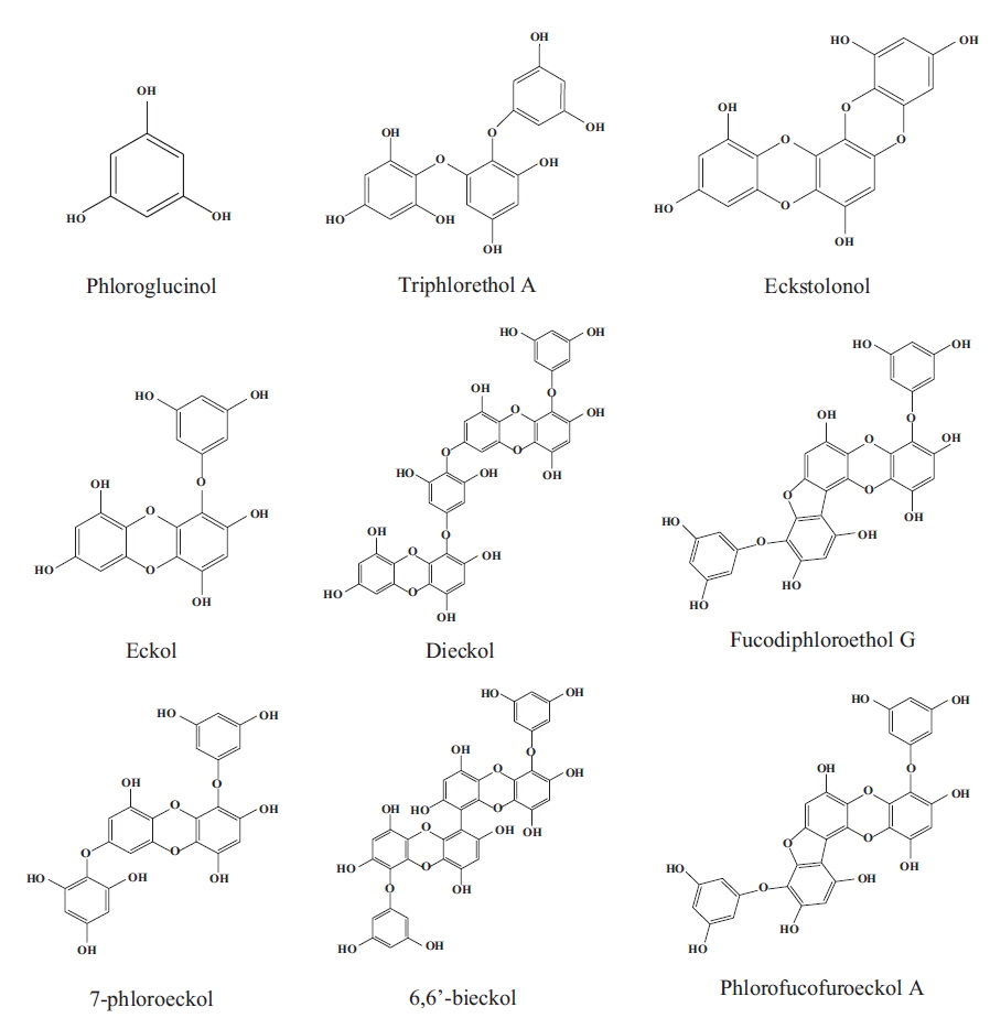
    Red algae are known as the largest species among algae, present the greates chemical diversity recorded to date in the literature, and supply many useful substances for the treatment diseases that affect living beings. These algae are a source of variable bioactive components, such as polyphenols, alkaloids, and terpenes, and particularly polyphenols, which considered one of the most numerous and widely distributed chemical groups. Based on their structures, red algae are classified into different families with different contents of phenolic acids, phenylpropanoids, flavonoids, and the less-common lignans and stilbenes and have been the object of intense interest due to their various pharmacological activities such as antioxidative, α-glucosidase inhibitory, antimicrobial, aldose reductase inhibitory, antitumor, and anti-inflammatory activities (Wang et al. 2005, Ma et al. 2006, Lee et al. 2007a, Kim et al. 2008b, Li et al. 2009, 2010, Xu et al. 2009).

    C. japonica is a red alga, and its ethanol extract has been intensely studied for antioxidant activity based on evaluations of intracellular reactive oxygen species and free radical and lipid peroxidation scavenging activities (Kang et al. 2005). Moreover, the hexane and ethyl acetate extracts of C. japonica exhibit radioprotective activity, as determined by their protection of blood progenitor cells in gamma-radiated mice (Kim et al. 2008a). Some studies have investigated the effects of the anti-obesity potential of the CJE in mice fed a high-fat diet and have revealed the effects of treatment with the C. japonica ethyl acetate extract on adipogenesis in murine 3T3-L1 preadipocytes (Kang et al. 2012). Polyphenols are well-known potent biologically active agents, but their wide diversity and chemical complexity makes it challenging to correlate their inflammation potency in vitro with specific biological activity in vivo (Wang et al. 2005, Chen et al. 2007, Park et al. 2007). In our previous study (Park et al. 2013), the polyphenol content of CJE was determined to be 39.8 mg g−1. However, low amounts of carbohydrate (1.2 mg g−1) were detected in the CJE. The high phenolic and low carbohydrate content in the CJE is consistent with previously published results for an extract of the red alga (Laurencia undulata) (Jung et al. 2009). Ethanol extracts of C. japonica and L. undulata containing a large amount of polyphenols have been shown to exert potent asthma-improving effects in a murine model. Therefore, red algae provide a rich source of polyphenols that can be used to design and develop novel potentially useful therapeutic agents. The biological effects of red alga C. japonica used in this study have been rarely. Additionally, the anti-inflammatory effect of C. japonica has not yet been reported. Thus, the CJE may have the potential to inhibit or minimize tissue injury and maintain homeostasis in the immune system. Therefore, in this study, we investigated the effects of the CJE on abnormal macrophage inflammation.

    Inflammation is the first response of the immune system to infection and plays a pivotal role in many diseases (Kim et al. 2010, Heo et al. 2010a, 2010b). NO is one of the major factors relevant to inflammationin macrophages and is generated from L-arginine by NO synthase, a three-member enzyme family that includes iNOS (Park et al. 2011). NO plays animportant role in various body functions, but its overproduction in macrophages, in particular, can lead to inflammation and the development of autoimmune disorders (Liu and Hotchkiss 1995). Additionally, COX-2 is an important mediator of inflammation involved in NO generation (Kim et al. 2014). Thus, the inhibition of these inflammatory mediators has been shown to be important in the treatment of inflammation. The results of this study demonstrated that the CJE inhibits NO production via the suppression of iNOS and COX-2 in an LPS-activated macrophage culture system.

    In addition, the CJE also significantly reduced the LPS-induced mRNA transcription of TNF-α, IL-1β, and IL-6. According to previous reports, the expression of iNOS is stimulated by pro-inflammatory cytokines, including TNF-α and IL-1β, which play key roles in the induction of inflammation in macrophages (De Nardin 2001, Marcus et al. 2003). TNF-α exhibits its pro-inflammatory activity by regulating several intracellular pathways and can stimulate the production or expression of IL-6 and IL-1β. It elicits a number of physiological effects, including septic shock, inflammation, and cytotoxicity (Aggarwal and Natarajan 1996). IL-6 is a multifunctional cytokine released by LPS-activated monocytes that plays a major role in the immune and inflammatory responses (Ghosh and Karin 2002). Additionally, IL-1β is a key pro-inflammatory cytokine that is involved in various pathological conditions, such as rheumatoid arthritis and fever (Van et al. 2009, Lee et al. 2012). Our results demostrate that the CJE significantly inhibits the transcription of the pro-inflammatory cytokines TNF-α, IL-1β, and IL-6 in macrophages stimulated by LPS, suggesting that the blockade of the iNOS/NO pathway by the CJE may be associated with attenuation of the production of TNF-α, IL-1β, and IL-6.

    Mechanical signals are important for maintenance of the normal immune system, and mechanical stress contributes significantly to disease initiation and progression. One of the most extensively investigated transduction mechanims involved in the inflammatory process is the MAPK pathway. Furthermore, MAPKs (ERK, p38, and JNK) have previously been implicated in the signaling pathways relevant to LPS-induced inflammation. The three major MAPKs have been shown to regulate iNOS and COX-2 expression and pro-inflammatory cytokine production in LPS-stimulated macrophages (Kim et al. 2013). It has been suggested that p38 performs a pivotal function in the regulation of iNOS and TNF-α expression (Bhat et al. 1998). Additionally, ERK activation is hypothesized to be involved in LPS-induced macrophage responses, including increases in the production of proinflammatory cytokines and iNOS (Bhat et al. 1998, Ajizian et al. 1999, Kim et al. 2013). Furthermore, JNK plays a significant role in the regulation of the expression of LPS-induced iNOS (Uto et al. 2005). Thus, the suppression of MAPKs activation or function is a key mechanism. In the present study, we examined the effects of the CJE on the activation of these MAPKs and found that the CJE suppressed the phosphorylation of MAPKs. The CJE attenuated LPS-induced phosphorylation of ERK, p38, and JNK. Taken together, these results suggest the potential role of ERK, p38, and JNK in the CJE-induced suppression of NO and pro-inflammatory cytokines in activated macrophages.

    In conclusion, the present study demonstrates that the CJE is a potent inhibitor of NO, TNF-α, IL-1β, and IL-6 production at the transcriptional level in LPS-stimulated RAW 264.7 cells. The mechanism underlying the inhibition of NO production appears to involve the down-regulation of iNOS and COX-2 protein and mRNA expression, which may be associated with the attenuation of TNF-α, IL-1β, and IL-6 production. Furthermore, the levels of phosphorylated MAPKs were significantly decreased due to pretreatment with the CJE in LPS-stimulated macrophage cells. Taken together, these results indicate that the CJE exerts its anti-inflammatory actions through a blockade of the MAPK pathways in RAW 264.7 macrophages.

참고문헌
  • 1. Aggarwal B. B., Natarajan K. 1996 Tumor necrosis factors: developments during the last decade [Eur. Cytokine Netw.] Vol.7 P.93-124 google
  • 2. Ajizian S. J., English B. K., Meals E. A. 1999 Specific inhibitors of p38 and extracellular signal-regulated kinase mitogen-activated protein kinase pathways block inducible nitric oxide synthase and tumor necrosis factor accumulation in murine macrophages stimulated with lipopolysaccharide and interferon-gamma [J. Infect. Dis.] Vol.179 P.939-944 google cross ref
  • 3. Akira S., Takeda K., Kaisho T. 2001 Toll-like receptors: critical proteins linking innate and acquired immunity [Nat. Immunol.] Vol.2 P.675-680 google cross ref
  • 4. Athanasiadis A. 2003 Taxonomy of Rhodophyta with particular reference to Mediterranean species [Bocconea] Vol.16 P.193-198 google
  • 5. Bhat N. R., Zhang P., Lee J. C., Hogan E. L. 1998 Extracellular signal-regulated kinase and p38 subgroups of mitogen-activated protein kinases regulate inducible nitric oxide synthase and tumor necrosis factor-alpha gene expression in endotoxin-stimulated primary glial cultures [J. Neurosci.] Vol.18 P.1633-1641 google
  • 6. Chen H.-Q., Jin Z.-Y., Li G.-H. 2007 Biochanin A protects dopaminergic neurons against lipopolysaccharide-induced damage through inhibition of microglia activation and proinflammatory factors generation [Neurosci. Lett.] Vol.417 P.112-117 google cross ref
  • 7. Coker R. K., Laurent G. J. 1998 Pulmonary fibrosis: cytokines in the balance [Eur. Resp. J.] Vol.11 P.1218-1221 google cross ref
  • 8. De Nardin E. 2001 The role of inflammatory and immunological mediators in periodontitis and cardiovascular disease [Ann. Periodontol.] Vol.6 P.30-40 google cross ref
  • 9. Ghosh S., Karin M. 2002 Missing pieces in the NF-κB puzzle [Cell] Vol.109 P.S81-96 google cross ref
  • 10. Hansen M. B., Nielsen S. E., Berg K. 1989 Re-examination and further development of a precise and rapid dye method for measuring cell growth/cell kill [J. Immunol. Methods] Vol.119 P.203-210 google cross ref
  • 11. Heo S.-J., Hwang J.-Y., Choi J.-I., Lee S.-H., Park P.-J., Kang D.-H., Oh C., Kim D.-W., Han J.-S., Jeon J.-Y., Kim H.-J., Choi I.-W. 2010 Protective effect of diphlorethohydroxycarmalol isolated from Ishige okamurae against high glucose-induced-oxidative stress in human umbilical vein endothelial cells [Food Chem. Toxicol.] Vol.48 P.1448-1454 google cross ref
  • 12. Heo S.-J., Yoon W.-J., Kim K.-N., Ahn G.-N., Kang S.-M., Kang D.-H., Affan A., Oh C., Jung W.-K., Jeon Y.-J. 2010 Evaluation of anti-inflammatory effect of fucoxanthin isolated from brown algae in lipopolysaccharide-stimulated RAW 264.7 macrophages [Food Chem. Toxicol.] Vol.48 P.2045-2051 google cross ref
  • 13. Jung W.-K., Choi I., Oh S., Park S.-G., Seo S.-K., Lee S. -W., Lee D.-S., Heo S.-J., Jeon Y.-J., Je J.-Y., Ahn C.-B., Kim J. S., Oh K. S., Kim Y.-M., Moon J., Choi I.-W. 2009 Anti-asthmatic effect of marine red alga (Laurencia undulata) polyphenolic extracts in a murine model of asthma [Food Chem. Toxicol.] Vol.47 P.293-297 google cross ref
  • 14. Kang K. A., Bu H. D., Park D. S., Go G. M., Jee Y., Shin T., Hyun J. W. 2005 Antioxidant activity of ethanol extract of Callophyllis japonica [Phytother. Res.] Vol.19 P.506-510 google cross ref
  • 15. Kang S.-I., Shin H.-S., Kim H.-M., Yoon S.-A., Kang S.-W., Ko H.-C., Kim S.-J. 2012 Callophyllis japonica extract improves high-fat diet-induced obesity and inhibits adipogenesis in 3T3-L1 cells [Anim. Cells Syst.] Vol.16 P.447-454 google cross ref
  • 16. Kelman D., Posner E. K., McDermid K. J., Tabandera N. K., Wright P. R., Wright A. D. 2012 Antioxidant activity of Hawaiian marine algae [Mar. Drugs] Vol.10 P.403-416 google cross ref
  • 17. Kim J., Moon C., Kim H., Jeong J., Lee J., Kim J., Hyun J. W., Park J. W., Moon M. Y., Lee N. H., Kim S. H., Jee Y., Shin T. 2008 The radioprotective effects of the hex-ane and ethyl acetate extracts of Callophyllis japonica in mice that undergo whole body irradiation [J. Vet. Sci.] Vol.9 P.281-284 google cross ref
  • 18. Kim K.-N., Heo S.-J., Yoon W.-J., Kang S.-M., Ahn G., Yi T.-H., Jeon Y.-J. 2010 Fucoxanthin inhibits the inflammatory response by suppressing the activation of NF-κB and MAPKs in lipopolysaccharide-induced RAW 264.7 macrophages [Eur. J. Pharmacol.] Vol.649 P.369-375 google cross ref
  • 19. Kim K.-N., Ko Y.-J., Kang M.-C., Yang H.-M., Roh S. W., Oda T., Jeon Y.-J., Jung W.-K., Heo S.-J., Yoon W.-J., Kim D. 2013 Anti-inflammatory effects of trans-1,3-diphenyl-2,3-epoxypropane-1-one mediated by suppression of inflammatory mediators in LPS-stimulated RAW 264.7 macrophages [Food Chem. Toxicol.] Vol.53 P.371-375 google cross ref
  • 20. Kim K. Y., Nam K.A., Kurihara H., Kim S. M. 2008 Potent α-glucosidase inhibitors purified from the red alga Grateloupia elliptica [Phytochemistry] Vol.69 P.2820-2825 google cross ref
  • 21. Kim S.-Y., Kim E.-A., Kang M.-C., Lee J.-H., Yang H.-W., Lee J.-S., Lim T. I., Jeon Y.-J. 2014 Polyphenol-rich fraction from Ecklonia cava (a brown alga) processing by-product reduces LPS-induced inflammation in vitro and in vivo in a zebrafish model [Alage] Vol.29 P.165-174 google cross ref
  • 22. Kim Y. A., Kong C. S., Um Y. R., Lee J. I., Nam T. J., Seo Y. 2008 Antioxidant efficacy of extracts from a variety of seaweeds in a cellular system [Ocean Sci. J.] Vol.43 P.31-37 google cross ref
  • 23. Lee H.-S., Ryu D.-S., Lee G.-S., Lee D.-S. 2012 Anti-inflammatory effects of dichloromethane fraction from Orostachys japonicus in RAW 264.7 cells: suppression of NF-κB activation and MAPK signaling [J. Ethnopharmacol.] Vol.140 P.271-276 google cross ref
  • 24. Lee J. H., Lee T.-K., Kang R.-S., Shin H. J., Lee H.-S. 2007 The in vitro antioxidant activities of the bromophenols from the red alga Tichocarpus crinitus and phenolic derivatives [J. Korean Magn. Reson. Soc.] Vol.11 P.56-63 google
  • 25. Li Y. X., Li Y., Lee S.-H., Qian Z.-J., Kim S.-K. 2010 Inhibitors of oxidation and matrix metalloproteinases, floridoside, and D-isofloridoside from marine red alga Laurencia undulata [J. Agric. Food. Chem.] Vol.58 P.578-586 google cross ref
  • 26. Li Y.-X., Li Y., Qian Z.-J., Kim M.-M., Kim S.-K. 2009 In vitro antioxidant activity of 5-HMF isolated from marine red alga Laurencia undulata in free-radical-mediated oxidative systems [J. Microbiol. Biotechnol.] Vol.19 P.1319-1327 google
  • 27. Liu R. H., Hotchkiss J. H. 1995 Potential genotoxicity of chronically elevated nitric oxide: a review [Mutat. Res.] Vol.339 P.73-89 google cross ref
  • 28. Ma M., Zhao J., Wang S., Li S., Yang Y., Shi J., Fan X., He L. 2006 Bromophenols coupled with methyl gamma-ureidobutyrate and bromophenol sulfates from the red alga Rhodomela confervoides [J. Nat. Prod.] Vol.69 P.206-210 google cross ref
  • 29. Marcus J. S., Karackattu S. L., Fleegal M. A., Sumners C. 2003 Cytokine-stimulated inducible nitric oxide synthase expression in astroglia: role of Erk mitogen-activated protein kinase and NF-kappaB [Glia] Vol.41 P.152-160 google cross ref
  • 30. Okai Y., Higashi-Okai K., Nakamura S., Yano Y., Otani S. 1994 Suppressive effects of the extracts of Japanese edible seaweeds on mutagen-induced umu C gene expression in Salmonella typhimurium (TA 1535/pSK 1002) and tumor promotor-dependent ornithine decarboxylase induction in BALB/c 3T3 fibroblast cells [Cancer Lett.] Vol.87 P.25-32 google cross ref
  • 31. Park D.-S., Lee K.-H., Kim H.-C., Ahn M.-J., Moon C.-J., Ko M.-S., Lee K.-K., Go G.-M., Shin T.-K. 2005 Effects of Callophyllis japonica powder on carbon tetrachloride-induced liver injury in rats [Orient. Pharm. Exp. Med.] Vol.5 P.231-235 google cross ref
  • 32. Park H. Y., Han M. H., Park C., Jin C.-Y., Kim G.-Y., Choi I.-W., Kim N. D., Nam T.-J., Kwon T. K., Choi Y. H. 2011 Anti-inflammatory effects of fucoidan through inhibition of NF-kB, MAPK and Akt activation in lipopolysaccharide-induced BV2 microglia cells [Food Chem. Toxicol.] Vol.49 P.1745-1752 google cross ref
  • 33. Park J.-S., Woo M.-S., Kim D.-H., Hyun J.-W., Kim W.-K., Lee J.-C., Kim H.-S. 2007 Anti-inflammatory mechanisms of isoflavone metabolites in lipopolysaccharide-stimulated microglial cells [J. Pharmacol. Exp. Ther.] Vol.320 P.1237-1245 google
  • 34. Park W. S., Lee K. S., Chun J. H., Urm S. H., Lee D.-S., Lee D.-Y., Park S.-G., Seo S.-K., Heo S.-J., Qian Z.-J., Jung W.-K., Choi I.-W. 2013 Investigation of the antiasthmatic properties of ethanol extract of Callophyllis japonica in mice [Trop. J. Pharm. Res.] Vol.12 P.981-987 google
  • 35. Samarakoon K. W., Ko J.-Y., Shah M. M. R., Lee J.-H., Kang M.-C., Kwon O.-N., Lee J.-B., Jeon Y.-J. 2013 In vitro studies of anti-inflammatory and anticancer activities of organic solvent extracts from cultured marine microalgae [Algae] Vol.28 P.111-119 google cross ref
  • 36. Ulevitch R. J., Tobias P. S. 1999 Recognition of gram-negative bacteria and endotoxin by the innate immune system [Curr. Opin. Immunol.] Vol.11 P.19-22 google cross ref
  • 37. Uto T., Fujii M., Hou D. X. 2005 6-(Methylsulfinyl)hexyl isothiocyanate suppresses inducible nitric oxide synthase expression through the inhibition of Janus kinase 2-mediated JNK pathway in lipopolysaccharide-activated murine macrophages [Biochem. Pharmacol.] Vol.70 P.1211-1221 google cross ref
  • 38. Van Q., Nayak B. N., Reimer M., Jones P. J. H., Fulcher R. G., Rempel C. B. 2009 Anti-inflammatory effect of Inonotus obliquus, Polygala senega L., and Viburnum trilobum in a cell screening assay [J. Ethnopharmacol.] Vol.125 P.487-493 google cross ref
  • 39. Wang W., Okada Y., Shi H., Wang Y., Okuyama T. 2005 Structures and aldose reductase inhibitory effects of bromophenols from the red alga Symphyocladia latiuscula [J. Nat. Prod.] Vol.68 P.620-622 google cross ref
  • 40. Xu N., Fan X., Yan X., Li X., Niu R., Tseng C. K. 2003 Antibacterial bromophenols from the marine red alga Rhodomela confervoides [Phytochemistry] Vol.62 P.1221-1224 google cross ref
  • 41. Xu X., Song F., Fan X., Fang N., Shi J. 2009 A novel bromophenol from marine red alga Symphyocladia latiuscula [Chem. Nat. Compd.] Vol.45 P.811-813 google cross ref
  • 42. Yamamoto I., Maruyama H. 1985 Effect of dietary seaweed preparations on 1,2-dimethylhydrazine-induced intestinal carcinogenesis in rats [Cancer Lett] Vol.26 P.241-251 google cross ref
OAK XML 통계
이미지 / 테이블
  • [ Fig. 1. ]  Chemical structures of phlorotannins derived from marine algae.
    Chemical structures of phlorotannins derived from marine algae.
  • [ Fig. 2. ]  Effects of the Callophyllis japonica extract (CJE) on cytotoxicity (A) and nitric oxide (NO) production (B) in lipopolysaccharide (LPS)-induced RAW 264.7 macrophages. The cells were pretreated for 1 h with different concentrations (25, 50, 100, and 200 μg mL?1) of the CJE. LPS (1 μg mL?1) was then added, and the cells were incubated for 24 h. The cytotoxicity was determined using the MTT assay. The values are expressed as the means ± standard deviation of triplicate experiments. *p < 0.05 and **p < 0.01 indicate significant differences compared with the LPS-stimulated group.
    Effects of the Callophyllis japonica extract (CJE) on cytotoxicity (A) and nitric oxide (NO) production (B) in lipopolysaccharide (LPS)-induced RAW 264.7 macrophages. The cells were pretreated for 1 h with different concentrations (25, 50, 100, and 200 μg mL?1) of the CJE. LPS (1 μg mL?1) was then added, and the cells were incubated for 24 h. The cytotoxicity was determined using the MTT assay. The values are expressed as the means ± standard deviation of triplicate experiments. *p < 0.05 and **p < 0.01 indicate significant differences compared with the LPS-stimulated group.
  • [ Fig. 3. ]  Inhibitory effect of the Callophyllis japonica extract (CJE) in lipopolysaccharide (LPS)-induced inducible nitric oxide synthase (iNOS) and cyclooxygenase-2 (COX-2) protein (A) and mRNA (B) expression in RAW 264.7 macrophages. (A) RAW 264.7 macrophages were pre-incubated for 18 h and stimulated with LPS (1 μg mL?1) for 24 h in the presence of the CJE (25, 50, and 100 μg mL?1). The cell lysates were electrophoresed, and the expression levels of iNOS and COX-2 were detected with specific antibodies. (B) After LPS treatment, the total RNA from RAW 264.7 macrophages was prepared, and reverse transcription-polymerase chain reaction (RT-PCR) was performed for detection of the iNOS and COX-2 genes. β-Actin and glyceraldehyde 3-phosphate dehydrogenase (GAPDH) were used as internal controls for the western blot analysis and RT-PCR assays, respectively. The values are expressed as mean ± standard deviation of triplicate experiments. *p < 0.05 and **p < 0.01 indicate significant differences compared with the LPS-stimulated group.
    Inhibitory effect of the Callophyllis japonica extract (CJE) in lipopolysaccharide (LPS)-induced inducible nitric oxide synthase (iNOS) and cyclooxygenase-2 (COX-2) protein (A) and mRNA (B) expression in RAW 264.7 macrophages. (A) RAW 264.7 macrophages were pre-incubated for 18 h and stimulated with LPS (1 μg mL?1) for 24 h in the presence of the CJE (25, 50, and 100 μg mL?1). The cell lysates were electrophoresed, and the expression levels of iNOS and COX-2 were detected with specific antibodies. (B) After LPS treatment, the total RNA from RAW 264.7 macrophages was prepared, and reverse transcription-polymerase chain reaction (RT-PCR) was performed for detection of the iNOS and COX-2 genes. β-Actin and glyceraldehyde 3-phosphate dehydrogenase (GAPDH) were used as internal controls for the western blot analysis and RT-PCR assays, respectively. The values are expressed as mean ± standard deviation of triplicate experiments. *p < 0.05 and **p < 0.01 indicate significant differences compared with the LPS-stimulated group.
  • [ Fig. 4. ]  Inhibitory effect of the Callophyllis japonica extract (CJE) on lipopolysaccharide (LPS)-induced interleukin (IL)-1β, IL-6, and tumor necrosis factor-α (TNF-α) production in RAW 264.7 macrophages. RAW 264.7 macrophages were pre-incubated for 18 h and stimulated with LPS (1 μg mL?1) for 24 h in the presence of the CJE (25, 50, and 100 μg mL?1). The levels of IL-1β, IL-6, and TNF-α mRNA were determined by reverse transcription-polymerase chain reaction. The values are expressed as the mean ± standard deviation of triplicate experiments. GAPDH, glyceraldehyde 3-phosphate dehydrogenase. *p < 0.05 and **p < 0.01 indicate significant differences compared with the LPS-stimulated group.
    Inhibitory effect of the Callophyllis japonica extract (CJE) on lipopolysaccharide (LPS)-induced interleukin (IL)-1β, IL-6, and tumor necrosis factor-α (TNF-α) production in RAW 264.7 macrophages. RAW 264.7 macrophages were pre-incubated for 18 h and stimulated with LPS (1 μg mL?1) for 24 h in the presence of the CJE (25, 50, and 100 μg mL?1). The levels of IL-1β, IL-6, and TNF-α mRNA were determined by reverse transcription-polymerase chain reaction. The values are expressed as the mean ± standard deviation of triplicate experiments. GAPDH, glyceraldehyde 3-phosphate dehydrogenase. *p < 0.05 and **p < 0.01 indicate significant differences compared with the LPS-stimulated group.
  • [ Fig. 5. ]  Inhibitory effect of the Callophyllis japonica extract (CJE) on the protein level of p38, extracellular signal-regulated kinase (ERK), and c-Jun N-terminal kinase (JNK) in RAW 264.7 macrophages. RAW 264.7 macrophages were pre-incubated for 18 h stimulated with lipopolysaccharide (LPS) (1 μg mL?1) for 20 min in the presence of the CJE (25, 50, and 100 μg mL?1). The levels of p-ERK, ERK, p-JNK, JNK, p-p38, and p-38 were determined by western blotting. The values are expressed as the mean ± standard deviation of triplicate experiments. *p < 0.05 and **p < 0.01 indicate significant differences compared with the LPS-stimulated group.
    Inhibitory effect of the Callophyllis japonica extract (CJE) on the protein level of p38, extracellular signal-regulated kinase (ERK), and c-Jun N-terminal kinase (JNK) in RAW 264.7 macrophages. RAW 264.7 macrophages were pre-incubated for 18 h stimulated with lipopolysaccharide (LPS) (1 μg mL?1) for 20 min in the presence of the CJE (25, 50, and 100 μg mL?1). The levels of p-ERK, ERK, p-JNK, JNK, p-p38, and p-38 were determined by western blotting. The values are expressed as the mean ± standard deviation of triplicate experiments. *p < 0.05 and **p < 0.01 indicate significant differences compared with the LPS-stimulated group.
(우)06579 서울시 서초구 반포대로 201(반포동)
Tel. 02-537-6389 | Fax. 02-590-0571 | 문의 : oak2014@korea.kr
Copyright(c) National Library of Korea. All rights reserved.