검색 전체 메뉴
PDF
맨 위로
OA 학술지
Ontogenetic Development of the Digestive System in Chub Mackerel Scomber japonicus Larvae and Juveniles
  • 비영리 CC BY-NC
  • 비영리 CC BY-NC
ABSTRACT
Ontogenetic Development of the Digestive System in Chub Mackerel Scomber japonicus Larvae and Juveniles
KEYWORD
Chub mackerel , Scomber japonicus , Digestive system , Larvae , Juveniles
  • Introduction

    The chub mackerel, Scomber japonicus, has high growth potential; it has a large head containing a large mouth and big eyes, there is a posterior shift of the anus with growth, and it has well-developed preopercular spines (Hunter, 1981). These morphological traits are indicative of the survival strategy of scombrid larvae, which includes large prey-fast growth. In addition, its feeding is opportunistic and non-selective, with the adult diet ranging from copepods and other crustaceans to fish and squid (FAO, 1983). The precocious digestive system of scombrid larvae supports their fast growth during their early life history. In common with marine fishes, the juvenile-type digestive system is established at the same time as the transformation to the juvenile stage (Tanaka, 1973). In bluefin tuna larvae, however, differentiation of the gastric glands, followed by the appearance of pyloric caeca, was observed at the flexion phase, suggesting that the digestive system attained a juvenile-type structure long before metamorphosis (Kaji, 1996; Miyashita, 1998). In addition, striped bonito (Harada et al., 1974) and Japanese Spanish mackerel (Fukunaga et al., 1982) larvae grew to 106 and 90 mm in length, respectively, during the first 30 days after hatching (DAH). This growth was the fastest among marine fish larvae hatched from pelagic eggs and explained the development of the precocious digestive system with a well-developed gastric blind sac (Tanaka et al., 1996; Kaji, 2002).

    The chub mackerel is a candidate for aquaculture development in Korea, with high market prices due to a strong demand for live aquaculture products. Although artificial rearing techniques of the chub mackerel have recently been reported in Korea, the early development of the scombrid larvae is poorly understood, primarily due to difficulties in rearing the larvae. Culturing of the early stages is considered the most critical phase for successful aquaculture (Tanaka, 1973). In particular, a lack of information of the nutritional requirements of the fish larvae and the development of their digestive system is thought to play a major role in the limited success of larval culture of many species. Hence, to develop successful aquaculture methods, species-specific information on the early development of larvae is required to provide the best feeding regimes to match the ontogenetic status of the larvae. However, there is no documented ontogeny of the digestive system in S. japonicus. The aim of this study was to describe the histological changes and organogenesis of S. japonicus during early ontogeny, from hatching to the early juvenile stages.

    Materials and Methods

      >  Rearing

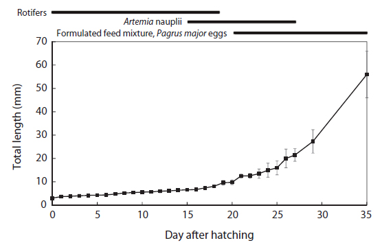
    Fertilized eggs of chub mackerel were obtained from a broodstock maintained by Gyongsangnam-do Fisheries Resources Research Institute, Korea and stocked in a 20-ton rearing tank. The water was maintained at an average temperature of 19.1 ± 2.2°C. Rotifers were fed immediately after hatching until 19 DAH, and Artemia nauplii (3–4 individuals/mL) were supplied from 15 to 27 DAH. Red sea bream (Pagrus major) eggs and a formulated feed mixture (Otohime; 53% protein) were supplied at a ratio of 1:3 from 20 to 35 DAH (Fig. 1).

      >  Sample collection

    Eighty individuals were collected from hatching on a daily basis for 35 days from 31 May 2011 until 5 July 2011. Fifty individuals were fixed in 10% natural formaldehyde for morphological investigation (Olympus DP20, Olympus Co., Japan). Total length (TL) and standard length (SL) were measured to the nearest 0.1 mm. The developmental phases were classified into 5 stages (the yolk-sac larvae, 0–2 DAH; preflexion larvae, 3–12 DAH; flexion larvae, 13–16 DAH; postflexion larvae, 17–26 DAH; and juvenile, 27–35 DAH) based on Kendall et al. (1984). The criteria for classification were as follows: the yolk-sac larval stage, a transitional stage (from hatching to yolk-sac absorption); the larval period, divided into three phases (preflexion phase: first feeding to onset of notochord flexion: flexion phase: to completion of notochord flexion; postflexion phase: to completion of fin formation) depending on the flexion of the notochord that accompanied the hypochordal development of the homocercal caudal fin, and the juvenile stage (completion of fin ray counts and beginning of squamation until the fish entered the adult population or attained sexual maturity).

    Individual fish were placed on pre-weighted pieces of foil and dried at 60°C for 48 h to obtain the dry weight measurements. The remaining fish were fixed in Bouin’s solution for 24 h and preserved in 80% ethanol for subsequent histological preparation. The larvae were dehydrated and embedded in paraffin. Serial sections were made with a microtome at a thickness of 5–7 μm. Sections were stained with Mayer’s hematoxylin and eosin and then observed under a microscope system (Olympus BX51, Japan, and Leica DM2500, Germany).

    Results

      >  Growth

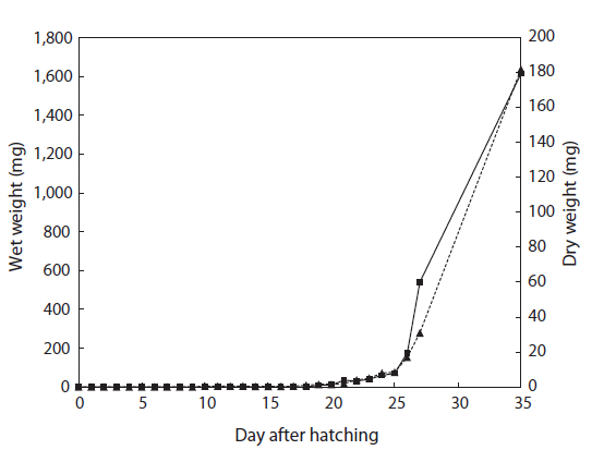
    The total length of larval fish averaged 3.35 ± 0.14 mm just after hatching, 3.91 ± 0.20 mm at first feeding (3 DAH), 5.91 ± 0.45 mm at the flexion phase (13 DAH), 7.09 ± 0.61 mm at the postflexion phase (17 DAH), 21.46 ± 2.45 mm at the juvenile phase (27 DAH), and 56.64 ± 9.27 mm at the experimental end date (35 DAH). The total length of the chub mackerel larvae increased markedly from the postflexion phase to the juvenile phase (Fig. 1). The wet and dry weights were 4.6 and 0.4 mg, respectively, at 17 DAH; by 27 DAH, the wet and dry body weights had increased >60 fold, to 277.1 mg and 59.1 mg, respectively (Fig. 2). The average growth rate during 35 days of rearing was approximately 1.52 mm/day.

      >  Development of the Digestive System

    Yolk-sac larvae

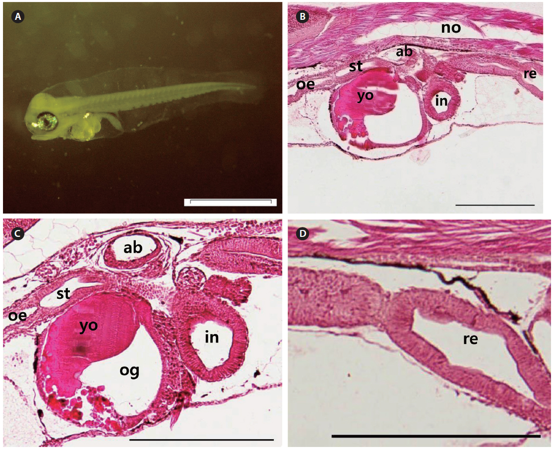
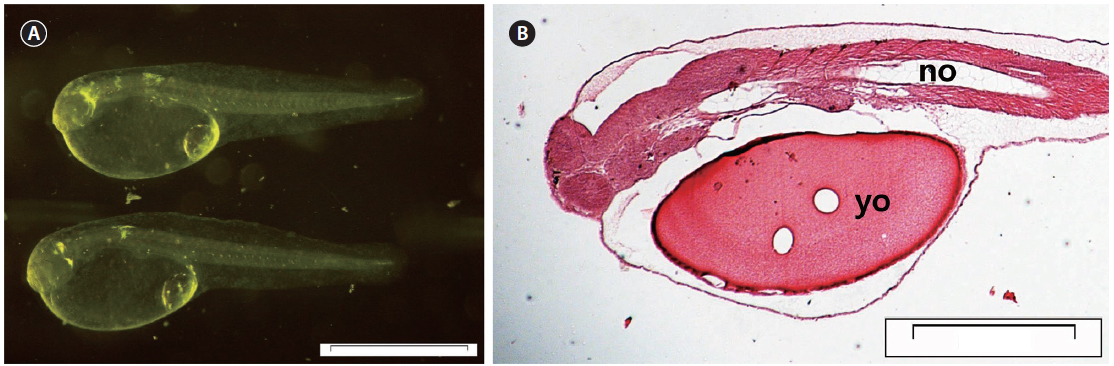
    At 0 DAH (3.35±0.15 mm TL), the mouth and anus were unopened, and the melanosome in the eyes was not yet colored (Fig. 3A). Most of the body cavity was occupied with yolk (Fig. 3B). The digestive tract was a simple straight tube observed along the dorsal wall of the yolk. At 1 DAH (3.69 ± 0.16 mm TL), the lumen of the gut was slightly enlarged, and the digestive system of the chub mackerel was opened to the anus, although the mouth had not yet opened. The intestines and rectum were differentiated, and the mouth opened completely. The rudimentary swim bladder began to differentiate from the dorsal wall of the anterior gut at 2 DAH (3.91 ± 0.17 mm TL).

    Preflexion larvae

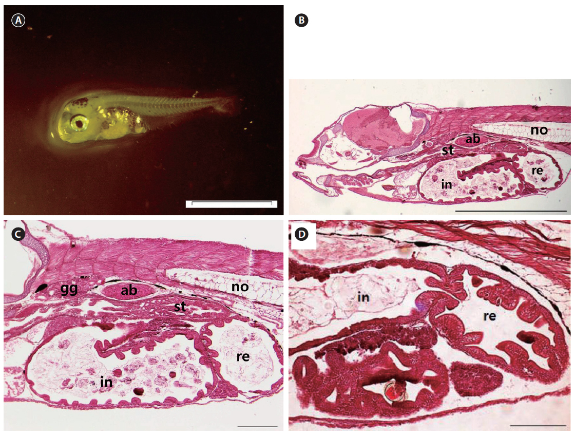
    The mouth and anus were completely open, and the eyes fully pigmented at 3 DAH (3.91 ± 0.20 mm TL). The primordial fin fold, which had developed by 3 DAH, formed the external structure that was able to find and consume food. The digestive tract had expanded, with a rudimentary stomach apparent between the esophagus and the intestines at 3 DAH (Fig. 4A). The primitive structure of the digestive system was divided into an oropharyngeal cavity, esophagus, rudimentary stomach, swim bladder, intestines, and rectum. The gastric gland and blind sac were not observed in the stomach mucosa. Rotifers first appeared in the digestive tract at 3 DAH, and the basic structure of the digestive system, which enabled the larvae to ingest and digest exogenous nutrients, was established (Fig. 4B). The yolk was mostly absorbed, and the intestinal epithelium had thickened (Fig. 4C). The rectum epithelium was composed of columnar absorptive cells, with well-developed, striated borders (Fig. 4D). By 5 DAH (4.11 ± 0.23 mm TL) the intestines had become increasingly inflated, and the epithelium was composed of vacuoles. The rectum epithelium from which the mucosal fold does not develop, became thick, and acidophil granules were observed in great numbers. The rectum was completely separated from the intestines by a well-developed rectal valve. The first appearance of the gastric gland was noted at 5 DAH. By 7 DAH (4.67 ± 0.29 mm TL) the intestines had enlarged, and the mucosal fold of the rectum had started to developed. The mucosal folds of the stomach developed, the dorso-posterior section had enlarged to form the blind sac, and the pyloric caeca was differentiated. The mucosal folds of the intestines and rectum developed, and the intestines were divided into the anterior and posterior intestines by 9 DAH (5.24 ± 0.37 mm TL). The esophagus was differentiated at 11 DAH (5.53 ± 0.37 mm TL), with the stratified squamous epithelium in the front and simple squamous epithelium in the back. The number of gastric glands increased, and the stomach elongated posteriorly to form the blind sac at 11 DAH.

    Flexion larvae

    The head became larger, and the body depth increased. The notochord tip began to turn upward with the appearance of urophysial bones at 13 DAH (5.91 ± 0.45 mm TL) (Fig. 5A). Development of the digestive system as a whole took place at this time. The mucosal folds of the stomach in particular were developed (Fig. 5B). The mucosal epithelium became thicker and noticeably developed (Fig. 5C, D).

    Postflexion larvae

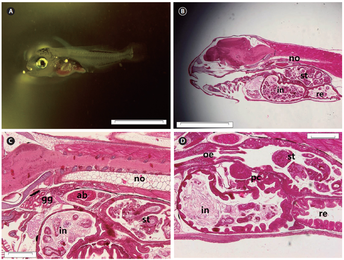
    At 17 DAH (7.09 ± 0.61 mm TL), the notochord tip upturn was complete, along with a nearly complete caudal fin (Fig. 6A). The stomach had separated into the cardiac and pyloric portions and the blind sac. The blind sac extended posteroventrally (Fig. 6B). The number of gastric glands had increased (Fig. 6C). The number of jaw and pharyngeal teeth, gastric glands, and pyloric caeca had also increased. The pyloric caeca were well formed at the anterior-most part of the intestines. There were few mucous cells on the oral cavity epithelium (Fig. 6D).

    Juveniles

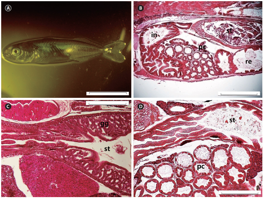
    At 27 DAH, all the fins were fundamentally established (Fig. 7A), and the histological characteristics of the digestive organ were similar to those of adult chub mackerel (Fig. 7B). The gastric glands had increased in number (Fig. 7C) and were distributed throughout the stomach, with the exception of the pyloric portion (Fig. 7D). The gastric lumen contained a large volume of food, resulting in the expansion of the blind sac toward the posterior edge of the abdominal cavity. Intestinal epithelial cells were stained dark with hematoxylin in all the fish examined.

    Discussion

    The development of the digestive system in S. japonicus larvae is similar to that reported for other scombrid species, particularly the striped bonito Sarda orientalis (Kaji et al., 2002), the bluefin tuna Thunnus thynnus, and the yellowfin tuna Thunnus albacares (Kaji et al., 1996; 1999). These all possessed an undifferentiated and rudimentary digestive system, which later differentiated into the buccopharynx, esophagus, intestines, and rectum at 3 DAH. Tanaka (1973) reported that Japanese sea bass (Lateolabrax japonicus), red sea bream, and black sea bream (Acanthopagrus schlegelii) formed a primitive larval-type digestive system at 4 DAH. These early larval-type digestive systems are established to enable the larvae to ingest, digest, and assimilate its first exogenous food when the yolk reserves have been completely absorbed. However, the assimilation efficiency may be lower in larvae than that in adult fish due to the lack of a morphological and functional stomach in the larvae.

    Pinocytosis has been suggested as an alternative pathway for the digestion of proteins in teleost larvae, as their enzymatic digestive system is poorly developed (Watanabe, 1982). This study showed that vacuoles appeared evenly on the intestinal epithelium of chub mackerel larvae, and acidophil granules were observed in the rectal epithelium at 3 DAH. Iwai and Tanaka (1968) and Tanaka (1973) reported that the vacuoles and acidophil granules were indicative of larval digestion and absorption mechanisms that existed before the development of the functionality of the early divided stomach and gastric gland of larval fish. In most marine fishes, the development of a digestive system consisting of a functional stomach with well-developed gastric glands and pyloric caeca occurs in association with the transformation to the juvenile stage. Accordingly, the typical development pattern of marine fishes involves a mechanism for digestion, and absorption changes from pinocytosis and intracellular digestion to extracellular digestion and membrane transport with the development of the gastric gland (Govoni et al., 1986).

    The first appearance of the gastric gland was reported at 21 DAH in red sea bream, 15 DAH in rainbow trout (Oncorhynchus mykiss), 25 DAH in black sea bream, and 90–120 DAH in Plecoglossus altivelis (Tanaka, 1973). However, in chub mackerel larvae, the gastric gland appeared at 5 DAH during the preflexion phase. Interestingly, the striped bonito (Kaji et al., 2002) and Spanish mackerel Scomberomorus niphonius (Tanaka et al., 1996), which are both members of the Scombridae, developed gastric glands at 3 and 4 DAH, respectively. In contrast, in bluefin and yellowfin tuna, the gastric glands emerged at 11 and 14 DAH, which was 2–3 times later than in chub mackerel (Kaji et al., 1996; 1999). The appearance of pyloric caeca followed a similar pattern to the gastric glands in the scombrid larvae. In the striped bonito (Kaji et al., 2002) and Japanese Spanish mackerel (Fukunaga et al., 1982), pyloric caeca appeared earlier (at 7 DAH) than in the chub mackerel, and these emerged at 14 and 16 DAH in bluefin and yellowfin tuna, respectively (Kaji et al., 1996; 1999).

    Although five scombrid larvae, including the chub mackerel, showed species-specific timing in the appearance of the gastric glands and pyloric caeca, their digestive system attained the juvenile-type structure long before metamorphosis. On the other hand, a juvenile-type digestive system formed during the juvenile period, at 35 DAH, in black sea bream and false kelpfish (Sebastiscus marmoratus), at 135 DAH in sweet fish (Tanaka, 1973), and at 21 DAH in large yellow croakers (Mai et al., 2005). Consequently, the development of the digestive system of chub mackerel larvae can be considered precocious compared to common marine fishes.

    Scombrids can grow very rapidly during their early life stages, and this may be related to the precocious development of the digestive system in this group of fishes (Tanaka et al., 1996). In chub mackerel, the total length and body weight increased gradually from 17 DAH; thereafter, there was a rapid increase from 25 DAH. As in other scombrids, chub mackerel larvae also undergo marked, rapid growth during the early life stages, which is supported by their precocious digestive system. In addition, Shoji et al. (2001) reported that the absolute growth of wild chub mackerel increased from 0.298 mm/day (post-first-feeding stage) to 0.712 mm/day (later larval and early juvenile stages), suggesting that the increase in growth rate of the chub mackerel may result from the shift in feeding to piscivory.

    Tanaka et al. (1996) suggested that the precocious development of the digestive system with differentiation of the functional stomach in scombrid larvae also supported the appearance of piscivory. The increase in pepsin-like enzyme activity measured from the whole body extract is likely to be concomitant with gastric gland differentiation in the scombrid larvae, as in many other species (Govoni et al., 1986). This physiological advancement, as well as the formation of a blind sac that enables the larvae to store larger prey items, would support the appearance of piscivory in scombrid larvae, including chub mackerel larvae.

    The peak occurrence of wild Spanish mackerel larvae is synchronized with the peak abundance of their prey larvae, suggesting the ecological advantage of early piscivory with the precocious development of the digestive system (Shoji et al., 1999). In contrast, the bluefin and yellowfin tuna larvae attained juvenile-type digestive systems during the flexion phase and underwent a larval period of planktivory with a larval-type digestive system, suggesting that they may have adapted to their offshore feeding habitat where the density of prey fish larvae may be low (Tanaka et al., 1996). Kohno et al. (1984) reported that swimming and feeding abilities develop rapidly after the development of the jaw and pharyngeal teeth, as well as the caudal fin formation in chub mackerel larvae of 5–7 mm SL. In addition, 25% of wild chub mackerel larvae measuring 5–6 mm SL contained fish in their stomachs; this increased to 80% at 9–10 mm SL (Shoji et al., 2001). As in the Spanish mackerel, the early morphological developments and precocious digestive system in chub mackerel larvae were likely associated with the early onset of piscivory. Stomach contents analysis of wild chub mackerel larvae of 4–9 mm TL showed that the main prey were the larvae of the sea squirt, crustaceans, Evadne sp., and water fleas (Ozawa et al., 1991). However, as the chub mackerel larvae grew, the amount of fish larvae in the diet increased, as the consumption of crustacean larvae and Evadne decreased. The size range (4–9 mm TL) of wild chub mackerel was estimated between 6 and 18 DAH in this study; this period coincides with the establishment of the juvenile-type digestive system during the larval period. Therefore, it could be predicted that the precocious development of the digestive system during the larval period of chub mackerel is related to the early appearance of piscivory, which is able to support the high growth potential of the larvae. Consequently, the precocious digestive system in chub mackerel larvae described in this study will provide useful information to improve current larval rearing practices and feeding protocols, and will reduce weaning costs for this fish species.

참고문헌
  • 1. FAO Species Catalogue. Scombrids of the world - An annotated and illustrated catalogue of tunas, mackerels, bonitos and related species known to date. 125 P.56-57 google
  • 2. Fukunaga T, Ishibashi N, Mitsuhashi N 1982 Artificial fertilization and seeding production of Spanish mackerel [Saibai-giken] Vol.11 P.29-48 google
  • 3. Govoni JJ, Boehlert GW, Watanabe Y 1986 The physiology of digestion in fish larvae [Environ Biol Fish] Vol.16 P.59-77 google cross ref
  • 4. Harada T, Murata O, Miyashita S 1974 On the artificial fertilization and rearing of larvae in bonito [Mem Fac Agri Kinki Univ] Vol.7 P.1-4 google
  • 5. Hunter JR, Lasker R 1981 Feeding ecology and predation of marine fish larvae P.33-77 google
  • 6. Iwai T, Tanaka M 1968 The comparative study of the digestive tracts of teleost larvae-III. Epithelial cells in the posterior gut of halfbeak larvae [Bull Japan Soc Sci Fish] Vol.34 P.44-48 google cross ref
  • 7. Kaji T, Tanaka M, Takahashi Y, Oka M, Ishibashi N 1996 Preliminary observations on development of pacific bluefin tuna Thunnus thynnus (Scombridae) larvae reared in the laboratory, with special reference to the digestive system [Mar Freshwater Res] Vol.47 P.261-269 google cross ref
  • 8. Kaji T, Tanaka M, Oka M, Takeuchi H, Ohsumi S, Teruya K, Hirokawa J 1999 Growth and morphological development of laboratory-reared yellowfin tuna Thunnus albacares larvae and early juveniles, with special emphasis on the digestive system [Fish Sci] Vol.65 P.700-707 google
  • 9. Kaji T, Kodama M, Arai H, Tagawa M, Tanaka M 2002 Precocious development of the digestive system in relation to early appearance of piscivory in striped bonito Sarda orientalis larvae [Fish Sci] Vol.68 P.1212-1218 google cross ref
  • 10. Kendall AW, Ahlstrom EH, Moser HG, Moser HG, Richards WJ, Cohen DM, Fahay MP, Kendall AW, Richardson SL 1984 Early life stages of fishes and their characters P.11-22 google
  • 11. Kohno H, Shimizu M, Nose Y 1984 Morphological aspects of the development of swimming and feeding functions in larval Scomber japonicus [Nip Sui Gak] Vol.50 P.1125-1137 google cross ref
  • 12. Mai K, Yo H, Ma H, Duan Q, Gisbert E, Zambonino-Infante JL, Cahu CL 2005 A histological study on the development of the digestive system of Pseudosciaena crocea larvae and juveniles [J Fish Biol] Vol.67 P.1094-1106 google cross ref
  • 13. Miyashita S, Kato K, Sawada Y, Murata O, Ishitani Y, Shimizu K, Yamamoto S, Kumai H 1998 Development of digestive system and digestive enzyme activities of laeval and juvenile bluefin tuna, Thunnus thynnus, reared in the laboratory [Suisanzoshoku] Vol.46 P.111-120 google
  • 14. Ozawa T, Kawai K, Uotani I 1991 Stomach content analysis of chub mackerel Scomber japonicus by quantification I method [Bull Jap Soc Sci Fish] Vol.57 P.1241-1245 google cross ref
  • 15. Shoji J, Maehara T, Tanaka M 1999 Short-term occurrence and rapid growth of Spanish mackerel larvae in the central waters of the Seto Inland Sea, Japan [Fish Sci] Vol.65 P.68-72 google
  • 16. Shoji J, Tanaka M, Maehara T 2001 Comparative diets and growth of two Scombrid larvae, chub mackerel Scomber japonicus and Japanese Spanish mackerel Scomberomorus niphonius in the central Seto Inland Sea, Japan [UJNR Technical Report] Vol.30 P.93-103 google
  • 17. Tanaka M 1973 Studies on the structure and function of the digestive system of teleost larvae. Ph.D. Dissertation google
  • 18. Tanaka M, Kaji T, Nakamura Y, Takahashi Y, Watanabe Y, Yamashita Y, Oozeki Y 1996 Development strategy of scombrid larvae: high growth potential related to food habits and precocious digestive system development P.125-139 google
  • 19. Watanabe Y 1982 Intracellular digestion of horseradish peroxidase by the intestinal cells of teleost larvae and juveniles [Bull Japan Soc Sci Fish] Vol.48 P.37-42 google cross ref
OAK XML 통계
이미지 / 테이블
  • [ Fig. 1. ]  Total length (mean ± S.D.) and feeding schedule of the chub mackerel Scomber japonicus larvae and juveniles.
    Total length (mean ± S.D.) and feeding schedule of the chub mackerel Scomber japonicus larvae and juveniles.
  • [ Fig. 2. ]  Developmental changes in wet (▲) and dry weight (■) of Scomber japonicus during larval and juvenile period.
    Developmental changes in wet (▲) and dry weight (■) of Scomber japonicus during larval and juvenile period.
  • [ Fig. 3. ]  Photomicrographs of a Scomber japonicus larva on the day just after hatching. (A) Whole-body photograph. (B) Sagittal section of the body axis of larva just after hatching showing large yolk. The scale bars indicate 1 mm for (A); 200 μm for (B). yo, yolk; no, notochord.
    Photomicrographs of a Scomber japonicus larva on the day just after hatching. (A) Whole-body photograph. (B) Sagittal section of the body axis of larva just after hatching showing large yolk. The scale bars indicate 1 mm for (A); 200 μm for (B). yo, yolk; no, notochord.
  • [ Fig. 4. ]  Photomicrographs of Scomber japonicus larva at first feeding stage on 3 DAH. (A) Whole-body photograph. (B) Sagittal section of the body axis showing the differentiated digestive tract. (C) Sagittal section of the rudimentary stomach, oil globule, intestine and air bladder. (D) Sagittal section of the rectum. The scale bars indicate 1 mm for (A); 100 μm for (B)-(D). st, stomach; og, oil globule; in, intestine; ab, air bladder; re, rectum; no, notochord; oe, oesophagus.
    Photomicrographs of Scomber japonicus larva at first feeding stage on 3 DAH. (A) Whole-body photograph. (B) Sagittal section of the body axis showing the differentiated digestive tract. (C) Sagittal section of the rudimentary stomach, oil globule, intestine and air bladder. (D) Sagittal section of the rectum. The scale bars indicate 1 mm for (A); 100 μm for (B)-(D). st, stomach; og, oil globule; in, intestine; ab, air bladder; re, rectum; no, notochord; oe, oesophagus.
  • [ Fig. 5. ]  Photomicrographs of a Scomber japonicus larva at the flexion phase on 13 DAH. (A) Whole-body photograph. (B) Sagittal section of the body axis showing the development of stomach, intestine and rectum. (C) Sagittal section of stomach showing the early appearance of the gastric glands. (D) Sagittal section of the rectum and intestine. The scale bars indicate 2 mm for (A); 500 μm for (B); 100 μm for (C) and (D). st, stomach; in, intestine; re, rectum; gg, gastric glands; no, notochord.
    Photomicrographs of a Scomber japonicus larva at the flexion phase on 13 DAH. (A) Whole-body photograph. (B) Sagittal section of the body axis showing the development of stomach, intestine and rectum. (C) Sagittal section of stomach showing the early appearance of the gastric glands. (D) Sagittal section of the rectum and intestine. The scale bars indicate 2 mm for (A); 500 μm for (B); 100 μm for (C) and (D). st, stomach; in, intestine; re, rectum; gg, gastric glands; no, notochord.
  • [ Fig. 6. ]  Photomicrographs of Scomber japonicus larva at the postflexion phase on 17 DAH. (A) Whole-body photograph. (B) Sagittal section of the body axis showing the development of stomach, intestine and rectum. (C) Sagittal section of the stomach showing the gastric glands. (D) Pyloric caecum differentiated from the anterior intestine. The scale bars indicate 3 mm for (A); 1 mm for (B); 100 μm for (C) and (D). st, stomach; in, intestine; re, rectum; gg, gastric glands; pc, pyloric caecum; no, notochord.
    Photomicrographs of Scomber japonicus larva at the postflexion phase on 17 DAH. (A) Whole-body photograph. (B) Sagittal section of the body axis showing the development of stomach, intestine and rectum. (C) Sagittal section of the stomach showing the gastric glands. (D) Pyloric caecum differentiated from the anterior intestine. The scale bars indicate 3 mm for (A); 1 mm for (B); 100 μm for (C) and (D). st, stomach; in, intestine; re, rectum; gg, gastric glands; pc, pyloric caecum; no, notochord.
  • [ Fig. 7. ]  Photomicrographs of Scomber japonicus at early juvenile phase on 27 DAH. (A) Whole-body photograph. (B) Sagittal section of the body axis showing the development of stomach, intestine, pyloric caecum and rectum. (C) Densely distributed gastric glands. (D) Pyloric caecum differentiated from the anterior intestine. The scale bars indicate 7 mm for (A); 2 mm for (B); 100 μm for (C) and (D). st, stomach; in, intestine; re, rectum; gg, gastric glands; pc, pyloric caecum.
    Photomicrographs of Scomber japonicus at early juvenile phase on 27 DAH. (A) Whole-body photograph. (B) Sagittal section of the body axis showing the development of stomach, intestine, pyloric caecum and rectum. (C) Densely distributed gastric glands. (D) Pyloric caecum differentiated from the anterior intestine. The scale bars indicate 7 mm for (A); 2 mm for (B); 100 μm for (C) and (D). st, stomach; in, intestine; re, rectum; gg, gastric glands; pc, pyloric caecum.
(우)06579 서울시 서초구 반포대로 201(반포동)
Tel. 02-537-6389 | Fax. 02-590-0571 | 문의 : oak2014@korea.kr
Copyright(c) National Library of Korea. All rights reserved.