New Record of Scolecenchelys fuscogularis (Anguilliformes: Ophichthidae) Leptocephali from Korea, as Revealed by Morphological and Molecular Analyses
- DOI : 10.5657/FAS.2015.0109
- Author: Ji Hwan-Sung, Kim Jin-Koo, Lee Soo Jeong, Kimura Seishi, Hibino Yusuke
- Publish: Fisheries and aquatic sciences Volume 18, Issue1, p109~113, 31 March 2015
-
ABSTRACT
Three leptocephali (22.2, 22.7, 56.0 mm in total length) collected from the East/Japan Sea were identified by morphological and genetic analyses as belonging to the genus
Scolecenchelys (Anguilliformes, Ophichthidae). Morphologically, the specimens were characterized by 148-158 myomeres, 10 gut swellings, dorsal fin origin above middle of the body, and 6 postanal melanophores between the anus and the caudal margin. An analysis of an 849-base pair 12S rRNA sequence of mitochondrial DNA showed that sequences are concordant with those of adultScolecenchelys fuscogularis (genetic distance = 0.001). Furthermore total number of myomeres is consistent with the total number of vertebrae in adultS. fuscogularis . This study provides the first description of the morphological characteristics ofS. fuscogularis leptocephali and their variations with size. The Korean name ofS. fuscogularis is “Ga-neun-mul-baem”, established byJi et al. (2012) .
-
KEYWORD
Scolecenchelys fuscogularis , New record , Ophichthidae , Leptocephalus , mtDNA 12S rRNA
-
The genus
Scolecenchelys Ogilby, 1897, of the subfamily Myrophinae, family Ophichthidae, order Anguilliformes, is distributed in tropical and temperate areas of the Indo-Pacific (Hibino et al., 2013a). The genus includes 21 species (Hibino et al., 2013a; Eschmeyer, 2014), 7 of which are known in Japan (Hatooka, 2013; Hibino et al., 2013a, b). However, only one species,Scolecenchelys aoki (Jordan and Snyder, 1901) (known from adults and leptocephali) has been recorded from Korea (Ji et al., 2012; Ji et al., 2013). Species ofScolecenchelys in adults are defined by the following combination of morphological characters: absence of pectoral fin; center of the eye located behind the mid-jaw; presence of three preopercular sensory pores, and two infraorbital pores located between the anterior and posterior nostrils; and teeth on upper jaw and vomer conical, pointed, and arranged in single to triple rows (Castle and McCosker, 1999; McCosker et al., 2012). Castle and McCosker (1999) provided a taxonomic review ofScolecenchelys , and several new species of the genus have been recently described by McCosker (2006), McCosker et al. (2012) and Hibino et al. (2013a).During November 2009 and October 2010, three leptocephali collected from the East/Japan Sea, and were identified as
Scolecenchelys fuscogularis (Hibino, Kai and Kimura, 2013a) based on a combination of morphological and molecular traits. Our study provides the first report of this species in Korean waters. We here present a detailed morphological description of size-related variations in the leptocephali ofS. fuscogularis .Material examined : PKU 5999-6000, 2 specimens, 22.2 mm and 27.2 mm in total length (TL), southern part of the East/Japan Sea (35°40′43″N, 129°44′47″E), October 2010, RN80 net, collected by JK Kim and HS Ji. PKUI 205, 1 specimen, 56.0 mm TL, southern part of the East/Japan Sea (34°54′47″, 129°33′14″E), 22 November 2009, RN80 net, collected by JK Kim and HS Ji.
The specimens were immediately preserved in 99% ethanol on shipboard. Counts and measurements followed those of Fahay and Obenchain (1978) and Tabeta and Mochioka (1988). Each body part was measured to the nearest 0.01 mm using digital vernier calipers, with measurements performed under zoom stereomicroscope (Olympus SZX-16, Japan). The description of tooth sequences followed Castle (1984). The specimens collected in the study are deposited in the Ichthyoplankton Collection of Pukyong National University (PKUI), Korea.
Genomic DNA was extracted from the right eyeball of the
S. fuscogularis leptocephali (22.2–56.0 mm TL) using forceps and Chelex 100 resin (Bio-Rad, USA). We also analyzed DNA of the adultS. fuscogularis using muscle tissue obtained from the holotype of the species, FAKU (Faculty of Agriculture, Kyoto University, Japan) 132857. Later, mitochondrial 12S rRNA sequences were amplified and sequenced according to the method of Ji and Kim (2010). The DNA sequences were aligned and edited using Clustal W (Thompson et al., 1994) and BioEdit version 7.0.0. The molecular results were compared with data from five adult ophichthids (S. aoki, Muraenichthys gymnopterus, Echelus uropterus, Pisodonophis sangjuensis , andOphichthus asakusae ) and one outgroup (Conger myriaster ). Genetic divergences were calculated using the Kimura two-parameter model (Kimura, 1980) and MEGA 6 (Tamura et al., 2013); a neighbor-joining (NJ) tree was constructed using the Kimura two-parameter method and 10,000 bootstrap replications in MEGA 6.>
Scolecenchelys fuscogularis Hibino, Kai and Kimura, 2013 
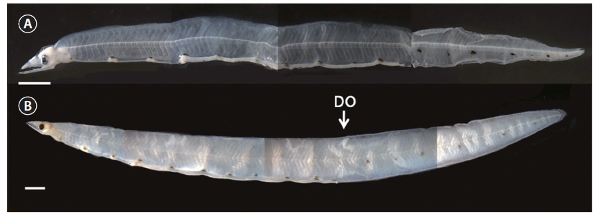
(Korean name: Ga-neun-mul-baem; Fig. 1, Table 1)
Scolecenchelys fuscogularis Hibino, Kai and Kimura, 2013: 43 (type locality: the Sea of Japan off the coast of Kyoto Prefecture, Japan).Scolecenchelys sp.: Ji et al., 2012: 415 (Korea).Morphological analysis
The present specimens have following characters: first and second liver lobe separated; strongly developed pterygiophores; and dorsal fin origin slightly above and behind the midpoint of the body. From these characters, the present leptocephali can be decided as a member of the subfamily Myrophinae (Castle, 1984). The present specimens also have following characters: 148-158 total myomeres; 10 gut swellings; dorsal fin origin above middle of the body; and 6 postanal melanophores between the anus and the caudal margin, and these characters are shared with a leptocephalus of
Scolecenchelys aoki (Ji et al., 2012).Mitochondrial DNA sequence analysis
The 849-base pair sequences of mtDNA 12S rRNA in the three leptocephali in this study were nearly identical to the sequences of adult
S. fuscogularis (genetic distance,d = 0.001), but were slightly distinct from adultS. aoki (d = 0.006) (Table 2). The neighbor joining tree showed that the three leptocephali were clustered with adultS. fuscogularis , being strongly supported by a 98% bootstrap value (Fig. 2).> Description of leptocephalus of
Scolecenchelys fuscogularis Precaudal fin developmental stage (22.2 mm and 27.2 mm TL) : Head relatively small; tip of snout highly acute. Fang-like teeth on both jaws. Two nostrils present in front of eyes. Anterior tip of lower jaw more acute than that of upper jaw. Caudal region short. Head length, 8.1-9.5% TL; body depth, 7.2-7.7% TL; preanal length, 65.4-78.0% TL (Table 1). Punctate and branched melanophores present on dorsal surfaces of all 10 gut swellings (Fig.1A); six developed melanophores present just below the lateral midline of the caudal region and extending to the caudal terminus (Fig. 1A).Caudal fin developmental stage (56.0 mm TL) : Number of teeth increasing from the preceding stage. Unlike in the preceding stage, both jaws coincident (Fig. 1B). Head length (6.8% TL) and preanal length (59.9% TL) becoming smaller than those of the preceding stage, whereas body depth (8.6 % TL) larger (Table 1). All fins developed; dorsal fin origin located above 74th myomere, anal, caudal, and pectoral fin rays developed. Unlike in the preceding stage, the 6 melanophores on the caudal region fading (Fig. 1B).East/Japan Sea (Ji et al., 2012; Hibino et al., 2013a; present study), and off the Pacific coast of Japan (Hibino et al., 2013a).
The total number of myomeres in the present
S. fuscogularis leptocephali (148-158) is approximate to the range of the number of vertebrae in adultS. fuscogularis (146-149) (Hibino et al., 2013a), and the number of predorsal myomeres in the present leptocephali (74) is also consistent with number of predorsal vertebrae in adultS. fuscogularis (72) (Table 1; Hibino et al., 2013). AlthoughS. fuscogulari are most similar toScolecenchelys australis (Macleay, 1881) andScolecenchelys chilensis (McCosker, 1970) in the number of vertebrae (146-149 inS. fuscogularis vs. 148-152 inS. australis vs.146-159 inS. chilensis ) (Hibino et al., 2013a), their distributional areas are different (northeast Asia forS. fuscogularis vs. the south eastern Pacific forS. australis vs. the south western Pacific forS. chilensis ) (McCosker, 1970; 1977; Hibino et al., 2013a).Scolecenchelys fuscogularis leptocephali differ fromS. aoki leptocephali in the number of myomeres (148-158 inS. fuscogularis vs. 135 inS. aoki ), and the number of preanal myomeres (71-74 inS. fuscogularis vs. 69 inS. aoki ) (Table 1; Ji et al., 2012). We adopted the Korean name forS. fuscogularis , “Ga-neun-mul-baem”, following Ji et al. (2012).-
[Fig. 1.] Leptocephalus of Scolecenchelys fuscogularis, PKU 5999, 22.2 mm TL (A); PKUI 205, 56.0 mm TL (B). Scale bars= 1.5 mm. DO: dorsal fin origin.
-
[Table 1.] 
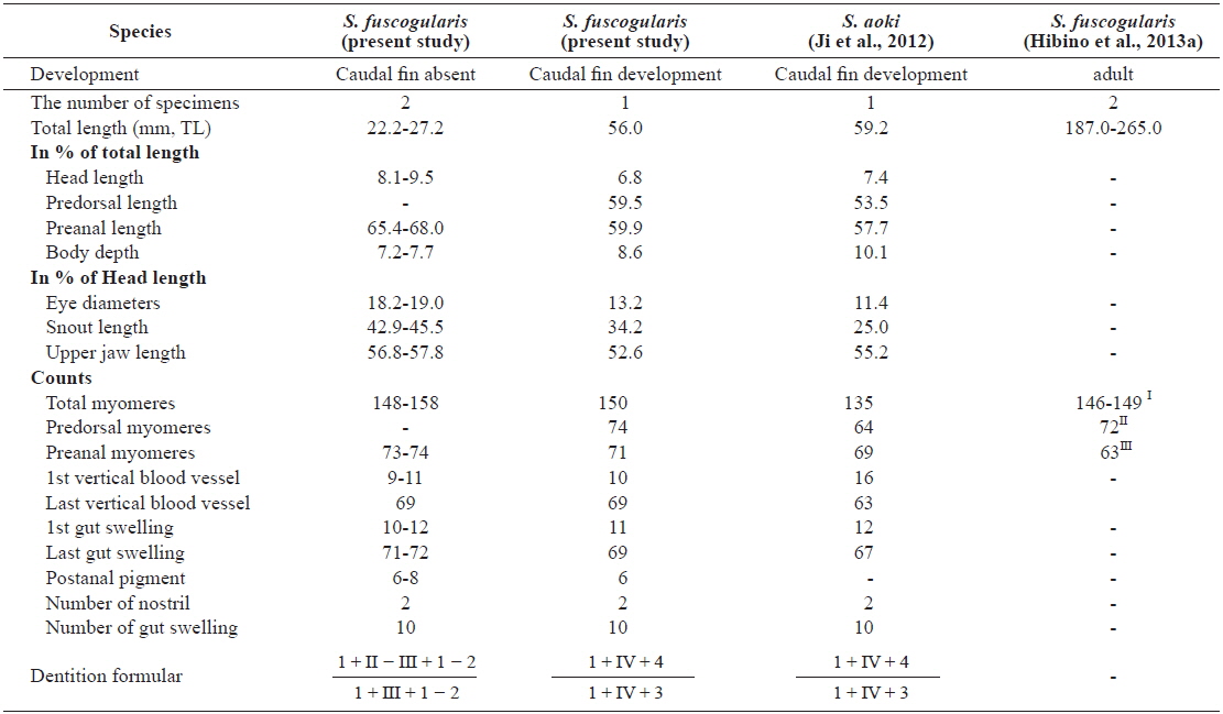
Comparisons of measurements and counts for Scolecenchelys fuscogularis and Scolecenchelys aoki
-
[Table 2.] 
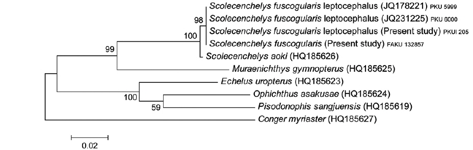
Genetic distance among Scolecencehelys fuscogularis leptocephali, 6 adult ophichthid species and 1 outgroup
-
[Fig. 2.] Neighbor-joining (NJ) tree based on partial mtDNA 12S rRNA sequences showing the relationships among the three Scolecenchelys fuscogularis leptocephali and 6 adult ophichthids with one outgroup Conger myriaster. The NJ tree was constructed using the Kimura-2-parameter distance model. 10,000 replications of bootstrap were conducted. Bar indicates genetic distance of 0.02.