검색 전체 메뉴
PDF
맨 위로
OA 학술지
임상에서 쓰이는 저 출력 초음파(Low Intensity Pulsed Ultrasound)가 조골세포에 미치는 영향 The Effect of Osteoblast like Cell on Low Intensity Pulsed Ultrasound Used in Current Clinical Practice
  • 비영리 CC BY-NC
ABSTRACT
임상에서 쓰이는 저 출력 초음파(Low Intensity Pulsed Ultrasound)가 조골세포에 미치는 영향

In this study, we investigated the proliferative and adhesional effect of human osteoblast like MG-63 cell treated with low intensity pulsed ultrasound (LIPUS). We tested the effectiveness of LIPUS in human osteoblast like MG-63 cells. Cell proliferation was measured using a water soluble tetrazolium salts-1 assay. The mRNA expression of alkaline phosphate, vascular endothelial growth factor (VEGF), integrin alpha 2, colla 1A1 were performed by reverse transcription-polymerase chain reaction. LIPUS was no cytotoxicity in human osteoblast like MG-63 cells. In addition, the data show that treatment with 1 MHz and 3 MHz LIPUS on increased proliferation 7 days after. There were significant increased in mRNA expression of alkaline phosphatase, osteocalcin, VEGF, integrin alpha 2 and colla 1A1 (p<0.05). Therefore, the LIPUS significantly increased differential expression of mRNA levels in osteoblast like MG-63 cell and new possibilities in dental clinical practice.

KEYWORD
Low-intensity focused ultrasound , Osteoblasts , Cell proliferation , Vascular endothelial growth factor A
  • 서 론

    현재 치과용 임플란트는 치료 후 상실된 무치악 부위에서 심미적, 기능적으로 많이 쓰여 오고 있다. 임플란트는 식립 후 골 유착이 매우 중요하며, 이는 임플란트와 골 조직 표면에 자연치아와 같은 치은 열구가 없고, 띠 형태로서 부착하기 때문이다. 이렇듯 골 유착은 임플란트의 초기 고정에 있어 중요하며, 임플란트와 골 조직은 골 유착 형태로서 결합하게 되며 임플란트와 치조골 사이의 골 유착은 임플란트의 성공률에 있어 큰 영향을 미치게 된다. 골 유착 시 임플란트와 주위 조직 간의 반응은 현재 여러 연구를 통해 발표되고 있는데, 임플란트 식립 시 뼈 유착에서 세포가 분비하는 인자들과 주위 조직은 임플란트의 표면에 반응하고 영향을 받게 된다1,2). 최근 연구에 따르면 저 출력 초음파를 이용하여 골절치유3)나 림프종, 암치료4), 골육종 세포5)에서의 연구와 함께 세포의 이동이나 뼈 치유 촉진 및 혈관 신생과 관련한 연구가 발표되고 있다6,7).

    최근 치과계에서 주목 받고 있는 치료용 저 출력 초음파는 여러 성장인자, 골 형성 단백질 등의 생물학적 자극뿐만 아니라 물리적인 자극에도 민감한 것으로 알려지고 있다2). 즉 낮은 전류, 저 출력 초음파 에너지 영역, 그리고 특수한 파장의 전자장에 의해 골 형성이 촉진될 수 있다고 보고되고 있는데, 이는 비침습적이고 임상적으로 제공이 용이한 장점이 있다. 이러한 이유로 인하여 최근 골절의 치유를 촉진하기 위하여 저 출력 초음파(low intensity pulsed ultrasounds, LIPUS) 등을 개발하고 사용하고 있다8). 치과계의 저 출력 초음파의 치유촉진에 대한 보고는 최근에 보고되기 시작하였으며, 초음파는 의료분야, 수술이나 진단 영역에서 폭넓게 사용되고 있다. 또한, 초음파는 각각의 사용 용도에 적합하게 출력 조절이 가능하여 각 영역에 맞게 조절되어 사용할 수 있는 장점이 있다2). Duarte8)와 Pilla 등9)의 연구에 따르면 초음파를 동물의 골 조직에 처리시 골 성장과 치유에 대한 효과가 보고되었으며, 저 출력 초음파 처리 후 골 치유 촉진에 대한 응용연구가 최근 활발히 이루어지고 있다2).

    이에 본 연구는 저 출력 초음파를 human osteoblast-like cell에 처리하였을 때, 뼈 치유 과정에서 발생하는 뼈 생성 촉진 단백질들의 발현 정도와 부착관련 인자들을 알아보고자 한다.

    연구대상 및 방법

       1. 세포 주 및 세포배양

    본 실험에 사용한 MG-63 세포는 human osteoblast-like cell로서 서울대학교 의과대학 한국세포주은행에서 분양 받아 본 실험에 사용하였다. 분양 받은 MG-63 세포는 10% fetal bovine serum (FBS), 100 IU/ml penicillin, 그리고 100 μg/ml streptomycin이 함유된 Dulbecco’s modified Eagle’s medium (DMEM) 배지에서 37℃, 5% CO2 배양기를 통하여 배양하였다. 본 실험에 사용할 때는 분화를 유도하 기 위해서 골 형성 유도인자인 50 μg/ ml L-ascorbic acid 2-phosphate, 10 nM dexamethasone, 그리고 10 mM β-glycerophosphate가 포함되고 10% FBS이 포함된 DMEM로 구성된 배지에서 배양하고 2∼3일 주기로 교체하였다.

       2. 초음파 처리

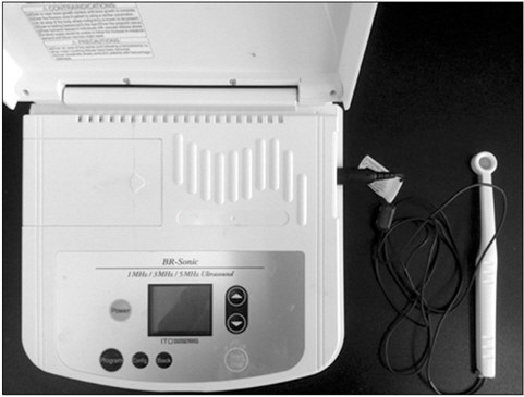
    Human osteoblast-like MG-63 세포는 6-well 배양용기에 3×10⁵으로 분주한 뒤 골 형성유도배지로 교체하였다. 실험에 사용된 저 출력 초음파는 임상에서 쓰이는 BR-Sonic(ITO Co., Tokyo, Japan) (Fig. 1)으로 human osteoblastlike cell에 저 출력 초음파를 처리하지 않은 대조군과 저 출력 초음파를 처리한 실험군으로 정하여 진행하였으며, 사용된 초음파 출력은 40 mW/cm2로, 각각 주파수를 1 MHz, 3 MHz로 나누어 15분씩 처리하였다.

       3. 세포 성장률 측정

    저 출력 초음파가 세포 성장률에 미치는 효과를 측정하기 위하여 water soluble tetrazolium salts (WST)-1 방법을 이용하였다. 6-well plate에서 3×10⁵으로 분주한 뒤 24시간 배양하였다. 24시간 뒤 세포의 상층 배지를 골 형성유도배지로 교환한 다음, 초음파를 1 MHz, 3 MHz로 처리하였다. 처리된 세포는 2일, 4일, 7일을 배양한 다음 각각의 well에 water soluble tetrazolium salts-1 용액을 처리하여 2시간 동안 37℃, 5% CO2 배양기에서 반응시켰다. 이후, ELISA Reader (Sunrise Remote Control, Tecan, Austria)로 450 nm 파장에서 흡광도를 측정하였으며, 각각의 분석은 독립적으로 세 번씩 시행되었다.

       4. Reverse transcription-polymerase chain reaction

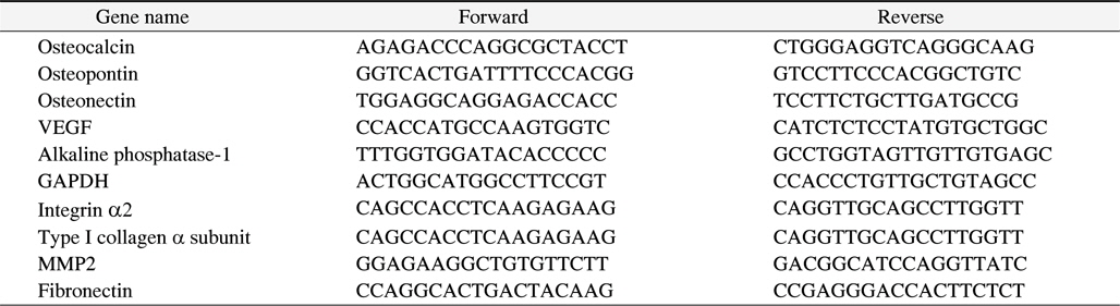
    총 mRNA를 저 출력 초음파가 처리된 세포에서 TRIzol reagent (Invitrogen, Carlsbad, CA, USA)를 처리하여 추출하였고 One-step PreMix kit (iNtRON Biotechnology Inc., Seongnam, Korea)를 이용하여 제조사의 지시대로 cDNA를 합성하였다. 합성된 cDNA로부터 2× polymerase chain reaction (PCR) master mix (Doctor Protein Corp., Seoul, Korea)를 이용하여 PCR 증폭을 실시하였다. 증폭한 산물은 1.5% agarose gel을 사용하여 전기영동으로 확인한 다음 mRNA 발현은 LAS (Fujifilm, Tokyo, Japan)를 통하여 나타내었다. PCR 실험에 사용한 primers는 Table 1과 같다.

    [Table 1.] Detailed Information of Primers

    label

    Detailed Information of Primers

       5. 통계 분석

    저 출력 초음파가 처리되지 않은 대조군과 처리된 실험군의 결과는 통계프로그램인 SPSS Window ver. 12.0 (SPSS Inc., Chicago, IL, USA)에 의해 paired t-test statistical method를 적용하여 분석하였다. 또한 p<0.05를 통계적으로 유의한 차이가 있는 것으로 판단하였다.

    결 과

       1. 저 출력 초음파가 조골세포 증식에 미치는 효과

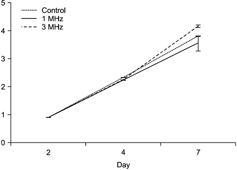
    저 출력 초음파가 조골세포에 처리 후 증식에 미치는 효과를 보기 위하여 WST-1 분석을 시행하였다. 저 출력 초음파를 처리 후 2일, 4일, 그리고 7일째 분석하였으며, 2일과 4일째에 분석된 대조군과 저 출력 초음파가 처리된 1 MHz, 3 MHz 실험군에서는 서로 별다른 변화가 없었다. 그러나 7일째부터 대조군과 비교 시 저 출력 초음파가 처리된 3 MHz 군이 유의하게 증식됨을 확인하였다(Fig. 2, p<0.05).

       2. 저 출력 초음파가 조골세포의 분화와 관련된 유전자 발현 분석

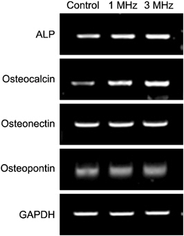
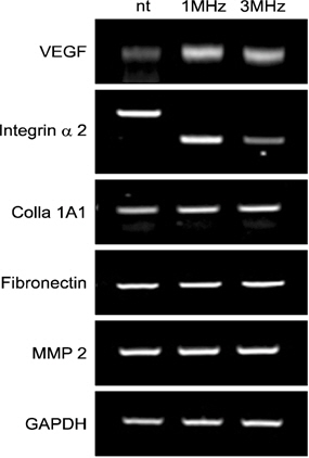
    저 출력 초음파가 조골세포의 분화와 관련된 발현의 정도를 확인하기 위하여 reverse transcription-PCR 분석을 시행하였다. 저 출력 초음파를 처리 후 7일 째에 대조군, 1 MHz, 그리고 3 MHz 실험군을 분석하였다. 대조군에 비하여 1 MHz, 3 MHz 실험군에서 alkaline phosphatase (ALP), osteocalcin, vascular endothelial growth factor (VEGF), colla 1A1은 모두 유전자의 발현이 증가함을 확인했으며, integrin alpha 2의 경우 3 MHz 실험군에서의 발현량은 적었으나, 대조군과 1 MHz 실험군에서 유사한 발현의 양을 보이는 것을 확인하였다. 반면 osteonectin, osteopontin, fibronectin 그리고 matrix metalloproteinase (MMP)2의 유전자 발현의 차이는 없었다(Fig. 3, 4).

    고 찰

    저 출력 초음파에 대한 골절치유 장애의 치료와 치유촉진에 대한 연구는 정형외과에서 오래전부터 보고되어 왔다10). 또한 치과계에서도 뼈의 골절이나 손상 시 자연치유 만으로는 회복과정이 매우 오래 걸리므로 치유속도를 증진시키기 위해 비침습적인 뼈의 재생 촉진을 위한 연구가 보고되고 있다. 이러한 많은 수의 임상 증례를 통하여11-13) 치과분야에서 저 출력 초음파의 효과에 주목하고 있으며, 임플란트 등 의 매식 시 가해지는 하악골 및 상악골의 손상 부위에서도 본격적으로 연구가 이루어지고 있다. 임플란트 매식 시 골의 치유속도는 임플란트 성공에 매우 큰 영향을 미치므로 골 치유가 적절히 진행되지 않을 시 골의 흡수, 골 유착의 실패, 그리고 치유의 지연이 발생할 수 있다.

    저 출력 초음파는 세포의 자극이 적고 비침습적 창상 치유 방법으로서, 주로 1 MHz, 3 MHz의 주파수를 주로 사용하고 있다8). 본 연구에서는 human osteoblast-like MG-63 세포에 1 MHz, 3 MHz 강도의 초음파를 처리하여 조골세포 의 증식과 분화와 관련된 발현 정도를 파악하였다. 본 연구에서 사용한 저 출력 초음파 장치를 human osteoblast-like MG-63 세포에 처리 후 7일째부터 3 MHz 실험군에서 유의하게 조골세포가 증식하는 것은 골 치유를 촉진시킴에 있어 3 MHz의 주파수가 효과적이며 이때 발생하는 열적, 전기적 효과가 세포증식에 효과적임을 알 수 있다14). 이에 본 연구는 human osteoblast-like MG-63 세포에 초음파 처리 시 발현되는 조골세포의 분화와 관련된 인자를 검증하고자 하였다.

    조골세포는 Type I collagen, ALP, osteocalcin, osteonectin, osteoopontin 등의 뼈기질 성분들을 합성하고 효소를 분비하며 골과 관련된 여러 유전자들의 연속적인 발현에 의해 조절된다15-17). 즉, 경조직의 중요한 구성요소이며 광화 조직의 증식 분화 시에 발현이 증가되는 효소로서18) 저 강도 초음파 처리시 조골세포의 분화와 관련된 ALP의 활성의 증가와 뼈의 석회화에 관여하는 뼈의 단백질 성분인 osteocalcin 19)의 발현이 증가가 확인되었으며, 이는 저 강도 초음파 장치가 뼈생성 인자들을 촉진시키는 것으로 생각된다. 또한, 혈관 형성에 관여하는 혈관내피 성장인자인 VEGF는 뼈 파괴 세포를 유도하여 뼈의 재형성을 촉진하는 것으로 보고되고 있으며20), 저 출력 초음파 처리 시 VEGF 발현의 증가는 이처럼 혈관의 신생은 골의 치유와 밀접한 연관이 있을 것으로 보인다.

    Integrin은 두 소단위의 이형이합체로 구성되는 당 단백질로서21) 세포부착에 관여를 하고 세포와 세포 간의 접착에 중심적인 역할을 하는 것으로 알려져 있다. 또한 integrin은 세포 외 기질(extracellular matrix)과 세포 골격인 액틴 섬유 와 결합할 수 있도록 하며 세포의 성장 및 분화를 조절하는 역할을 한다22). 본 연구에서 3 MHz를 처리한 군에서 integrin alpha 2의 발현량은 다른 군에 비하여 낮은 발현을 보인 것으로 관찰되었다. 반면 1 MHz의 초음파 처리 시 대조 군에서 나타나는 integrin alpha 2와의 발현 차이가 나지 않는 것으로 보아 1 MHz 처리 시 3 MHz를 처리한 군보다 세포의 부착과 이동에 밀접한 관련이 있는 것으로 생각된다.

    생체 고분자인 colla 1A1은 세포 외 기질 중 가장 많은 단백 부분을 차지하며, 골을 형성 및 무기질을 침착시키는 역할23)을 하며, 본 연구 결과 저 출력 초음파 처리 시 증가되어 발현되는 것을 확인하였다. 즉, ALP, osteocalcin, VEGF, integrin alpha 2, colla 1A1 등의 인자 등은 골 형성을 촉진하는 것으로 보고되고 있으며, 본 연구에서도 저 출력의 초 음파를 osteoblast-like MG-63 세포에 7일간 처리 후 세포의 증식이 유의하게 증가하는 것을 알 수 있다(p<0.05). 또한 연골기질 분해 효소인 MMP는 연골에서 세포 외 기질을 구성하는 콜라겐과 proteoglycan을 직접적으로 분해하는 효소로서 연골의 퇴행성 관절염의 원인 인자로 알려져 있으며24), 본 연구에서 저 출력 초음파 처리 시 변화가 없음을 증명하였으며 이는 저 출력 초음파 장치가 골의 손상을 억제시키고 골세포의 증식 및 분화를 촉진시키는 것으로 생각된다. 반면 뼈에 존재하는 비 콜라겐성 단백질인 osteonectin 과 osteiopontin, 세포 외 기질 단백질인 fibronectin 인자들은 변화가 없었으며 이에 대한 추가적인 연구가 필요할 것 으로 생각된다. 본 결과를 종합하여 볼 때 임플란트 식립 후 저 출력 초음파 처리 시 초기 치유 과정에서 활발하게 뼈의 활성이 일어날 수 있음을 유추할 수 있었으며, 골다공증 환자 등의 골질환 환자들에게 적용시 치유의 촉진과 초기 뼈 세포의 활성과 골 형성 촉진을 증가시킬 수 있을 것으로 생각된다. 그러나 in vitro에서 실험을 진행하여 동물실험을 통한 in vivo 실험 시 미칠 수 있는 부작용이나 활성인자들에 대해 추가적인 연구가 필요하며, 저 출력 초음파 명확한 mechanism에 대한 지속적인 연구가 필요할 것으로 생각된다.

참고문헌
  • 1. Boyan BD, Lohmann CH, Dean DD, Sylvia VL, Cochran DL, Schwartz Z 2001 Mechanism involved in osteoblast response to implant surface morphology [Annu Rev Mater Res] Vol.31 P.357-371 google
  • 2. Hwang YS, Jeon HJ, Shin SH 2011 Effect of low intensity pulsed ultrasound (LIPUS) on bone healing around a titanium implant in the tibia of osteoporosis-induced rats [J Korean Assoc Oral Maxillofac Surg] Vol.37 P.386-395 google
  • 3. Pounder NM, Harrison AJ 2008 Low intensity pulsed ultrasound for fracture healing: a review of the clinical evidence and the associated biological mechanism of action [Ultrasonics] Vol.48 P.330-338 google
  • 4. Kondo T, Yoshida T, Ogawa R 2009 Low-intensity ultrasound adjuvant therapy: enhancement of doxorubicin induced cytotoxicity and the acoustic mechanisms involved [J Med Ultrasonics] Vol.36 P.61-68 google
  • 5. Sawai Y, Murata H, Koto K 2012 Effects of low-intensity pulsed ultrasound on osteosarcoma and cancer cells [Oncol Rep] Vol.28 P.481-486 google
  • 6. Wang FS, Kuo YR, Wang CJ 2004 Nitric oxide mediates ultrasound-induced hypoxia-inducible factor-1alpha activation and vascular endothelial growth factor-A expression in human osteoblasts [Bone] Vol.35 P.114-123 google
  • 7. Tang CH, Lu DY, Tan TW, Fu WM, Yang RS 2007 Ultrasound induces hypoxia-inducible factor-1 activation and inducible nitric-oxide synthase expression through the integrin/integrin-linked kinase/Akt/mammalian target of rapamycin pathway in osteoblasts [J Biol Chem] Vol.282 P.25406-25415 google
  • 8. Duarte LR 1983 The stimulation of bone growth by ultrasound [Arch Orthop Trauma Surg] Vol.101 P.153-159 google
  • 9. Pilla AA, Mont MA, Nasser PR 1990 Non-invasive low-intensity pulsed ultrasound accelerates bone healing in the rabbit [J Orthop Trauma] Vol.4 P.246-253 google
  • 10. Jeon IH, Oh CW, Kim SJ 2003 Treatment of nonunion in the long bone with low intensity pulsed ultrasound (LIPUS) and LASER [J Korean Fract Soc] Vol.16 P.177-185 google
  • 11. Kristiansen TK, Ryaby JP, McCabe J, Frey JJ, Roe LR 1997 Accelerated healing of distal radial fractures with the use of specific, low-intensity ultrasound. A multicenter, prospective, randomized, double-blind, placebo-controlled study [J Bone Joint Surg Am] Vol.79 P.961-973 google
  • 12. Fujioka H, Tsunoda M, Noda M, Matsui N, Mizuno K 2000 Treatment of ununited fracture of the hook of hamate by low-intensity pulsed ultrasound: a case report [J Hand Surg Am] Vol.25 P.77-79 google
  • 13. Rubin C, Bolander M, Ryaby JP, Hadjiargyrou M 2001 The use of low-intensity ultrasound to accelerate the healing of fractures [J Bone Joint Surg Am] Vol.83 P.259-270 google
  • 14. Machen M, Tis JE, Inoue N 2002 The effect of low intensity pulsed ultrasound on regenerate bone in a lessthan-rigid biomechanical environment [Biomed Mater Eng] Vol.12 P.239-247 google
  • 15. Choi JY, Lee BH, Song KB 1996 Expression patterns of bone-related proteins during osteoblastic differentiation in MC3T3-E1 cells [Cell Biochem] Vol.61 P.609-618 google
  • 16. Quarles LD, Yohay DA, Lever LW 1992 Distinct proliferative and differentiated stages of murine MC3T3-E1 cells in culture: an in vitro model of osteoblast development [J Bone Miner Res] Vol.7 P.683-692 google
  • 17. Cho SB, Ha SY, Lee SR, Lee SR, Koh KJ 2003 Effects of irradiation on the mRNA expression of the osteocalcin and osteopontin in MC3T3-E1 osteoblastic cell line [Korean J Oral Maxillofac Radiol] Vol.33 P.179-185 google
  • 18. Choung SY, Park JB, Park KK, Kwon YH, Kim SJ 2001 Therapeutic effects of safflower seed extract and chitosan on hard tissue regeneration [Korean Soc Appl Pharmacol] Vol.9 P.244-248 google
  • 19. Choi JY 2005 Bone/vascular calcification: signal transduction pathway and calcification related genes [Endocrinol Metab] Vol.20 P.597-607 google
  • 20. Kim JJ, Shon HC, Chang JS, Kim JH, Lee KS, Kim SW 2008 HIF-1α and VEGF expression in fracture healing [J Korean Orthop Assoc] Vol.43 P.479-487 google
  • 21. Choi JR, Park DW, Choi DS, Min CK 2006 Role of integrin, FAK (Focal Adhesion Kinase) and ERK (Extracellular Signal Regulated Kinase) on the suppressed cell proliferation of endometrial cancer cells by GnRH (Gonadotropin-Releasing Hormone) [Korean J Fertil Steril] Vol.33 P.115-123 google
  • 22. Lee MS 2012 Therapeutic effect of a recombinant Big-h3 Fragment-RGD peptide for chronic inflammatory arthritis [J Rheum Dis] Vol.19 P.115-117 google
  • 23. Lee J, Jang JD 2009 Effect of type I collagen, osteoblast and PTH mixture on bone formation of the bony defects around implant in minipigs [Tissue Eng Regen Med] Vol.6 P.700-710 google
  • 24. Jin SY, Kang SS, Lee DW, Yim SJ, Yun YH, Lee BI 2001 Expression of matrix metalloproteinase (MMP)-1, MMP-3 nitrotyrosine and apoptosis in articular cartilage of human osteoarthritis [Korean J Pathol] Vol.35 P.319-329 google
OAK XML 통계
이미지 / 테이블
  • [ Fig. 1. ]  Low intensity pulsed ultrasound picture used in the experiment.
    Low intensity pulsed ultrasound picture used in the experiment.
  • [ Table 1. ]  Detailed Information of Primers
    Detailed Information of Primers
  • [ Fig. 2. ]  The results of WST-1 assay. Effects of low intensity pulsed ultrasound (1 MHz and 3 MHz) on the growth of MG-63 cells.
    The results of WST-1 assay. Effects of low intensity pulsed ultrasound (1 MHz and 3 MHz) on the growth of MG-63 cells.
  • [ Fig. 3. ]  The results of reverse transcription-polymerase chain reaction analysis. Expression of alkaline phosphatase (ALP), osteocalcin, osteonectin and osteopontin during the osteoblastic differentiation of MG-63 cells. GAPDH: glyceraldehyde 3-phosphate dehydrogenase.
    The results of reverse transcription-polymerase chain reaction analysis. Expression of alkaline phosphatase (ALP), osteocalcin, osteonectin and osteopontin during the osteoblastic differentiation of MG-63 cells. GAPDH: glyceraldehyde 3-phosphate dehydrogenase.
  • [ Fig. 4. ]  The results of reverse transcription-polymerase chain reaction analysis. Expression of vascular endothelial growth factor (VEGF), integrin α2, colla 1A1, fibronectin and matrix metalloproteinase (MMP) 2 during the osteoblastic differentiation of MG-63 cells. GAPDH: glyceraldehyde 3-phosphate dehydrogenase.
    The results of reverse transcription-polymerase chain reaction analysis. Expression of vascular endothelial growth factor (VEGF), integrin α2, colla 1A1, fibronectin and matrix metalloproteinase (MMP) 2 during the osteoblastic differentiation of MG-63 cells. GAPDH: glyceraldehyde 3-phosphate dehydrogenase.
(우)06579 서울시 서초구 반포대로 201(반포동)
Tel. 02-537-6389 | Fax. 02-590-0571 | 문의 : oak2014@korea.kr
Copyright(c) National Library of Korea. All rights reserved.