In mammals, growth of long bones occurs at the growth plate, a cartilaginous structure present in tubular bones and vertebrae (Abad et al., 2002; Lui and Baron, 2011). The growth plate is a layer of cartilage present in growing long bones between the epiphysis and the metaphysis (Abad et al., 2002). The rate of longitudinal bone growth at the growth plate is controlled by a system of endocrine signals including growth hormone (GH), insulin-like growth factor-I (IGF-I), thyroid hormone, androgens and estrogens. If a child is undernourished, circulating IGF-I and thyroid hormone decline. In adolescents, undernutrition also causes decline in sex steroids. These endocrine changes suppress the bone growth (Lui and Baron, 2011). To reproduce the undernourishment, a low protein diet was used in mice. Protein-energy malnutrition (PEM) is a major form of malnutrition and is defined as an imbalance between food intake (protein and energy) and the amount that the body requires to ensure the most favorable growth and function (Fock et al., 2010).
GH is a major regulator of growth and development (de Vos et al., 1992). Actions of GH are initiated by binding to the GH receptor (GHR) on the cell surface (Brooks and Waters, 2010). Binding of GH to GHR activates receptor-associated intracellular tyrosine protein kinase Janus kinase 2 (JAK2), which phosphorylates signal transducer and activator of transcription 5 (STAT5). The phosphorylated STAT proteins translocate to the nucleus, where they bind to specific DNA sequences and regulate gene transcription (Le Roith et al., 2001). Among the signal cascades from the GHR, the JAK2-STAT5 pathway is regarded as a major pathway that mediates the action of GH on gene transcription in the liver (Davey et al., 1999; Rabkin et al., 2005). This pathway was shown to be responsible for the transcriptional action of GH on IGF-I (Woelfle et al., 2003). IGF-I is a mitogenic factor for various cells and plays an important role in cell growth and survival, and the majority of plasma IGF-I is biosynthesized in the liver (King et al., 2013; Weng et al., 2009; Youreva et al., 2013; Zhang et al., 2006).
KH-BaRoKer-SeongJangTang (KBS) is a recently developed formulation by using traditional drugs considering traditional medical theory of Oriental books such as ShinNongBonChoGyeong and JuRye, which has been used to improve the growth of child in Korea. Although KBS is usually prescribed to many children who are in retard for their age, its pharmacological effects have not been fully understood in experimental models. Thus, this study used a murine model to investigate how KBS and its constituents, arginine (Arg) and glutamine (Glu) affect the bone growth.
Arg, avidin-peroxidase, and bovine serum albumin were purchased from Sigma Chemical Co. (St. Louis, MO, USA). Anti-mouse IGF-I antibody,biotinylated anti- mouse IGF-I antibody, and recombinant mouse IGF-I were purchased from R&D Systems (Minneapolis, MN, USA). Phosphorylated (p)STAT5 antibody was purchased from Invitrogen Corp. (Camarillo, CA, USA). STAT5 antibody was purchased from Santa Cruz Biotechnology (Santa Cruz, CA, USA).
Male ICR mice (4 weeks old) and diets were purchased from Dae-Han Experimental Animal Center (Eumsung, Republic of Korea), acclimated for 7 days, and then randomly assigned for 2 weeks to adequate protein (CON, 20% protein) or low protein diet (PEM, 4% protein) (Reeves et al., 1993). The protein source used was casein. Except for the protein content, the two diets were identical and isocaloric (Table 1). After 2 weeks, mice were divided into five groups, CON (adequate protein diet + distilled water (DW)-administered group); PEM (low protein diet + DW-administered group); KBS (low protein diet + KBSadministered group); Arg (low protein diet + Arg-administered group); Glu (low protein diet + Glu-administered group). The mice were fed indicated diet, administered each material three times a week for 12 weeks, housed four to six per cage in a laminar air-flow room, and maintained at a temperature of 22 ± 1℃, a relative humidity of 55 ± 1% throughout the study. The research was conducted in accordance with internationally accepted principles for laboratory animal use and care as found in US guidelines (NIH publication #85-23, revised in 1985).
[Table 1.] Composition of Experimental Diets.a

Composition of Experimental Diets.a
KBS obtained from the Kyungheebaroker Clinic of Korea. Traditional Medicine (Seoul, Republic of Korea) consisted of 15 Korean medicinal drugs (Table 2). Among 15 drugs, Hominis Placenta extract is the most abundant component in KBS. UNICENTA®(UNIMED PHARM, INC., Seoul, Republic of Korea) was substitubed for Hominis Placenta extract, because UNICENTA® is a manufactured medicine using Hominis Placenta collected from hospital. UNICENTA® contains Glu and Arg (manufacturer’s specifications). Dilutions were made in distilled water then filtered through a 0.45-μm syringe filter.
[Table 2.] The Ratio of the Components in KBS

The Ratio of the Components in KBS
>
Enzyme-linked immunosorbent assay (ELISA)
To measure the level of IGF-I in serum, a modified sandwich ELISA method was used (Moon and Kim, 2011; 2012). In brief, 100 μl aliquots of anti-mouse IGF-I monoclonal antibody in PBS were coated in 96-well microplate. The plate was incubated overnight at 4℃, washed with PBS containing 0.05 % tween-20 (Sigma, St. Louis, MO, USA), and blocked for 1 h with PBS containing 1% BSA, 5% sucrose and 0.05% NaN3. After washing the plates again, avidin-peroxidase was added and the plate was incubated for 30 min at 37℃. The well was again washed and TMB substrate (Pharmingen) was added. Color developmentwasmeasured at 405 nm using an automated microplate ELISA reader. A standard curve was run on the plate using recombinant mouse IGF-I in serial dilutions.
>
Polymerase chain reaction (PCR) analysis
Using an easy-BLUETM RNA extraction kit (iNtRON Biotech, Republic of Korea), total RNA was isolated from liver tissues according to the manufacturer’s specifications, as previously described (Han et al., 2011; Moon et al., 2012). The concentrations of total RNA in the final elutes were determined by a spectrophotometer. Total RNA (1 μg) was heated at 65℃ for 10 min and then chilled on ice. Each sample was reversetranscribed to cDNA for 90 min at 37℃ using a cDNA synthesis kit (Amersham Pharmacia Biotech, Piscataway, NJ, USA). The PCR was performed with the following primer for mouse IGF-I (5’ CCG GAC CAG AGA CCC TTT G3’; 5’ CCT GTG GGC TTG TTG AAG TAA AA3’); GAPDH (5’GGC AAA TTC AAC GGC ACA3’; 5’ GTT AGT GGG GTC TCG CTC CTG3’). Quantitative real-time PCR was performed using a SYBR Green master mix and the detection of mRNA was analyzed using an ABI StepOne real-time PCR System (Applied Biosystems, Foster City, CA, USA). The level of the target mRNA was normalized to the level of the GAPDH and compared with the control. All data were analyzed using the ΔΔCT method.
The supernatants of homogenized liver tissues were prepared in a sample buffer containing sodium dodecyl sulfate (SDS). The samples were heated at 95℃ for 5 min and briefly cooled on ice. Following the centrifugation at 15,000 ×
Tissue samples were immediately fixed with 4% formaldehyde and embedded in paraffin. After dewaxing and dehydration, sections were blocked with bovine serum albumin followed by 60 min of incubation with anti–mouse pSTAT5 (Santacruz, CA, USA) at a concentration of 1 μg/ml. The secondary antibody, TRITC-conjugated anti-rabbit IgG (invitrogen), was added to the sections for 30 min. All specimens were examined with a confocal laser-scanning microscope. Sections were coded and randomly analyzed by three blinded observers.
High-resolution microcomputed tomography (μCT) was used to provide the 2D and 3D information on bone geometry. The mice were sacrificed; femora and tibiae were dissected, cleaned of soft tissue, and fixed in 4% formaldehyde before storage. The source of the open tube type and the minimum focal spot size was 8 μm. Reconstruction was carried out using a modified Feldkamp algorithm using the Sky Scan Nrecon software (Sky Scan, Ltd., Kartuizersweg, Kontich, Belgium). The x-ray source was set at 75 kV and 100 μA. Four hundred projections were acquired over an angle of 180°. The image slices were reconstructed using cone-beam reconstruction software based on the Feldkamp algorithm (Dataviewer; Skyscan, Belgium). The trabecular bone was extracted by drawing ellipsoid contours with the CT analyzer software. Trabecular bone volume (BV/TV; percentage) and trabecular number (Tb.N) of femur epiphysis and proximal tibial metaphysis were calculated by the mean intercept length method. Trabecular thickness (Tb.Th; mm) was calculated according to the method of Hildebrand and Ruegsegger (Ngueguim et al., 2012). 3D parameters were based on analysis of a Marching cubes-type model with a rendered surface. To analyze 3D parameters intibias, whole bone was scanned, and 600 slices of 8 μm in thickness were placed through the former area. Bone volume/ tissue volume (BV/TV), trabecular thickness (Tb.Th), trabecular number (Tb.N), connection density (Conn.D), and total porosity were recorded. The thickness of excised bone growth plate was determined using the Sky Scan 1076 as described in a previously published protocol (Sharan et al., 2011).
Statistical analysis was performed using SPSS software (version 14.0, SPSS Inc, Chicago, IL, USA). All results are expressed as the mean ± SEM. The statistical evaluation of the results was performed by an independent
>
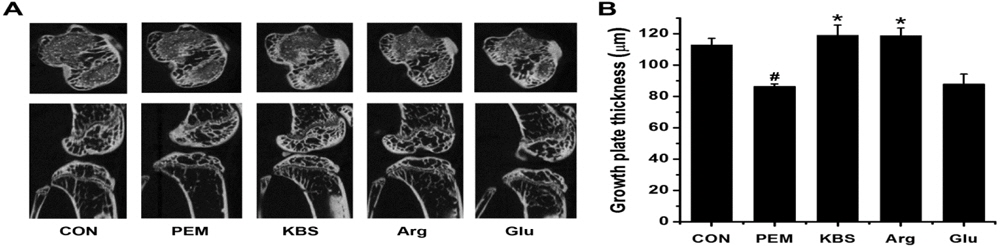
Effect of KBS or Arg or Glu on growth plate in mice
The proximal tibia growth plate length was measured to examine the longitudinal bone growth using a μCT. The lengths of proximal tibia growth plate in the CON and PEM groups were 112.82 ± 4.18 and 86.43 ± 1.47, respectively. The growth plate lengths in the KBS, Arg, and Glu groups were 119.05 ± 6.48, 118.75 ± 4.81, and 87.82 ± 6.38, respectively. KBS and Arg significantly enhanced the longitudinal bone growth, whereas Glu did not (
>
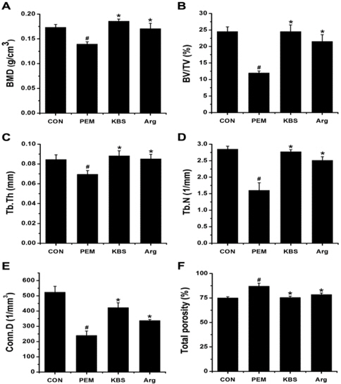
Effect of KBS or Arg on trabecular bone parameters
We performed μCT measurements to evaluate whether KBS or Arg could affect developmental bone growth. The KBS or Argadministered mice showed an increase in the bone mineral density (BMD) (Fig. 2A). The 3D μCT reconstruction of trabecular bone images, converted into parameters representing trabecular connectivity, revealed an increase in the BV/TV, Tb.Th, Tb.N, and Conn.D, and a fall in the total porosity at tibia (Fig. 2B-F).
>
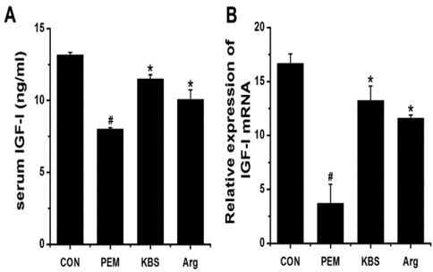
Effect of KBS or Arg on the regulation of IGF-I
To examine whether KBS or Arg can elevate IGF-I level in the serum, we measured the level of IGF-I by means of sandwich ELISA method. The serum IGF-I level was significantly upregulated by KBS or Arg (
>
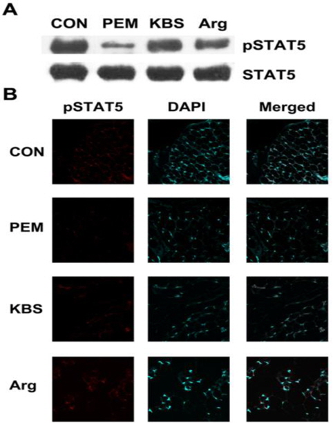
Effect of KBS or Arg on the phosphorylation of STAT5
To determine whether the longitudinal bone growth by KBS or Arg is mediated by the phosphorylation of STAT5, we performed Western blot analysis and immunohistochemistry. As shown in Fig. 4A, the phosphorylation of STAT5 in the liver decreased by malnutrition. However, the decreased STAT5 phosphorylation was up-regulated by KBS or Arg. Furthermore, STAT5 phosphorylation increased by KBS or Arg in the tibia (Fig. 4B).
In this study, we have shown that KBS increases the length of growth plate and improves the bone parameters such as BV/TV, Tb.Th, Tb.N, Conn.D, and total porosity. KBS also elevated the level of serum IGF-I through the phosphorylation of STAT5.
Generally, endochondral bone growth initiates at the growth plate cartilage. During normal bone growth, chondrocytes continually mature and produce cartilage at the growth plate to allow bones to lengthen (endochondral bone formation) (Harris et al., 2009). In the present study, KBS increased the thickness of growth plate (Fig. 1). Thus, we can speculate that KBS could be helpful to children who are in retard for their age.
Glucocorticoids are a class of steroid hormones that bind to intracellular glucocorticoid receptors (GRs). In turn, the GR complex migrates to the nucleus where it inhibits nuclear factor (NF)-κB and activator protein (AP)-1 driven gene expression (Barnes, 2006). Moreover, glucocorticoids may suppress granulocyte activation and recruitment, preserve endothelial cell integrity and control vascular permeability (Thompson, 2003). Dexamethasone (Dex) is a synthetic glucocorticoid which shows a 20 to 30 times higher potency to evoke antiinflammatory effects relative to the endogenously produced cortisol (Blaser et al., 2011). It has been reported that the glucocorticoid Dex has an antiallergic activity and clinical efficacy in allergic diseases (Puigneró et al., 1995; Wershil et al., 1995). Thus, Dex has been proposed as a medication to treat various diseases such as edema, headaches, acute otitis externa, and ocular toxoplasmosis (Brynskov et al., 2013; Giuliano et al., 2012; Rahman et al., 2007; Soheilian et al., 2011). However, Kugelberg et al. (2005) reported that Dex eye drops inhibit growth in the newborn rabbit. Study by Chrysis et al. (2003) demonstrated that systemic Dex treatment led to a pronounced reduction of the size of the growth plate in tibiae.
Furthremore, Baron et al. (1992) reported that glucocorticoids act locally in the growth plate to inhibit bone growth. It’s fortunate that IGF-1 has antiapoptotic actions in lots of tissues and may therefore have a potential to prevent glucocorticoid-induced chondrocyte apoptosis. It was reported that IGF-I prevents dexamethasone-induced apoptosis and suppression of chondrocyte proliferation (Chrysis et al., 2005). Our results showed that KBS increases the serum IGF-I level (Fig. 3). Thus, we can presume that KBS could partially recover the growth retardation by Dex through the elevation of IGF-I level.
KBS is consisted of 15 Korean medicinal drugs. Among 15 drugs, Hominis Placenta protected osteoporosis in ovariectomized rats (Chae et al., 2006). Carthami Tinctorii Fructus increased the level of serum IGF-I and lengths of femur and tibia, however, its effect was very small and transient (Lee et al., 2009). Kim et al. (2002) reported that Carthami Tinctorii Fructus partially prevents ovariectomy-induced bone loss. However, the effect of individual drug was less than those of all drugs, prescription KBS.
In conclusion, the present study demonstrated that KBS increases the length of growth plate and improves the bone parameters. KBS also elevated the level of serum IGF-I through the phosphorylation of STAT5. Overall, this study suggests that KBS would be helpful to children who are in retard for their age through the elevation of IGF-I.