검색 전체 메뉴
PDF
맨 위로
OA 학술지
치아우식증 유발 균주에 대한 패러다임의 변화: 생태학적 관점 Change of Paradigms in Caries-Associated Bacteria in the Caries Process: Ecological Perspectives
  • 비영리 CC BY-NC
ABSTRACT
치아우식증 유발 균주에 대한 패러다임의 변화: 생태학적 관점

Dental plaque resides passively at a site and makes an active contribution to the maintenance of health. The bacterial composition of plaque remains relatively stable despite regular exposure to minor environmental stress. This stability, homeostasis is due to a dynamic balance of microbial interactions. However, the homeostasis can break down, leading to shifts in the balance of the microflora. This change can be a sign of initial dental caries. It is proposed that disease can be prevented or treated not only by targeting the putative pathogens but also by interfering with the processes that drive the breakdown in homeostasis. It is essential to understand the plaque as a mixed species biofilm. In this essay I reviewed an extension of the caries ecological hypothesis to explain the relation between dynamic changes in the phenotypic/genotypic properties of plaque bacteria and the demineralization and remineralization balance of the dental caries process. We will have the strategies to impact significantly on clinical practice as understanding dental biofilm.

KEYWORD
Biofilm , Dental caries , Dental plaque , Ecology , Hydrogen-Ion concentration
  • 서 론

    구강의 양대 질환인 치아우식증과 치주질환을 잘 관리하기 위해서는 치면세균막(dental plaque)에 대한 정확한 이해가 필수적이다. 과거에는 구강 질환의 발생 기전을 밝히기 위해 발병에 영향을 미치는 특정 미생물을 동정하는 데 주력하였다. 그 결과, 치아우식증에 관여하는 Streptococcus species (sp.)와 Lactobacillus sp. 등이 동정되었고, 치주질환과 밀접한 관련성이 있는 Porphyromonas sp.와 Treponema sp. 등이 동정되었지만 특정 미생물의 개체수나 특성만으로 구강 질환의 병적 변화를 명확히 규명하기에는 한계가 있었다. 이후 특정 미생물의 역할보다는 치면에 침착된 치면세균막의 전체 양이 문제가 된다는 “비특이적 치면세균막에 대한 가설(non-specific plaque hypothesis)”이 주목을 받으면서 치면세균막 관리를 강조하게 되었다. 1960년대 이후 다시 “특이적 치면세균막에 대한 가설(specific plaque hypothesis)”이 대두되면서 특히 치주질환에 영향을 미치는 특정 원인균을 알아내고자 하는 연구가 활발히 진행되었다. 그러나 1990년도 후반 이후 등장한 “바이오필름(biofilm)”이라는 개념에 의해 치면세균막을 새롭게 인식하게 되는 전환점을 맞이하게 되었다(Fig. 1)1).

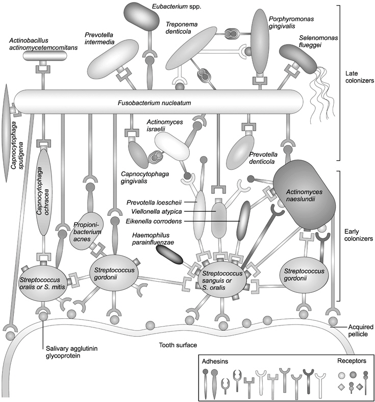
    자연계에 존재하는 대부분의 표면은 액체성 물질과 닿는 순간 세균성 바이오필름(microbial biofilm)을 형성하기 시작한다. 바이오필름은 단단한 표면 위에 다양한 종류의 미생물들이 축적된 복합체이다2). 세균 부착과 관련된 바이오필름 내의 생리학적 및 생화학적 상호 작용은 구강 내에서 치면세균막이 형성되는 과정 중에도 재현된다. 치면세균막은 수백여 종의 미생물이 구강 내 표면 위에 부착하면서 지속적으로 상호작용을 하고, 숙주 및 외부 환경에 따라 끊임없이 변화하며 구강 건강에 영향을 미친다(Fig. 2)3).

    바이오필름을 형성한 세균 집단은 부유성 세균(planktonic cell)에 비해 영양분의 고갈, 산성도의 변화, 삼투압의 증감, 온도의 변화, 독성물질의 노출 등 여러 환경적인 스트레스에 대한 저항성이 현저히 증가된다고 알려져 있다4,5). 따라서 환경 조건의 변화에 따른 바이오필름의 성격 변화는 질병 발생 요인에 결정적 역할을 할 것으로 추론되었다. 최근 선행 연구는 바이오필름의 특성을 고려한 미생물 생태계의 총체적인 변화를 기반으로 하는 우식 생태학적 가설 (extended caries ecological hypothesis)을 활용하여 치아우식증의 발생 기전을 재조명하고 있다6). 그러므로 본 연구에서는 치면세균막이 바이오필름으로서 가지는 특성에 대해 알아보고, 치아우식증을 유발하는 균주에 대한 변화된 관점을 생태학적으로 이해하고자 한다.

    본 론

       1. 미생물 생태계의 항상성

    구강뿐만 아니라 인체 내의 모든 점막 표면에는 광범위하게 미생물들이 정착하고 있다. 그럼에도 불구하고 우리 인간의 건강은 정상적으로 유지된다. 이것은 인체와 정착 미생물이 서로 양생(amphibiosis)하기에 나타날 수 있는 결과이다7). 구강 내 환경을 예로 들어보면, 인간은 지속적으로 분비하는 타액과 섭취하는 음식을 통하여 미생물이 생장하는데 필요한 당분과 아미노산 등의 영양분을 제공하고 구강내 정착 미생물은 병인으로 작용할 수 있는 해로운 미생물이 인체 내로 침입하는 것을 방어하는 보호작용을 한다. 이러한 양생관계가 잘 유지되기 위해서는 생태계의 균형 관계(equilibrium)가 매우 중요하다. 구강 내에는 약 700 여종의 미생물이 존재하는데, 이것들은 구강 내의 서로 다른 미세환경(micro-environments) 내에서 복잡 다양한 환경 조건에 적응하며 서식하고 있다. 사람은 항상 식사를 하고 치아를 닦는 등 일상적인 행동패턴으로 구강 미생물에게 간헐적인 스트레스를 가하지만, 구강 내에 서식하는 미생물의 종류나 그 구성 비율은 항상 일정하게 유지되려고 하는 항상성(homeostasis)을 지닌다. 이러한 미생물 생태계의 항상성은 인체의 면역시스템이 정상적으로 가동되면서 미생물 집단에 과도한 스트레스 요인만 제공하지 않는다면 구강 상태를 건강하게 유지하면서 지속될 수 있다.

       2. 치아우식증의 진행 단계와 구강 미생물의 구성

    치아우식증은 앞서 설명하였듯이, 미생물계의 항상성이 파괴되었을 때 나타날 수 있는 대표적인 구강질환이다. 잦은 발효성 탄수화물 섭취와 더불어 적절한 구강 관리가 수행되지 못하고, 여기에 타액 분비량까지 감소된다면 구강내 산도(pH)는 낮아질 수 밖에 없다. 이러한 환경에 처하게 되면 치면의 탈회가 시작되고 가속화되어 결국은 치아우식증의 임상적 증상이 나타나기 시작한다.

    수많은 선행연구들은 mutans streptococci (MS)가 치아우식증 발생 초기에 매우 결정적인 역할을 하는 것으로 보고하였다8). 이것은 거의 대부분의 와동형 치아우식증에서 MS를 동정할 수 있었기 때문이었다. 그러나, 최근 분자생물학적 분석 기법의 괄목할 만한 성장으로 인해, MS균의 출현과 치아우식증의 발병이 반드시 정비례 관계에 있지 않다는 것이 밝혀졌다9,10). 선행연구에 의하면, 임상적으로 건전한 치면 위의 초기 치면세균막 중에서 MS는 구강 내 총 세균의 2% 미만이었고11), 주로 S. sanguinis, S. oralis, S. gordonii, S. mitis 등의 non-MS가 분포한다고 보고하고 있다12). 치면의 백색 반점(white spot) 부위의 성숙된 치면세균막에서는 MS는 단지 0.001∼10%에 불과했고, 여전히 non-MS나 Actinomyces가 우세하였다10,13,14). 따라서 오히려 이러한 균종들이 치아우식증의 시작과 관련이 있을 것으로 추론되었다15). 한편 와동형 우식병소에서는 MS가 총 세균수의 약 30%까지 증가하는 것을 관찰할 수 있었다16-18). 그러나 이보다 더 특이한 것은 오히려 MS보다 lactobacilli (LB)나 Bifidobacterium이 더 많이 발견되었고, MS보다 우식 진행에 더 큰 영향을 미치는 것으로 보고되었다19-21). 이러한 연구결과들에 주목하기 시작하면서, 우식학계에서는 치면세균막과 치아우식증 간의 관계를 구강 내 환경조건의 변화에 따른 통합된 생태학적 관점으로 재조명할 필요성을 제기하게 되었다.

       3. 바이오필름으로서 치면세균막의 형성 과정

    일반적으로 세균은 자연환경에서 고립된 세포로 존재하는 것이 아니라, 유기적으로 조직된 공동체(community)를 형성하며 성장한다. 고체 또는 공기와 액체 간의 계면에서 형성되는 이러한 미생물 공동체를 우리는 바이오필름(biofilm)이라고 부른다. 바이오필름의 가장 큰 특징은 생물 고분자 물질의 이동 및 확산을 가능하게 하는 개방형 구조를 유지하며 공동체를 이루는 것인데, 각 개체 간에 매우 상이한 대사조건을 필요로 하는 세균들이 이 공동체 안에서 미세 환경을 구축하며 상호 공존한다22). 이와 더불어 바이오필름은 부유성 세균에 비해 외부 환경 변화에 대한 방어력이 월등히 우수하고 항생물질(antimicrobials)에 대한 내성 또한 뛰어나다22).

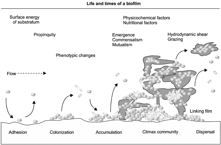
    구강 바이오필름 즉, 치면세균막의 형성 과정은 발달 주기로 설명할 수 있다(Fig. 3)23). 발달주기의 각 단계는 물리적, 생물학적 및 환경적인 요소에 의해 좌우된다. 가장 첫 단계는 타액에서 유래된 당단백질(glycoprotein)에 의해 1 μm 미만의 무세포성 막인 획득 피막(acquired pellicle)이 치면에 형성되는 것이다. 이러한 조건이 갖추어지면 구강 내 미생물들의 초기 부착이 시작된다. 이후 지속적으로 새로운 세균들이 부착하고 상호응집하면서 연결 막(linking film)을 형성하고, 동시에 초기 집락체 내의 세균은 표현형에 변화를 일으키며 바이오필름 내부 환경에 적응하게 된다. 이 연결 막 위로 새로운 세균들이 상호부착하게 되고, 계속해서 다른 종의 세균들이 상호응집하면서 후기 집락체가 형성되기 시작한다. 이러한 증식 과정은 일정한 미생물 구성을 갖는 세균의 공동체가 형성되는 가장 기본적인 원리이다. 하나의 공동체를 형성하는 세균 종들은 서로 간에 분자신호를 주고 받으며 그 안에서 상호 경쟁하거나 협력하는데, 이러한 과정(정족수 감지, quorum sensing)을 통해 영양소를 확산시키고, 최종 대사 산물인 노폐물을 용이하게 제거할 수 있도록 적정 표면적ㆍ부피 비율을 유지하며 자신들의 생태계를 유지한다22). 이렇게 형성된 구강 바이오필름은 세포외기질(extracellular matrix)을 형성하여 외부 환경 변화에 대한 물리적ㆍ전기적 보호막 작용을 증진시킨다22). 따라서 부유성 세균과 비교했을 때, 치약이나 구강 양치액에 함유되어 있는 항생물질에 대한 내성이 바이오필름에서 증가하는 것이다. 구강 영역에 이용하는 대표적인 물질인 클로르헥시딘(chlorhexidine digluconate)에 대해서는 300배, 불화아민(amine fluoride)에 대하여는 75배의 저항성을 보인다22).

       4. 치아우식증 유발 균주에 대한 이해의 변화: 우식 생태학적 가설

    구강 질환은 세포 혹은 분자 수준에서 숙주와 미생물 간균형이 깨질 때 발생한다. 이러한 과정에는 미생물학적인 영향뿐만 아니라 생화학적이고 생태학적인 영향 요인들까지 관여한다. 따라서 최근 10여 년 간에 걸친 미생물군(microbiome)에 대한 혁신적인 연구 성과에 힘입어, 우식학자들은 치아우식증의 원인을 이러한 총체적인 관점에서 규명하기 위해 우식 생태학적 가설(extended caries ecological hypothesis)에 주목하기 시작했다(Fig. 4)6,24). 이 가설은 바이오필름 내의 생태계의 변화, 즉 구성 미생물들의 상대적 비율 변화가 치아우식증 발생의 신호가 될 수 있다는 것을 시사한다. 즉, 치면세균막 내의 세균이 나타내는 표현형(phenotype)과 유전자형(genotype)이 변화됨에 따라 치아우식병소의 진행 단계가 설명될 수 있는 것이다.

    1) 동적 안정상태(dynamic stability stage)

    발효성 탄수화물을 섭취하면 구강 내 미생물은 발효성 대사 과정을 거쳐 구강 내 pH를 떨어뜨린다. 그러나 탄수화물의 섭취 횟수가 빈번하지 않고 보통의 수준이라면 치면세균막 내의 항상성은 파괴되지 않는다. 따라서 구강 내 치면에서 무기질의 동적 평형상태는 안정적으로 유지될 수 있다. 이 때, 치면세균막 내의 미생물은 주로 non-MS나 Actinomyces가 우세하다. 이들은 획득피막에 부착할 수 있는 부착 소를 가지고 있고, 또한 타액 내의 당단백질인 뮤신(mucin)을 분해하여 이들의 성장에 이용할 수 있는 능력을 가진다.

    2) 산생성 상태(acidogenic stage)

    구강 내에 당이 더 자주 공급되거나 타액 분비가 원활하지 않으면, non-MS나 Actinomyces들의 산생성능이 증가하면서 치면세균막 내의 pH는 점점 낮아진다. 이러한 환경적 변화는 non-MS균의 산생성능이나 내산성을 좀 더 강화시킨다. 특히, S. sanguinis, S. oralis, S. gordonii, S. mitis가 산성 환경에 노출되면 그들의 산생성능이 더욱 증가된다고 보고되었다. 따라서 이러한 환경적 변화에 적응하기 위해이들 세균은 자신들의 표현형을 변화시키게 된다. 그런데, 이러한 환경적 변화에 자주 노출되면 non-MS 균주들 중에서 특히 산성환경에 잘 견딜 수 있는 소수의 균종만이 선택적으로 살아남게 된다. 새로이 우위를 점한 균종들은 그들이 생존하기에 유리하도록 바이오필름 내 환경을 변화시키게 되고, 이 때 MS와 LB가 출현하게 된다. 치면세균막의 pH가 5.0 이하가 되면 non-MS나 Actinomyces는 감소하면서 MS와 LB는 증가하기 시작하고, pH가 4.5 이하로 떨어지면 치면세균막 내에서 MS와 LB가 훨씬 더 우위를 점하게 된다.

    3) 내산성 상태(aciduric stage)

    구강 내 환경이 점점 산성화되면 낮은 pH 상태에서도 견딜 수 있는 극소수의 non-MS나 Actinomyces가 산생성능이나 내산성을 증가시키지만, 결국은 이들의 성장은 지연되기 시작하고 이러한 산성 환경에 적응하여 생존할 수 있는 세균들로 서서히 구강 미생물들의 구성이 교체되기 시작한다. 이 때, 이러한 환경 변화에 가장 경쟁력 있는 MS와 LB가 선택되는 것이다. 성숙된 치면세균막의 평균 pH는 보통 4.0이하이다. 이 상태가 되면 non-MS균이나 Actinomyces는 생존능력을 상실하고 최종적으로 MS와 LB만이 살아남게 된다. 일반적으로 우식 와동의 평균 pH가 5.0 이하라는 것을 감안한다면, 왜 와동형 우식증 부위의 치면세균막에서 주로 MS와 LB가 동정되었는지에 대해 추론할 수 있게 된다. 결국 치면세균막 내의 미생물 성장능력을 좌우하는 것은 pH이다. 이러한 일련의 미생물 구성의 변화에 의해 치면에서는 탈회와 재광화 간의 균형이 깨지면서 무기질이 소실되기 시작하고 우식 병소가 출현하게 된다.

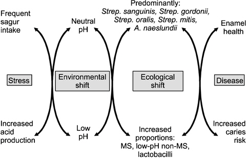
    이를 종합해 보면, 잦은 음식물 섭취, 타액 분비율의 저하, 국소적 자극 및 면역력 저하 등에 의해 구강 바이오필름의 생태계에 스트레스를 가하게 되면 결국은 이들 생태계 환경에 변화를 야기하게 되고, 세균들이 이 환경에 적응하기 위해 표현형 및 유전자형에 변화를 일으킴으로써 최종적으로 치면의 무기질이 소실되면서 치아우식증이 발병될 수 밖에 없다(Fig. 5)7,25,26).

    결 론

    아무리 치아를 깨끗이 닦는다 하더라도, 수 초 내에 치면에는 다시 치면세균막이 형성된다. 따라서 환자의 구강 건강을 최상으로 유지하기 위해서는 치면세균막의 생태 환경을 안정시키고, 치면세균막 내의 세균이 사람에게 이로운 영향을 미치는 균종으로 구성될 수 있도록 관리하여야 한다. 이렇게 치면세균막이 잘 관리된다면 초기 상태의 치아우식증은 특별한 치료 없이도 회복될 수 있다. 이러한 구강건강관리가 가능하기 위해서는 치아우식증의 발병에 영향을 미치는 요소를 일정 세균에만 한정시키지 말고, 환자와 미생물 간의 양생관계를 저해하는 통합적인 요인들에 관심을 가져야만 한다. 불소나 자일리톨을 이용하여 치면세균막내 pH가 산성화되는 것을 막고, 식이상담을 통해 당의 섭취횟수나 식이의 질을 조절하고, 타액이 잘 분비될 수 있도록 관리한다면, 환자의 구강환경은 건강한 상태를 유지할 수 있을 것이다. 또한 더 나아가 치면세균막을 구성하는 미생물 공동체 내의 신호 체계 기작과 주요 세균 간의 상호관계 등을 이해함으로써 세균 간의 응집이나 부착을 컨트롤할 수 있는 유효한 생화학적 제어 방법을 찾을 수 있다면, 치면세균막이 침착되는 것을 사전 차단함으로써 구강질환을 예방할 수 있을 것이다. 따라서 우리는 치면세균막을 바이오필름이 가지는 특징과 견주어 이해할 필요가 있으며, 이와 더불어 치아우식증 유발의 원인을 치면세균막의 생태학적 관점으로 접근함으로써 치아우식증의 관리 방법에 대한 새로운 지침을 마련할 수 있을 것으로 사료된다.

참고문헌
  • 1. Overman PR 2000 Biofilm: a new view of plaque [J Contemp Dent Pract] Vol.1 P.18-29 google
  • 2. Marsh PD, Bradshaw DJ 1995 Dental plaque as a biofilm [J Ind Microbiol] Vol.15 P.169-175 google
  • 3. Bakaletz LO 2004 Developing animal models for polymicrobial diseases [Nat Rev Microbiol] Vol.2 P.552-568 google
  • 4. Pratt LA, Kolter R 1999 Genetic analyses of bacterial biofilm formation [Curr Opin Microbiol] Vol.2 P.598-603 google
  • 5. Stoodley P, Sauer K, Davies DG, Costerton JW 2002 Biofilms as complex differentiated communities [Annu Rev Microbiol] Vol.56 P.187-209 google
  • 6. Takahashi N, Nyvad B 2011 The role of bacteria in the caries process: ecological perspectives [J Dent Res] Vol.90 P.294-303 google
  • 7. Marsh PD 1994 Microbial ecology of dental plaque and its significance in health and disease [Adv Dent Res] Vol.8 P.263-271 google
  • 8. Tanzer JM, Livingston J, Thompson AM 2001 The microbiology of primary dental caries in humans [J Dent Educ] Vol.65 P.1028-1037 google
  • 9. Van Houte J, Sansone C, Joshipura K, Kent R 1991 In vitro acidogenic potential and mutans streptococci of human smooth-surface plaque associated with initial caries lesions and sound enamel [J Dent Res] Vol.70 P.1497-1502 google
  • 10. Sansone C, Van Houte J, Joshipura K, Kent R, Margolis HC 1993 The association of mutans streptococci and non-mutans streptococci capable of acidogenesis at a low pH with dental caries on enamel and root surfaces [J Dent Res] Vol.72 P.508-516 google
  • 11. Nyvad B, Kilian M 1990 Comparison of the initial streptococcal microflora on dental enamel in caries-active and in cariesinactive individuals [Caries Res] Vol.24 P.267-272 google
  • 12. Nyvad B, Kilian M 1987 Microbiology of the early colonization of human enamel and root surfaces in vivo [Scand J Dent Res] Vol.95 P.369-380 google
  • 13. XimAnez-Fyvie LA, Haffajee AD, Socransky SS 2000 Microbial composition of supra- and subgingival plaque in subjects with adult periodontitis [J Clin Periodontol] Vol.27 P.722-732 google
  • 14. van Houte J, Lopman J, Kent R 1996 The final pH of bacteria comprising the predominant flora on sound and carious human root and enamel surfaces [J Dent Res] Vol.75 P.1008-1014 google
  • 15. Boyar RM, Thylstrup A, Holmen L, Bowden GH 1989 The microflora associated with the development of initial enamel decalcification below orthodontic bands in vivo in children living in a fluoridated-water area [J Dent Res] Vol.68 P.1734-1738 google
  • 16. Loesche WJ, Eklund S, Earnest R, Burt B 1984 Longitudinal investigation of bacteriology of human fissure decay: epidemiological studies in molars shortly after eruption [Infect Immun] Vol.46 P.765-772 google
  • 17. Milnes AR, Bowden GH 1985 The microflora associated with developing lesions of nursing caries [Caries Res] Vol.19 P.289-297 google
  • 18. Boue D, Armau E, Tiraby G 1987 A bacteriological study of rampant caries in children [J Dent Res] Vol.66 P.23-28 google
  • 19. Mantzourani M, Gilbert SC, Sulong HN, Sheehy EC, Tank S, Fenlon M 2009 The isolation of bifidobacteria from occlusal carious lesions in children and adults [Caries Res] Vol.43 P.308-313 google
  • 20. Aas JA, Griffen AL, Dardis SR, Lee AM, Olsen I, Dewhirst FE 2008 Bacteria of dental caries in primary and permanent teeth in children and young adults [J Clin Microbiol] Vol.46 P.1407-1417 google
  • 21. Chhour K, Nadkarni MA, Byun R, Martin FE, Jacques NA, Hunter N 2005 Molecular analysis of microbial diversity in advanced caries [J Clin Microbiol] Vol.43 P.843-849 google
  • 22. Marsh PD 2004 Dental plaque as a microbial biofilm [Caries Res] Vol.38 P.204-211 google
  • 23. Jenkinson HF, Lappin Scott HM 2001 Biofilms adhere to stay [Trends Microbiol] Vol.9 P.9-10 google
  • 24. Takahashi N, Nyvad B 2008 Caries ecology revisited: microbial dynamics and the caries process [Caries Res] Vol.42 P.409-418 google
  • 25. Marsh PD 2010 Microbiology of dental plaque biofilms and their role in oral health and caries [Dent Clin North Am] Vol.54 P.441-454 google
  • 26. Marsh PD, Bradshaw DJ 1997 Physiological approaches to the control of oral biofilms [Adv Dent Res] Vol.11 P.176-185 google
OAK XML 통계
이미지 / 테이블
  • [ Fig. 1. ]  The changing views of plaque and periodontal diseases. Adapted from Overman (J Contemp Dent Pract 1: 18-29, 2000)1).
    The changing views of plaque and periodontal diseases. Adapted from Overman (J Contemp Dent Pract 1: 18-29, 2000)1).
  • [ Fig. 2. ]  A model of oral biofilm formation. The tooth surface is covered by an acquired pellicle comprised oflipids and proteins, including salivary agglutinin glycoprotein. The pellicle is recognized by primarycolonizing bacteria (Streptococcus oralis, Streptococcus mitis, Streptococcus gordonii and Streptococcus sanguis) that express receptors for salivary agglutinin glycoprotein. Other bacteria then colonize in a spatialand temporal manner as shown, using receptors and adhesins to eventually form dental plaque. Adapted from Bakaletz (Nat Rev Microbiol 2: 552-568, 2004)3).
    A model of oral biofilm formation. The tooth surface is covered by an acquired pellicle comprised oflipids and proteins, including salivary agglutinin glycoprotein. The pellicle is recognized by primarycolonizing bacteria (Streptococcus oralis, Streptococcus mitis, Streptococcus gordonii and Streptococcus sanguis) that express receptors for salivary agglutinin glycoprotein. Other bacteria then colonize in a spatialand temporal manner as shown, using receptors and adhesins to eventually form dental plaque. Adapted from Bakaletz (Nat Rev Microbiol 2: 552-568, 2004)3).
  • [ Fig. 3. ]  The life and times of a biofilm, depicted as a developmental cycle, the various stages of which are determined by a range of physical, biological and environmental factors, as indicated at the top of the diagram. Initial adhesion to a surface is a random event influenced by surface free energy and propinquity (nearness) of bacterial cells. Adhered bacteria then undergo cell division, colonize the surface, and thus provide for further cell adhesion and accumulation to generate a ‘linking film’ upon which a climax community is generated. The biofilm is a dynamic entity: cells continually enter or leave the community, promoting diversification or dispersal, and protozoal grazing and shear forces in flow systems lead to remodelling of the biofilm structure. Adapted from Jenkinson and Lappin Scott (Trends Microbiol 9: 9-10, 2001)23).
    The life and times of a biofilm, depicted as a developmental cycle, the various stages of which are determined by a range of physical, biological and environmental factors, as indicated at the top of the diagram. Initial adhesion to a surface is a random event influenced by surface free energy and propinquity (nearness) of bacterial cells. Adhered bacteria then undergo cell division, colonize the surface, and thus provide for further cell adhesion and accumulation to generate a ‘linking film’ upon which a climax community is generated. The biofilm is a dynamic entity: cells continually enter or leave the community, promoting diversification or dispersal, and protozoal grazing and shear forces in flow systems lead to remodelling of the biofilm structure. Adapted from Jenkinson and Lappin Scott (Trends Microbiol 9: 9-10, 2001)23).
  • [ Fig. 4. ]  n extended caries ecological hypothesis explaining the relationship between acidogenic and aciduric shifts in the composition of the dental biofilm and changes in the mineral balance of the dental hard tissues. Note that the cascade of ecological events in the biofilm is reversible and is reflected in the surface features of the dental hard tissues at any stage of lesion formation. MS: Mutans streptococci. Adapted from Takahashi and Nyvad (Caries Res 42: 409-418, 2008)24).
    n extended caries ecological hypothesis explaining the relationship between acidogenic and aciduric shifts in the composition of the dental biofilm and changes in the mineral balance of the dental hard tissues. Note that the cascade of ecological events in the biofilm is reversible and is reflected in the surface features of the dental hard tissues at any stage of lesion formation. MS: Mutans streptococci. Adapted from Takahashi and Nyvad (Caries Res 42: 409-418, 2008)24).
  • [ Fig. 5. ]  The ecological plaque hypothesis. The diagram depicts the dynamic relationship that exists between the plaque biofilm and the local environment. When the frequency of intake of fermentable sugar increases, bacterial metabolism results in the biofilm spending more time at a low pH. An acidic pH inhibits the growth of many bacteria associated with enamel health while selecting for those bacteria with an acidogenic and acid-tolerating (aciduric) phenotype. Similar events occur if the flow of saliva is reduced. Under these conditions, demineralization is promoted, which increases the probability of a caries lesion developing. The caries process could be prevented not only by inhibiting the causative bacteria directly but also by interfering with the environmental changes that drive the deleterious shifts in the composition and metabolic activity of the biofilm. MS: Mutans streptococci. Adapted from Marsh (Dent Clin North Am 54: 441- 454, 2010)25).
    The ecological plaque hypothesis. The diagram depicts the dynamic relationship that exists between the plaque biofilm and the local environment. When the frequency of intake of fermentable sugar increases, bacterial metabolism results in the biofilm spending more time at a low pH. An acidic pH inhibits the growth of many bacteria associated with enamel health while selecting for those bacteria with an acidogenic and acid-tolerating (aciduric) phenotype. Similar events occur if the flow of saliva is reduced. Under these conditions, demineralization is promoted, which increases the probability of a caries lesion developing. The caries process could be prevented not only by inhibiting the causative bacteria directly but also by interfering with the environmental changes that drive the deleterious shifts in the composition and metabolic activity of the biofilm. MS: Mutans streptococci. Adapted from Marsh (Dent Clin North Am 54: 441- 454, 2010)25).
(우)06579 서울시 서초구 반포대로 201(반포동)
Tel. 02-537-6389 | Fax. 02-590-0571 | 문의 : oak2014@korea.kr
Copyright(c) National Library of Korea. All rights reserved.