검색 전체 메뉴
PDF
맨 위로
OA 학술지
Rhizoctonia solani AG-1 IB에 의한 Kentucky Bluegrass 갈색잎마름병 발생 Occurrence of Brown Patch on Kentucky Bluegrass Caused by Rhizoctonia solani AG-1 IB
ABSTRACT
Rhizoctonia solani AG-1 IB에 의한 Kentucky Bluegrass 갈색잎마름병 발생

Brown patch caused by Rhizoctonia solani AG-1 IB occurred on Kentucky bluegrass during late May through early October 2010 at golf course in Gyeongbuk Province, Korea. Disease symptoms on the turfgrass for spring season were leaf blights dying from the leaf tip, which appeared patches of brown color in the field. However, it appeared patches of dark brown color or gray brown color in fall. The fungus (B-7 isolate) of brown patch was isolated from the diseased leaf tissue and cultured on potato-dextrose agar (PDA) for identification. The young hyphae had acute angular branching and few septa and mature hyphal branches showed about 90-degree angles and development of monilioid cells, which were morphologically identical to Rhizoctonia solani AG-1 IB reported previously. DNA sequences of ribosomal RNA gene (internal transcribed spacer) of the fungus were homologous with similarity of 99% to those of Rhizoctonia solani AG-1 IB isolates in GenBank database, confirming the identity of the causal agent of the disease. Pathogenicity of the fungus was also confirmed on the creeping bentgrass and Kentucky bluegrass by Koch's postulates. This is the first report of brown patch on Kentucky bluegrass caused by Rhizoctonia solani AG-1 IB in Korea.

KEYWORD
갈색잎마름병 , 균사 , 동정
  • 병 발생과 증상

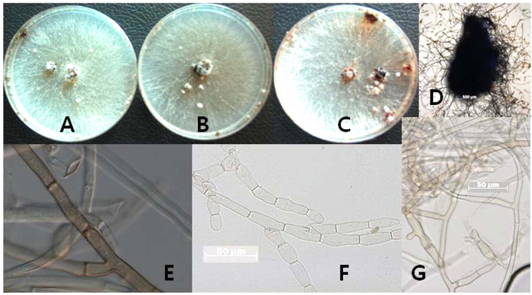
    2010년과 2011년에 경상북도 청도군 소재 골프장 그린에서 봄철인 5월중순과 가을철인 9월 하순에 전형적인 갈색잎마름병이 그린의 creeping bentgrass 종과 그린 주변에 식재 된 Kentucky bluegrass 혼합 품종에서 발생하였다(Fig.1 A). 5월 하순에 Kentucky bluegrass품종에서 발생한 병의 초기 증상은 잎의 선단부가 갈색으로 변색하여 갈색의 타원형 patch를 형성하고, 시간이 지나면서 갈색 잎 부분은 황색이 퇴화되어 잎의 끝이 마르며 patch의 가장자리로부터 밀짚 색으로 나타나고, 심한 부분은 고사하는 증상을 보였다(Fig. 1 B). 10월 상순에 Kentucky bluegrass품종에서 발병한 갈색잎마름병의 병징은 봄철과 다른 증상을 보였다(Fig. 1 C, D). 원형과 타원형의 갈색의 patch가 링의 형태로 나타나고, patch 가장자리는 회갈색과 검은 황갈색의 증상이 나타났다. Patch의 크기는 다양하였다. 이 병 잔디 주변이나 고사한 잔디에 균핵은 발견되지 않았다.

    R. solani AG-11B는 토양에 생존하는 곰팡이로 잎에 발병이 되어 잎이 고사하게 된다(Smiley et al., 1992). 갈색잎마름병은 일반적으로 여름철 온도가 25℃ 보다 낮고, 잎에 습도가 10시간이 유지되는 습한 조건에서 발생하며, 증상은 잎의 가장자리에서부터 진한갈색과 함께 밝은 갈색의 병반 증상이 나타난다. 증상이 진전되면, 잎은 괴사가 되어 일반적으로 둥근 원형이나 부정형의 병반이 링의 형태로 병징을 나타나지만(Smiley et al, 1992), 잔디의 종류, 예고 및 관리 정도(관주, 비료)에 따라서 다르게 나타날 수 도 있다(Burpee and Martine, 1992). Creeping bentgrass와annual bluegrass의 예고를 1.3 cm 이상으로 깎기를 하면 검은 회색의 링과 균사의 원형이 잔디가 젖을 때는 관찰이 됨으로 갈색잎마름병을 진단하는데 중요한 특징이 될 수 있지만 잔디의 예고를 1.3 cm보다 낮게 관리할 경우는 균사의 원형이나 링 또는 검은 링은 나타나지 않거나, 또한 잔디 잎의 밀도가 낮아도 검은 원형은 나타나지 않는다. 그리고 모든 감수성인 잔디는 갈색이 된 잎이 괴사가 되거나 밀짚 색의 patch의 링이 나타나는데 크기도 수cm에서 1 m 미만이 된다(Burpee and Martine, 1992).

    병원균의 형태적 특징

    본 연구에 사용된 병원균은 2010년과 2011년에 경상북도 청도군 소재 골프장 그린에서 봄철과 가을철에 전형적인 갈색잎마름병 증상을 보이는 creeping bentgrass “Penn cross” 품종과 Kentucky bluegrass의 잎과 줄기에서 채집하였다. 채집한 이병 잎과 줄기에서 병환부와 건전부 경계의 조직을 잘라 1% sodium hypochlorit에서 1분간 표면살균 후, 70% ethanol에서 다시 30초간 살균하였다. 표면 살균한 병든 조직은 멸균수로 세척하여, 멸균된 거름종이(Whatman No. 1)에 올려놓아 수분을 제거하고, water agar (WA)에 미리 준비한 항생제 용액 (살균증류수: 100 ml; pimaricin: 100 mg; ampicillin: 1000 mg)을 1%을 첨가하여 만든 배지에 치상하고, 25℃ 항온기에서 3일간 배양하였다. 치상한 조직에서 자라나온 균사 선단의 균사를 떼어서 PDA (Potato Dextrose Agar) 배지에 옮겨 25℃ 항온기에서 배양한 15개 균주 중 생리적 반응이 동일한 균주 B-7을 대표 균주로 보관하면서 본 실험을 수행하였다. 공시 균주의 보관은 25℃ 항온기에서 계대 배양을 하면서 시험하거나, 3일간 배양한 균총의 선단부에서 직경 5 mm의 균사 조각을 떼어내어 cap tube(직경: 1 cm; 높이: 10 cm)에 5조각씩 넣고, 보관용액 5 ml(살균수: 5 ml; streptomycin (3,000 μg): 0.5 ml)를 넣어 25℃ 항온기에 보관하며 실험에 사용하였다.

    PDA배지에서 배양한 결과 생육초기에는 균사 색깔은 흰색을 띠며 왕성하게 생장을 하고(Fig. 2 A, B) 배양시간이 지남에 따라서 균사는 갈색으로 변하고, 균사 위에 검은 균핵을 형성한다(Fig. 2 C, D). 광학현미경하에서 관찰한 균사는 clamp 연결을 가지고 있으며, 균사의 모양은 어린 균사와 성숙한 균사의 모양이 다른 것을 관찰할 수 있었다(Fig. 2 E, F, G). 어린 균사는 분지각이 90도 보다 작으며, 성숙한 균사는 분지각이 직각으로 꺾이고, 격막이 있으며, 뿌리 모양으로 자라는 것을 볼 수 있다.

    Rhizoctonia 속에 중요한 특성은 분생 포자가 없고, clamp 연결을 가지고 있으며, 균사는 뿌리모양을 하며, PDA 배지상에서 자란 균사의 모양으로는 종간에 차이를 관찰할 수 없다고 한다(Ogoshi, 1987). 한지형 잔디에 갈색잎마름병을 일으키는 병원균은 미국에서도 대부분 R. solani AG-1 IA에 의하여 brown patch 병을 일으키는 주요 병원균이지만, brown patch 병은 한지형 잔디와 난지형 잔디에서 발병하는 병으로 발병환경이나, 발병조건 및 잔디에 종류에 따라서 여러 종류의 Rhizoctonia spp.가 분리되기도 하고, 가끔씩은 원인 균이 분리가 되지 않을 경우도 있다 (Vargas, 1994; Watschke et al., 1995).

    R. solani은 PDA 배지에서 어린 균총은 흰색을 나타내지만 다자란 균총은 담황색에서 갈색을 형성 한다(Sneh et al., 1986). 균핵은 검은색으로 갈색잎마름병을 보이는 bentgrass류의 잎집과 잎에 붙은 것을 볼 수 있으며, 온실 에서 병원균을 접종한 잔디에서 균핵은 잎집의 배측면과 잔디 뿌리가 붙어있는 지제부(crown)에서 형성한다(Shurtleff,1953).

    배양적인 특성

    병원균의 균사의 형태적인 특징조사를 위하여 PDA배지에 접종 후 25℃항온기에서 5일간 자란 균사조각을 떼어내어 광학현미경하에서 격막 등을 관찰하였고, 생리적인 특성은 생육 온도와 배지종류에 대한 반응을 조사하였다. 생육온도는 PDA배지 중앙에 3일간 배양한 가장자리 균총의 직경 5 mm의 균사 조각을 떼어내어 접종한 후 20℃,23℃, 25℃, 28℃, 및 30℃에서 3일간 배양한 후 균총 직경을 조사하였다. 배지의 종류에 대한 균사생장 반응을 조사하기 위하여 생육온도와 같은 방법으로 균주를 PDA배지에 접종한 후 25℃에서 72시간 배양 후 균사 생장량을 측정하였다.

    균사의 생육에 대한 두 종간에서 최적 생육온도에 뚜렷 한 차이를 보였다(Fig. 3). 공시 균주인 B-7은 대조 균주인 R. solani AG-1 IB과의 생육온도에 대한 차이를 전혀 보 이지 않았다. 하지만 갈색퍼짐병(large patch)을 일으키는 R. solani AG2-2 (IV)과 생육온도를 조사한 결과 현저한 유의성의 차이를 보였다. 공시균은 20℃ 와 25℃에서 대 조 균주인 R. solani AG2-2 (IV)비하여 균사생육이 왕성 한 반면, 30℃에서는 생육이 크게 둔화 되는 것을 관찰할 수 있었다. 균핵은 20℃에서 30℃도까지 배양 7일 후부터 형성되기 시작하여, 배양 25일에 균핵 형성이 크게 증가 하였고, 공시 균주인 B-7 (R. solani AG-1 IB)과 대조 균 주인 R. solani AG-1 IB (KACC 40107)가 R. solani AG2- 2 (IV) 보다 균핵의 수가 많이 형성되었다(자료미제출).

    염기서열 분석

    분자생물학적 동정을 위하여 25℃ 항온기에서 3일간 배양한 후 DNA를 추출하여 ITS 영역의 DNA의 증폭을 위하여 universal primer인 ITS5(5'-GGA AGT AAA AGTCGT AAC AAG G-3)와 ITS4(5'-TCC TCC GCT TATTGA TAT GC G-3')를 사용하였다(White et al., 1990). DNA증폭을 위한 25 μl의 반응액(template로 사용할 병원 균의 DNA 5 μl; 10배의 Taq buffer 2.5 μl; dNTP 2.5 μl; ITS50.5 μl; ITS4 0.5 μl; Taq polymerase, 0.125 μl; 살균증류수 13.875 μl)을 만들어 반응시켰다. 반응조건은 94℃에 서 4분 동안 pre-denaturation을 실시하였고, 95℃에서 30 초간 denaturation, 50℃에서 30초간 annealing, 72℃에서 1분 동안 extension하는 것을 30회 수행한 후, 72℃에서 7분간 최종 반응시켰다. PCR산물은 2% agarose gel을 만들어 50V에서 120분 동안 전기영동하였다. 리보소옴 ITS 지역의 DNA 염기서열을 분석을 위하여 ITS1 (5'-TTC GTA GGT GAA CCT GCG G-3')과 ITS4 (5'-AAC ATG CGT GAG ATT GTA AGT-3') primers 사용하여 분석하였으며 (White et al., 1990), DNA 염기서열은 솔젠트(주)에서 실시하였다. ITS 염기서열은 Thompson et al. (1994) 방법을 따랐으며, phylogenetic tree는 MEGA version 4를 사용하여 Rhizoctonia alpina (CBS 309.35) 염기서열과 GenBank 의 염기서열을 참고로하여 작성하였다(Tamura et al., 2007). 거리는 Tamura-Nei parameter model을 이용하여 산출하였고, phylogenetic tree는 Neighbor-Joining (NJ) 분석을 통하여 구축하였다.

    공시 균주인 B-7을 DNA 리보소옴 ITS 지역과 RAPD 분석을 한 결과 R. solani AG-1 IB로 동정되었으며, R.solani AG-1은 3개의 서브그룹으로 완전히 구분된다는 것 을 알 수 있었다(Fig. 4).

    Hong et al. (1998)은 R. solani AG-1의 그룹 내 종을 동정하기 위하여 PCR-RFLP 17S 밴드패턴과 ITS 지역 Cfr13I 제한 효소로 잘라서 분석하였다. Rhizoctonia spp.의 분류 에는 균사융합그룹(AG)이 사용 된다(Ogoshi, 1987; Parmeter, et al., 1967). 동일한 균사융합그룹 안에 균주는 다른 균사융합그룹 안에 균주 보다 유전적으로 더 동질성을 갖고 있고, 배양적인 형태적 특성 및 AG-2의 서브그룹 사이에 균사융합의 빈도에서도 차이가 있다(Ogoshi, 1987). AG-1 은 3개의 서브그룹인 AG-1 IA, AG-1 IB, AG-1 IC을 포 함하고 있으며, 형태적이나 병을 유발하는 증상이 다르다 (Ogoshi, 1987; Yang, et al., 1990). R. Solani AG-1 IA는 벼(Oryza sativa)를 포함한 곡물류에서 잎집마름병을 일으 킨다(Kim, 1996). 국내에서 R. solani 에 대한 연구는 여 러 작물에서 분리 동정한 균주에 대한 배양적인 특성이 연구 되었으며(Kim et al., 1994), 리보소옴 DNA의 18SITS- 5.8S 지역을 PCR-RFLP 분석을 통하여 AG-1 그룹에 대한 IA 와 IB의 동정이 수행된 이후(Hong et al., 1998), 국내 여러 작물에서 수집된 46 균주들에 대한 분류학적인 지위 구분을 위하여 리보소옴 ITS 지역을 DNA 염기서열분석과 DNA을 이용한 RAPD분석을 통하여 6개 균사융합그룹과 5개 배양타입그룹으로 분석하였다(Jeon et al., 2010).

    R. solani은 1980년 이전에서 Rhizoctonia속 안에서 잔디 에 병을 유발하는 종은 1종으로 담자균류에 속하는 불완 전세대의 곰팡이로 보고된(Smiley, et al., 1992) 이후, 지금까지 R. solani은 AG-1그룹으로 미국에서는 대부분이 tall fescue종에서 분리 동정 되었으며, R. solani AG2-2균주는 난지형 잔디인 St. Augustinegrass의 줄기에서 분리되 었다(Hurd and Grisham, 1983). 일본에서는 갈색잎마름병을 유발하는 R. solani AG-1 또는 R. solani AG2-2 III은 갈색퍼짐병을 유발하는 R. solani AG2-2 (IV)와는 생태적 으로 현저하게 구별이 된다고 하였다(Oniki, et al., 1986). Rhizoctonia spp.의 분류에서 균사융합을 이용하여 균사융 합그룹을 사용하여 구분하였다(Ogoshi, 1987; Parmeter, et al., 1967).

    병원성 검정

    병원성을 검정하기 위하여 발병이 잘되는 creeping bentgrass품종인 “Penn Cross” 와 Kentucky bluegrass 혼용 품종(Midnight 33%, Moonlight 33%, Prosperity 33%)을 재배하는 경남 합천의 (주)한울스포츠잔디 농장에서 뗏장을 구입하였다. 잔디는 관행 관리에 의해 1년 이상 자란 잔디를 뗏장으로 떼어내어 그린용 홀 컷터로 잔디를 잘라내어 화분에 옮긴 후, 온실에서 20일간 관리한 잔디에 병원균을 접종하였다. 접종원은 Chang (2011)의 방법에 따라 수행되었다. PDA 배지에서 자란 활력이 양호한 균총의 가장자리 균사를 지름 5 mm의 크기로 자른 균총 1조각을 PDB (Potato Dextrose Broth) 배지에 접종한 뒤 25 ± 1℃ 암 상태에서 5일간 배양하였다. 배양된 균사는 수확하여 멸균된 거름종이에 올려놓아 수분을 제거하고, 무게를 단 후 다시 멸균수를 넣어 30초간 균일하게 균사를 마쇄하였다. 접종원은 균사 생체 중 0.1 g ml-1가 함유된 1 ml를 포트당 접종을 하고, 바닥에 1 cm 높이로 물을 채운 플라스틱에 포트를 넣고, 밀봉한 후 48시간 동안 25 ± 1℃ 암 상태의 생장상(growth chamber)에서 배양한 후 온실로 옮겨 발병 정도를 조사하였다.

    인공 접종한 병원균은 Kentucky bluegrass와 creeping bentgrass의 “Penn Cross” 품종에서 감염 초기증상을 잎의 선단부가 갈변하고, 이병 부위가 수침상으로 변하면서 점차 갈색이 퇴화하고, 시간이 경과됨에 따라 patch 형태를 유지하면서 잎과 줄기는 고사하여 최종 회갈색이 되었다 (Fig. 5). 인공 접종하여 발병된 병반부에서 다시 병원균을 재분리하여 PDA배지에서 배양한 후, 균핵 형성의 특성과 광학현미경에서 균사의 격막과 균사모양을 관찰한 결과 접종한 균과 일치하여 동일한 균으로 확인되었다.

    따라서 본 병은 한지형 잔디인 Kentucky bluegrass 품종에서 R. solani AG-1 IB에 의해 처음 국내에서 발생한 병으로 기존 병명과 동일한 갈색잎마름병으로 사용하기를 제안 하고자 한다.

참고문헌
  • 1. Burpee L., Martin B. 1992 Biology of Rhizoctonia species associated with turfgrass [Plant Dis] Vol.76 P.112-117 google cross ref
  • 2. Chang T.H., Lee Y.S. 2010 Occurrence of brown patch disease and turfgrass survival of cool season turfgras cultivar in species during summer season [Kor. Turfgrass Sci.] Vol.24 P.117-124 google
  • 3. Chang T.H. 2011 Difference of susceptibility on bentgrass cultivars to pink snow mold caused by Microdochium nivale [Asian J. Turfgrass Sci.] Vol.25 P.177-183 google
  • 4. Couch H.B 1985 Turfgrass (several cultivated spp.) [Plant Dis.] Vol.69 P.672-675 google
  • 5. Hurd B., Grisham M.P. 1983 Rhizoctonia spp. Associated with brown patch of Saint Augustinegrass [Phytopathology] Vol.73 P.1661-1665 google cross ref
  • 6. Kim W.G., Shim G.Y., Cho W.D., Lee Y.H. 1991 Anastomsis groups and pathogenicity of Rhizoctonia solani isolates casuing Rhizoctonia blight of turfgrass [Korean J. Plant Pathol.] Vol.7 P.257-259 google
  • 7. Kim W.G. 1996 Pathogenicity of anastomosis groups and cultural types of Rhizoctonia solani isolates on crops [Korean J. Plant Pathol.] Vol.12 P.21-32 google
  • 8. Hong S.B., Go S.J., Ryu J.C., Kim W.G., Kim I.S. 1998 Differentiation of intraspecific groups within Korean isolates of Rhizoctonia solani using PCR-RFLP of ribosomal DNA [Korean J. Plant Pathol.] Vol.14 P.157-163 google
  • 9. Kim W.G., Cho W.D., Lee Y.H. 1994 Anastomosis groups and cultural characteristics of Rhizoctonia solani isolates from crops in Korea [Kor. J. Mycol.] Vol.22 P.309-324 google
  • 10. Jeon Y.A., Kim W.G., Kim D.H., Kwon S.W., Hong S.B. 2010 Taxonomic position of Korean isolates of Rhizoctonia solani based on RAPD and ITS sequencing of ribosomal DNA [Plant Pathol. J.] Vol.26 P.83-89 google cross ref
  • 11. Martin S.B., Campbell C.L., Lucas L.T. 1983 Horizontal distribution and characterization of Rhizoctonia spp. in tall fescue turf [Phytopathology] Vol.73 P.1064-1068 google cross ref
  • 12. Martin S.B., Lucas L.T 1983 Pathogenicity of Rhizoctonia zeae on tall fescue and other turfgrass [Plant Dis.] Vol.67 P.676-678 google cross ref
  • 13. Martin S.B., Lucas L.T. 1984 Characterization and pathogenicity of Rhizoctonia spp. and binucleate Rhizoctonia- like fungi from turfgrass in North Carolina [Phytopathology] Vol.74 P.170-175 google cross ref
  • 14. Ogoshi A. 1985 Anastomosis and intraspecific groups of Rhizoctonia solani and binucleate Rhizoctonia spp [Fitopatol. Bras.] Vol.10 P.372-390 google
  • 15. Ogoshi A 1987 Ecology and pathogenicity of anastomosis and intraspecific groups of Rhizoctonia solani Kuhn [Annu. Rev. Phytopathol.] Vol.25 P.125-143 google cross ref
  • 16. Oniki M., Kobayashi K., Araki T., Ogoshi A. 1986 A new disease of turfgrass caused by binucleate Rhizoctonia AG-Q [Ann. Pathopathol. Soc. Jpn.] Vol.52 P.850-853 google cross ref
  • 17. Parmeter J.R. Jr., Whitney H.S., Platt W.D. 1967 Affinities of some Rhizoctonia species that resemble mycelium of thanatephorus cucumeris [Phytopathology] Vol.57 P.218-223 google
  • 18. Shim G.Y., Min G.Y., Shin H.D., Lee H.J. 2001 Ocureence of fungicide resistance isolate caused by Sclerotinia homoeocarpa [Kor. J. Turfgrass sci.] Vol.15 P.1-8 google
  • 19. Shurtleff M.C. 1953 Brown patch of turf caused by Rhizoctonia solani. Ph.D. thesis P.177 google
  • 20. Smiley R.W., Dernoeden P.H., Clarke B.B. 1992 Compendium of Turfgrass Diseases google
  • 21. Sneh B., Zeidan M., Ichielevich-Auster M., Barash I., Koltin Y. 1986 Increased growth responses induced by nonpathogenic Rhizoctonia solani [Can. J. Bot.] Vol.64 P.2372-2378 google cross ref
  • 22. Tamura K., Dudley J., Nei M., Kumar S. 2007 MEGA4: Molecular Evolutionary Genetics Analysis (MEGA) software version 4.0 [Mol. Biol. Evol.] Vol.24 P.1596-1599 google cross ref
  • 23. Thompson J.D., Higgins D.G., Gibson T.J. 1994 CLUSTAL W: Improving the sensitivity of progressive multiple sequence alignment through sequence weighting, position specific gap penalties and weight matrix choice [Nucl. Acids Res.] Vol.22 P.4673-4680 google cross ref
  • 24. Vargas J.M 1994 Management of Turfgrass Diseases google
  • 25. White T.J., Bruns T.D., Lee S., Taylor J.W 1990 Amplification and direct sequencing of fungal ribosomal RNA genes for phylogenetics P.315-322 google
  • 26. Watschke T.L., Dernoeden P.H., Shetlar D.J. 1995 Managing Turfgrass Pests google
  • 27. Yang X.B., Berggren G.T., Snow J.P. 1990 Types of Rhizoctonia foliar blight on soybean in Louisiana [Plant Dis] Vol.74 P.501-504 google cross ref
  • 28. Zarlengo P.J., Rothrock C.S., King J.W. 1994 Influence of shading on the response of tall fescue cultivars to Rhizoctonia solani AG1-IA [Plant Dis.] Vol.78 P.126-129 google cross ref
OAK XML 통계
이미지 / 테이블
  • [ Fig. 1. ]  Symptoms of brown patch on Kentucky bluegrass caused by B-7 isolate (Rhizoctonia solani AG-1 IB). A: Symptoms on early May was showed brown-colored patches; B: Leaf dries (blighted) on late May; C and D: Symptom on early October was showed dark brown-colored patches.
    Symptoms of brown patch on Kentucky bluegrass caused by B-7 isolate (Rhizoctonia solani AG-1 IB). A: Symptoms on early May was showed brown-colored patches; B: Leaf dries (blighted) on late May; C and D: Symptom on early October was showed dark brown-colored patches.
  • [ Fig. 2. ]  Morphological characters and variations in the hyphae of Rhizoctonia solani AG-1 IB. A: Rhizoctonia solani AG-1 IB (KACC 40107); B: B-7 isolate; C and D: Sclerotium of B-7 isolate; E: Young mycelium; F: Development of monilioid cells; G: Mature mycelium. Bars = 50 μm (E, F, G) with × 400 and 100 μm (D) with × 200.
    Morphological characters and variations in the hyphae of Rhizoctonia solani AG-1 IB. A: Rhizoctonia solani AG-1 IB (KACC 40107); B: B-7 isolate; C and D: Sclerotium of B-7 isolate; E: Young mycelium; F: Development of monilioid cells; G: Mature mycelium. Bars = 50 μm (E, F, G) with × 400 and 100 μm (D) with × 200.
  • [ Fig. 3. ]  Responses of temperature in the mycelium growth of Rhizoctonia spp. on PDA. Data collection was taken on 3day after mycelial plug inoculation. Bars represent standard deviations.
    Responses of temperature in the mycelium growth of Rhizoctonia spp. on PDA. Data collection was taken on 3day after mycelial plug inoculation. Bars represent standard deviations.
  • [ Fig. 4. ]  Taxonomic position of B-7 Isolate based on internal transcribed spacer (ITS) region of ribosomal DNA. ITS region (primer ITS1 and ITS4) sequences were first analyzed using the Tamura-Nei parameter distance calculation model with gamma-distributed substitution rates, which were then used to construct the Neighbor-Joining tree with MEGA version 4.0.2. Numbers below or above nodes are bootstrap values.
    Taxonomic position of B-7 Isolate based on internal transcribed spacer (ITS) region of ribosomal DNA. ITS region (primer ITS1 and ITS4) sequences were first analyzed using the Tamura-Nei parameter distance calculation model with gamma-distributed substitution rates, which were then used to construct the Neighbor-Joining tree with MEGA version 4.0.2. Numbers below or above nodes are bootstrap values.
  • [ Fig. 5. ]  Pathogenicity test on creeping bentgrass cultivar “Penn cross” (A) and Kentucky bluegrass mixed cultivars (Midnight 33%, Moonlight 33%, Prosperity 33%) (B). Inoculation was applied with mycelium suspension of 1 ml by injection in the pots.
    Pathogenicity test on creeping bentgrass cultivar “Penn cross” (A) and Kentucky bluegrass mixed cultivars (Midnight 33%, Moonlight 33%, Prosperity 33%) (B). Inoculation was applied with mycelium suspension of 1 ml by injection in the pots.
(우)06579 서울시 서초구 반포대로 201(반포동)
Tel. 02-537-6389 | Fax. 02-590-0571 | 문의 : oak2014@korea.kr
Copyright(c) National Library of Korea. All rights reserved.