The genus
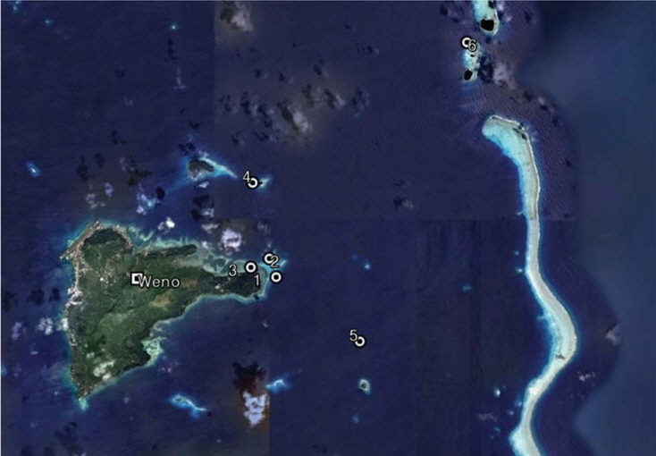
Sponges from Chuuk atoll were studied near Weno Island, 7˚25′N, 151˚47′E. They were taken from depths of 10-50m using scuba diving during 18-22 Feb 2010, 2-9 Feb 2012 (Table 1, Fig. 1). Collected specimens were frozen and some preserved in 95% ethyl alcohol and identified based on their morphological characters. The external features of sponges were observed with a stereo-microscope. The skeletal fibres were studied under a light microscope (Carl Zeiss, Axioscope).
[Table 1.] Geological information for collection sites
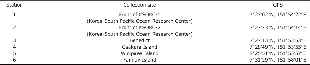
Geological information for collection sites
Phylum Porifera Grant, 1836Class Demospongiae Sollas, 1885Order Dictyoceratida Minehin, 1900Family Spongiidae Gray, 1867Genus Coscinoderma Carter, 1883
Type specimen. Holotype (MABIK IV00151582), front of KSORC-1, Chuuk, Micronesia, 17 Feb 2010, Kim HS, by scuba, depth 25 m, deposited in the MABIK, Seocheon, Korea.
Description. Large vase shape around 50×55×3-7 cm
Etymology. This species named after its foliose shape.
Remarks. This new species is a very large sponge. Primary fibres in the conules form loose fascicles and densely cored with large sand. Choanosomal, primary fibres are rarely cored over short distances. This new species is similar to the
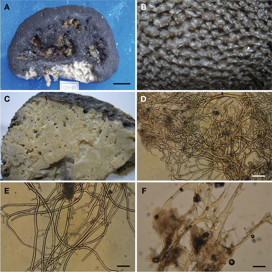
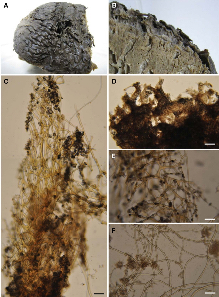
Type specimen. Holotype (MABIK IV00151583), Fannuk Island, Chuuk, Micronesia, 18 Feb 2010, Kim HS, by scuba, depth 25 m, deposited in the MABIK, Seocheon, Korea.
Description. Large subspherical sponge, 42×18 cm. Surface covered with a thick leathery two-layered membrane with very rare sand. Numerous dense small round conules 3 mm high especially on the top of the sponge. No oscules. Texture firm and compressible. Color in life dark black, underneath gray, beige internally. Center of sponge with a large hole, and side with a small hole. Skeleton. Primary fibres very well developed in the conules, 500 μm in diameter, lacey branched uncored primary fibres with a round cap, covered with a small amount of sand, but cored primary fibres at the base of the conule. Some primary fibres in choanosom 500-1,000 μm in diameter, rare cored but sand wrapped with numerous bridged secondary fibres. Secondary fibres in chonosome, 10-25 μm in diameter, easy to tear.
Etymology. This species named after the lace shape of primary fibres in conules.
Remarks. Primary fibres create a lace cap in short round conules on the top of the sponge but is covered with a small amount of sand, easy to separate from the fibres. Primary fibres in conules are no cored. This sponge has no ectosomal sand crust and choanosomal membrane very rarely has sand.
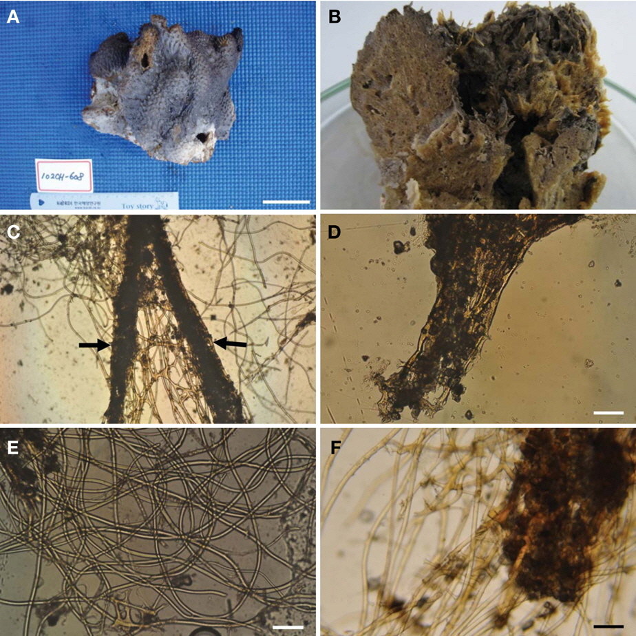
Type specimen. Holotype (MABIK IV00151584), Winipirea Island, Chuuk, Micronesia, 22 Feb 2010, Kim HS, by scuba, depth 25 m, deposited in the MABIK, Seocheon, Korea.
Description. Sponge massive with several short branches, size up to 16×20 cm and very cavernous. Surface very rough with long sharp conules with a thick sand crust, easily broken. Surface, covered two layered dermal membrane and sand crust. No oscules. Texture soft and compressible. Color in life gray on the surface and brown inside. Skeleton. Primary fibres in the conules mixed with sand grains and strong spongin, difficult to separate from fibres. Fasciculate primary fibres in conules measures, 300-400 μm in diameter, some branched (Fig. 4C). In the choanosome, primary fibres are rare. Secondary fibres 15-20 μm in diameter.
Etymology. This species name after its cavernous structure.
Remarks. This new species is very cavernous which is unique in the genus
Type specimen. Holotype (MABIK IV00151585), Winipirea Island, Chuuk, Micronesia, 22 Feb 2010, Kim HS, by scuba, depth 25 m, deposited in the MABIK, Seocheon, Korea.
Description. Sponge a subcylindrical mass 35×25×15 cm. Surface, three layered, thin dermal membrane with rare sand and space, second thin sand crust and third thick sand crust with long conules, with a stellate shape. Large space between ectosome and choanosome. No oscules. Texture firm and compressible. Color in life black outside, yellowish brown inside. Skeleton. Primary fibres in the conules loosely distributed and weakly adherent with sand. Ectosomal sand crust 0.1 mm thick, separate from surface. Primary fibres rarely cored with sand grains, only attached at the surface of thin primary fibres 600-700 μm in diameter, 5-7 mm long. Secondary fibres 10-15 μm in diameter, not easy to tear in the choanosome. Many thin membranes with sand in the choanosome.
Etymology. This new species name after the type locality, Weno Island, Chuuk, Micronesia.
Remarks. This new species has long conules and primary fibres are long in the choanosome. Surface sand membrane has space between ectosome and choanosome.
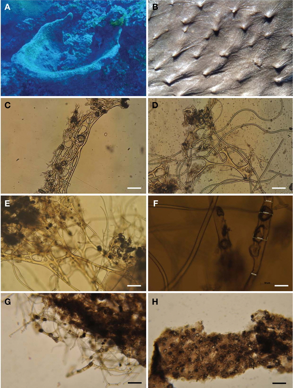
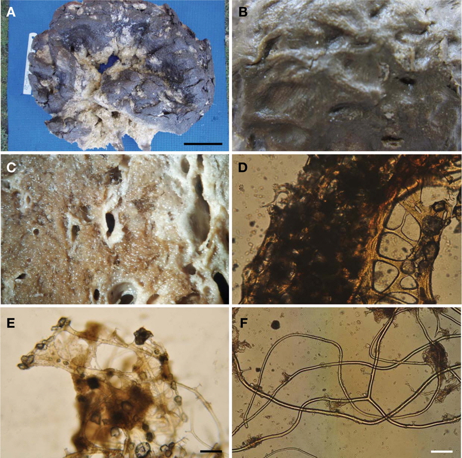
Type specimen. Holotype (MABIK IV00151586), Osakura Island, Chuuk, Micronesia, 20 Feb 2010, Kim HS, by scuba, depth 25 m, deposited in the MABIK, Seocheon, Korea.
Description. Large irregular sponge, size up to 45×22 cm. The surface a leathery thick membrane with conules. Under the membrane, sand crust 4-8 mm thick, tightly adherent with the choanosome. No oscules. Texture soft and compressible. Color in life black outside, beige inside. Skeleton. Primary fibres in the conules cored with large sand grains, creating large meshed fascicles. Ectosomal skeleton under surface membrane creating thick sand crust 5-7 mm thick, attached to the surface of the fibres 500-700 μm in diameter. Long fasciculate primary fibres with sand emerge from the choanosome. Secondary fibres 20-25 μm in diameter, loosely distributed in the choanosome, seldom connected with other secondary fibres. Sponge perpendicular section, choanocyte chambers surrounded with a sand crust 1-3 mm thick.
Etymology. This new species named after shape of mop.
Remarks. This new species is a very soft and compressible. In the choanosome, numerous thick sand crust cover choanocyte chamber (Fig. 6C).
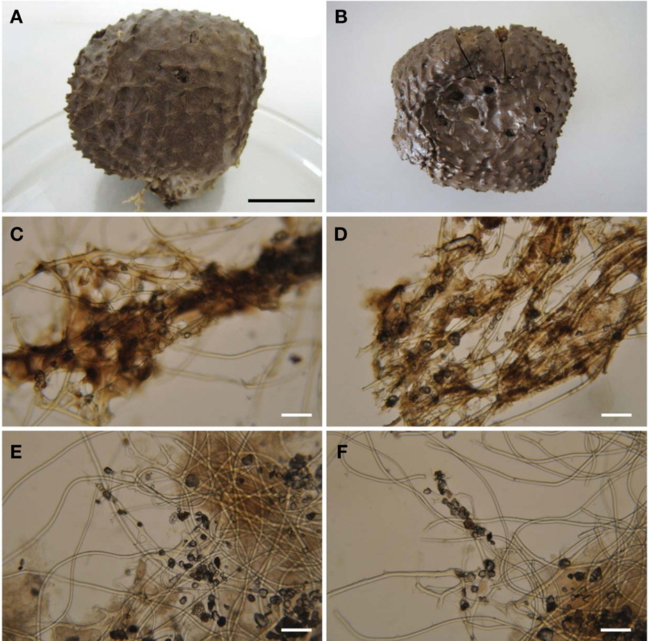
Type specimen. Holotype (MABIK IV00151587), Fannuk Island, Chuuk, Micronesia, 18 Feb 2010, Kim HS, by scuba, depth 25 m, deposited in the MABIK, Seocheon, Korea.
Description. Round mass shape, with the base narrow than top. Size up to 6×5 cm. Surface many distinct satellite conules with round tips, 900 μm high; 150 μm in diameter. Surface, one layer sand membrane. Many oscules 2-6 mm in diameter open on the top. Conule, cover a thick sand crust, easily separate from primary fibres in conules. Texture hard and compressible. Color change gray to dark brown. Skeleton. Cored primary fibres fascicles wrapped with many bridged secondary fibres tightly. Primary fibres 150 μm in diameter. No cored secondary fibres, 15-25 μm in diameter.
Etymology. This species name bakusi is named after the late Dr. Gerald J. Bakus who was a professor in the department of Biological Sciences, University of Southern California, a marine ecologist and a sponge taxonomist.
Remarks. This new species is characterized by primary fibre fascicle wrapped with bridged secondary fibres, conules with very thick sand crust, and color changed from live gray to preserved dark brown.
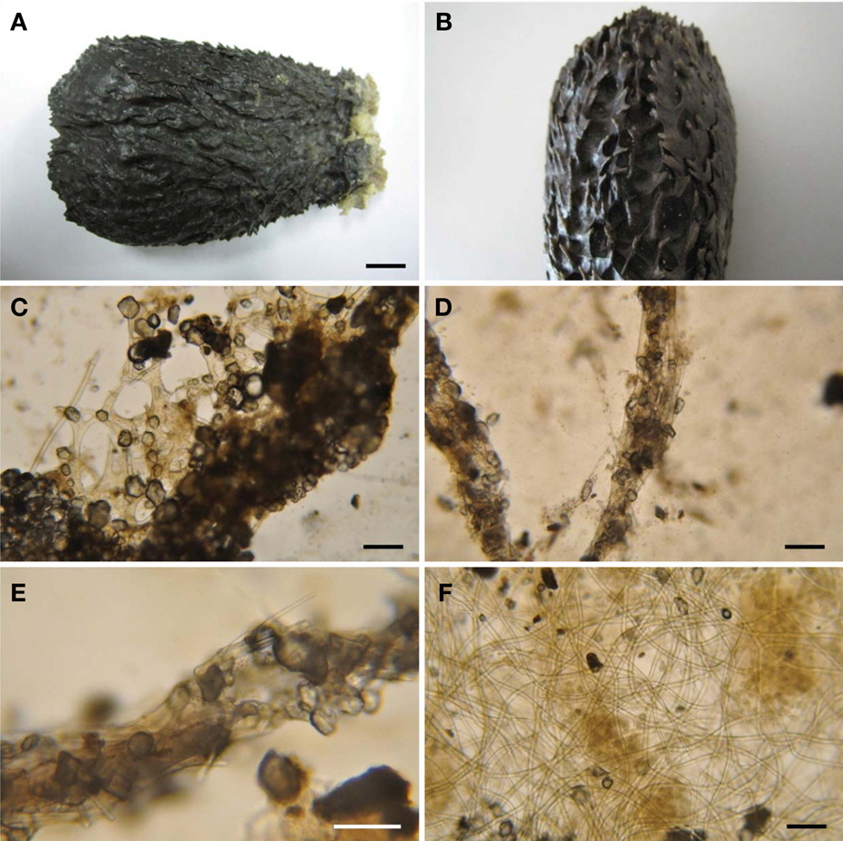
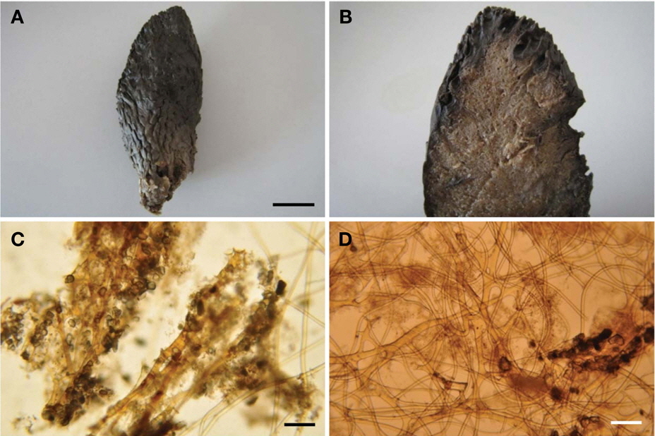
Type specimen. Holotype (MABIK IV00151588), Benedict, Chuuk, Micronesia, 8 Feb 2012, Kim HS, by scuba, depth 21 m, deposited in the MABIK, Seocheon, Korea.
Description. Small thumb shape, usually a single, 9×3.5× 2.5 cm. Surface smooth with low conules. One layer thin sand membrane. No oscules. Texture tough and compressible. Color in life dark gray outside, brown inside. Skeleton. Primary fibres, 300-400 μm in diameter in the conule. Height of conule 3.6 mm. Primary fibres in choanosome numerous between secondary fibres. No cored secondary fibres in choanosome, 10-25 μm in diameter.
Etymology. This new species is named after its thumb-like shape.
Remarks. This species is always small and thumb-shaped.
Type specimen. Holotype (MABIK IV00151589), front of KSORC-2, Chuuk, Micronesia, 8 Feb 2012, Kim HS, by scuba, depth 20 m, deposited in the MABIK, Seocheon, Korea.
Description. Small thumb shape, size up to 8×3×3 cm. Wider on the top than at the base. Surface rough with large sharp conules has one layered sand membrane. Texture hard and compressible, color in life black outside, beige inside. Skeleton. Primary fibres in conules. 4 mm in length, 20-40 μm in diameter without sand crust. Primary fibres in choanosome very rare. Cored primary fibres with many spicules and sand. No cored secondary fibres 8-20 μm in diameter.
Etymology. This species is named after type locality, Truk, old name of Chuuk, Micronesia.
Remarks. This new species is similar to
Eight new species of
Voultsiadou-Koudoura et al. (1991) concluded that species of
All our new species are different from