Copepods associated with marine invertebrates have been very poorly investigated in Thailand. Ho and Kim (1990) reported six species of the family Synapticolidae associated with holothurians from Phuket:
During a couple of international scientific conferences held in the past in Thailand, the authors could make short field works to collect copepods at vicinities of conference places. One of them is the 10th International Conference on Copepoda at Pattaya in July 2008, and the other being the First Asian Marine Biology Symposium at Phuket in December 2012. A total of 15 species of poecilostome copepods were collected, including ten new species, as described below.
The copepods examined in the present study were recovered by washing, sweeping, and sucking. On the tidal flat the host animals were dug out with a stick and collected in plastic bags, with different species being placed in different bags and later being fixed in alcohol. On the coral reef and rocky shore, the copepods on the hosts (mainly hard corals) living in the shallow water were swept into a hand net using a small brush. Some copepods were sucked up directly from the host’s burrows with a large pipette. The sucked material were filtered using a small hand net and later in the laboratory the copepods were sorted out from the filtrates.
Before dissection for microscopic observation, copepod specimens were immersed in lactic acid. Dissections were done using the reversed slide method (Humes and Gooding, 1964). Dissected specimens were mounted on glass slides with Hoyer’s mounting medium. Type specimens have been deposited in the Marine Biodiversity Institute of Korea (MABIK), Seocheon, Korea.
Order Cyclopoida Burmeister, 1834Family Clausidiidae Embleton, 1901Genus Hemicyclops Boeck, 1873
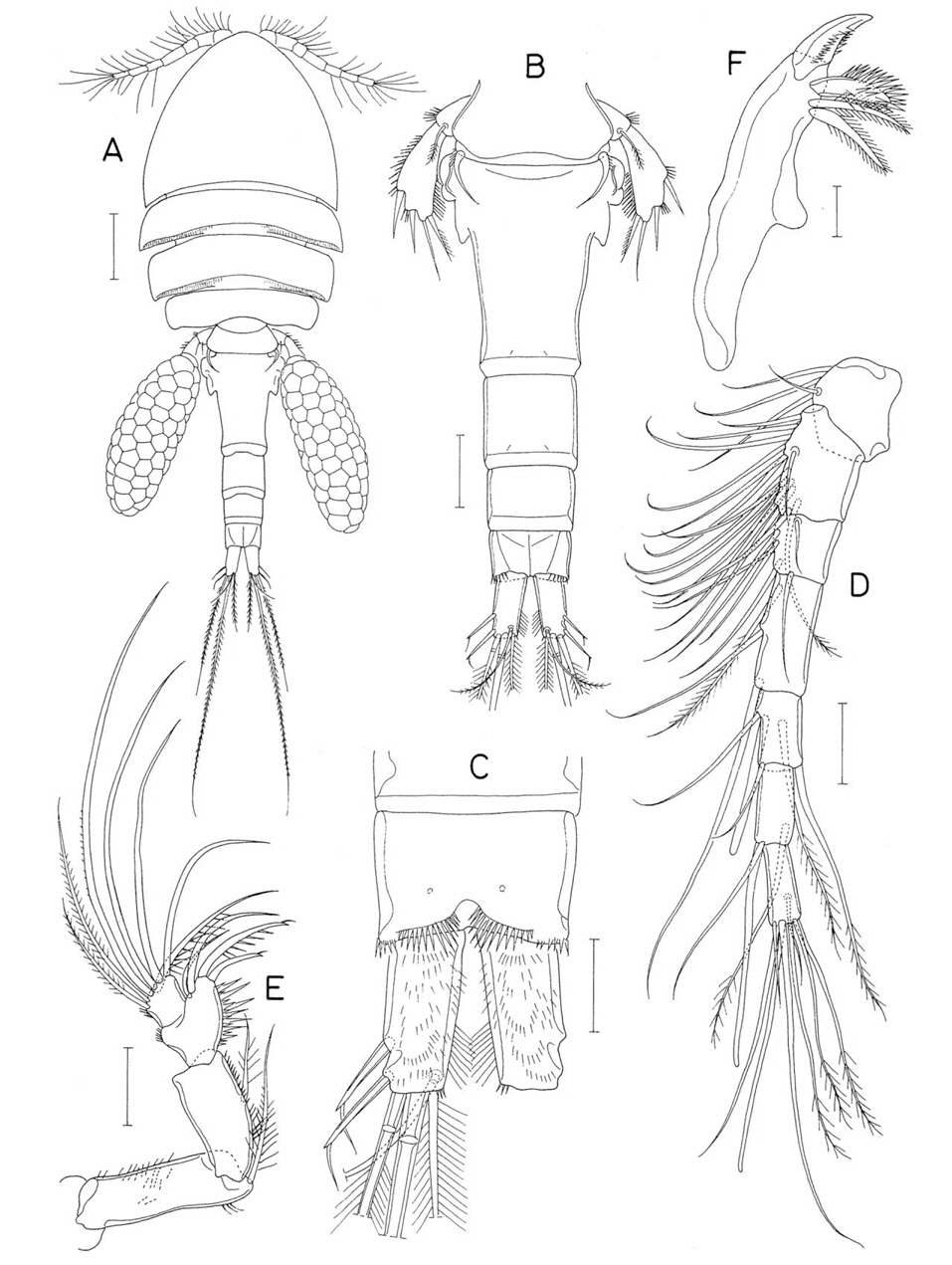
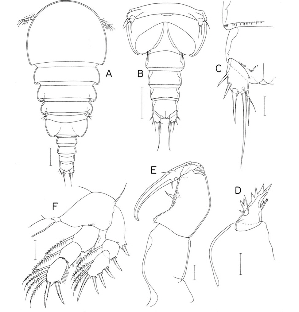
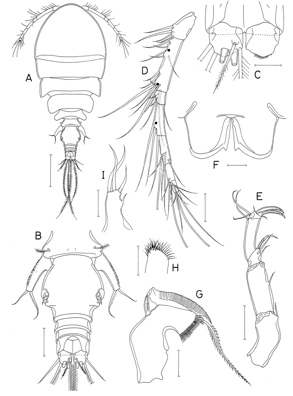
Hemicyclops cornutus n. sp. (Figs. 1-3)
Material examined. 1♀ (holotype) from invertebrate burrows on a tidal flat (Ban Pa Khok seagrass bed) on the northeastern coast of Phuket Province, Thailand, 16 Dec 2012. Holotype (CR00233151, dissected and mounted on a glass slide) has been deposited in the MABIK, Seocheon, Korea.
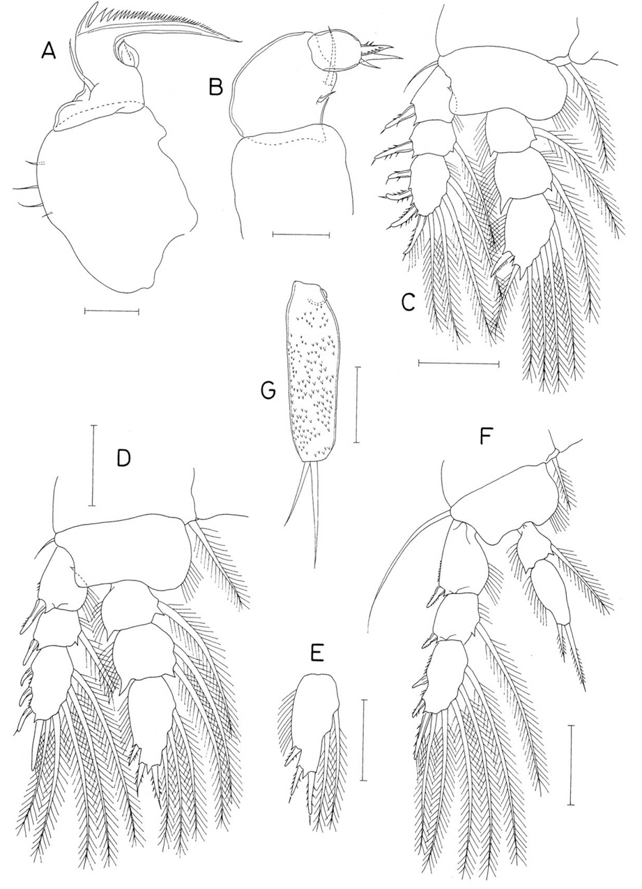
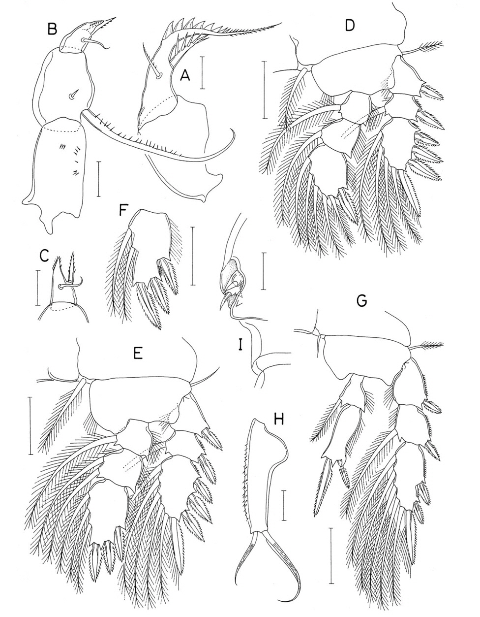
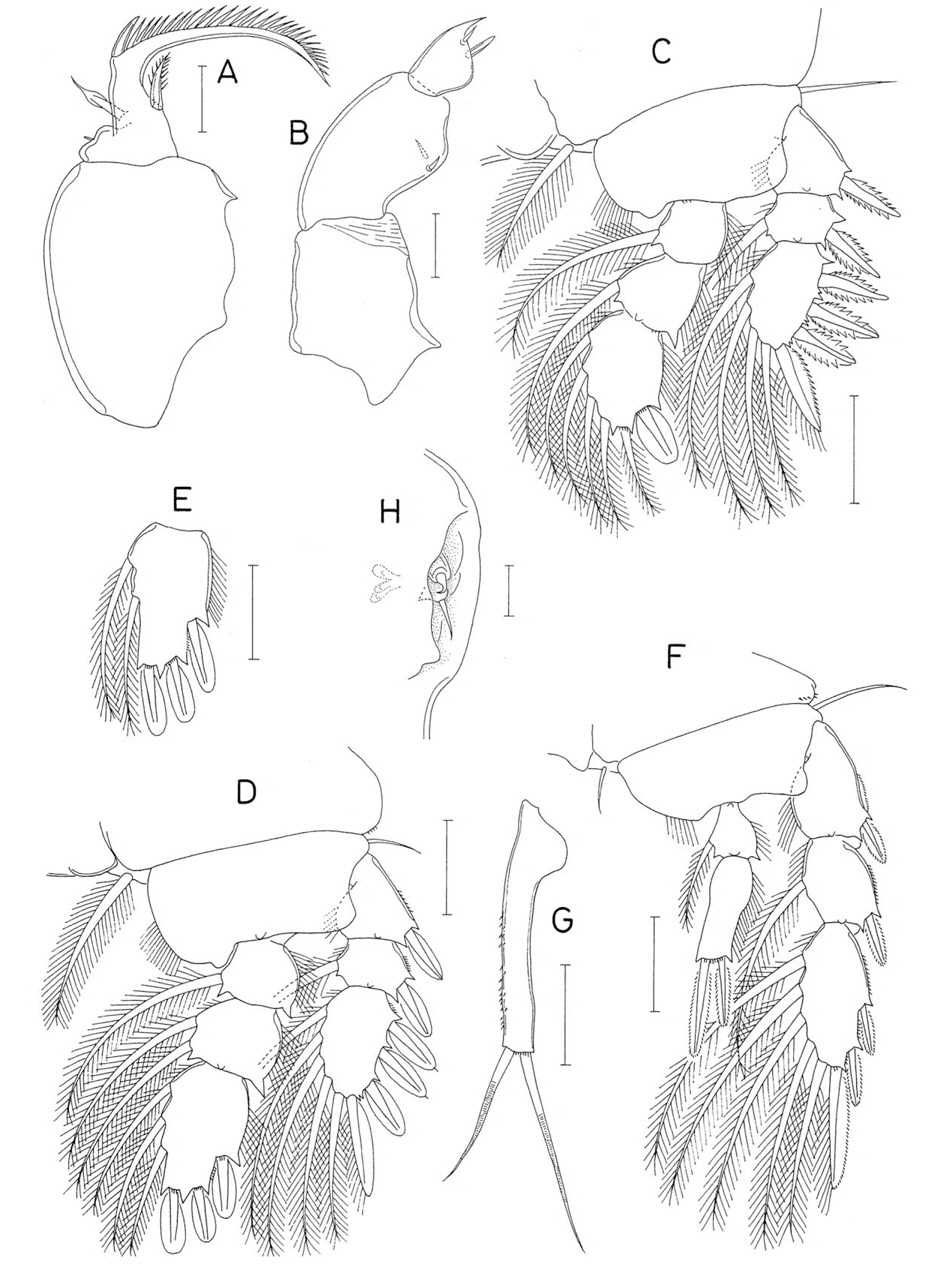
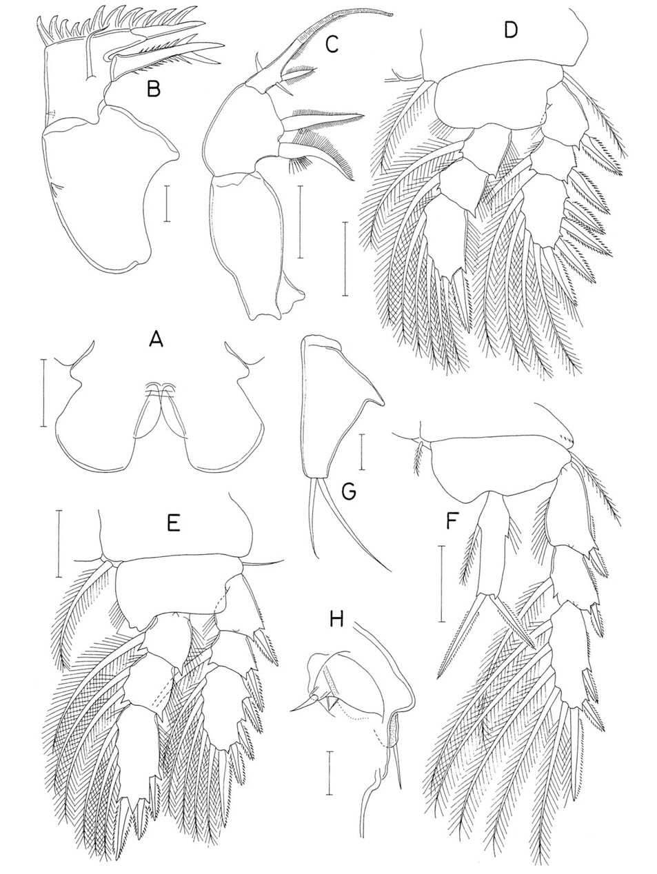
Female. Body (Fig. 1A) moderately broad, 1.77 mm long. Prosome fusiform, 931 μm long, and consisting of cephalothorax and 3 pedigerous somites. Greatest width of prosome 638 μm across second pedigerous somite. Cephalothorax 500×608 μm. Second and third pedigerous somites with angular posterolateral corners; their posterior margin fringed with narrow membrane. Third pedigerous somite with blunt posterolateral corners. Urosome (Fig. 1B) 5-segmented. Fifth pedigerous somite (first urosomite) 230 μm wide, with gently curved, horn-like spine (indicated by arrow in Fig. 2G) at each posterolateral corner of dorsal side; this spine spinulose at proximal outer margin, but smooth in remaining part. Genital doublesomite 308×258 μm (length to width ratio 1.19 : 1), with anterolateral expansion; lateral margins of this expanded region complicated, with 3 curvatures (Fig. 3E); genital aperture locating laterally at this expanded region; posterior part of somite weakly narrowing posteriorly, with width of 140 μm across posterior region. Three free abdominal somites 126×129, 92×123, and 71×114 μm, respectively. Anal somite (Fig. 1C) with row of spinules along all posterior margins. Caudal rami (Fig. 1C) slightly divergent; each ramus rectangular, 85×40 μm (ratio 2.13 : 1), covered with fine setules on dorsal and ventral surfaces, and armed with 6 setae; outer seta (seta II) and outermost distal seta (seta VI) spiniform, not flexible, naked, and tipped by setule; other 4 setae pinnate; inner margin of ramus with long setules along distal third. Egg sac fusiform, 577×207 μm, distinctly shorter than urosome.
Rostrum not examined. Antennule (Fig. 1D) 359 μm long and 7-segmented, with armature formula 4, 15, 6, 3, 4+aesthetasc, 2+aesthetasc, and 7+aesthetasc; setae generally long and naked, except for following pinnate setae: 1 on second, fourth, and sixth segments each, 2 on fifth, and 3 on last. Antenna (Fig. 1E) 4-segmented; first segment longest, with scattered spinules and 1 pinnate seta at inner distal corner; second segment with spinulose inner margin and 1 pinnate seta on inner margin; third segment with dense spinules on inner margin, 4 setae at distinctly projected inner distal corner; terminal segment as long as wide, 25×25 μm, and armed with 7 setae, of which 2 outer ones weakly pinnate and one of remainings spinulose.
Labrum (Fig. 2A) as in general of the genus, with broad proximal part, narrower, spinulose distal part, and straight posterior margin. Mandible (Fig. 1F) with 4 elements distally: 1 claw-like thick element bearing 2 rows of fine spinules, 1 plate-like, densely spinulose element, and 2 pinnate setae. Paragnath (Fig. 2B) as small, unarticulated lobe bearing setules in distal region. Maxillule (Fig. 2C) distally bilobed, with patch of fine spinules on inner margin, and armed with 3 setae on inner lobe and 5 setae on outer lobe; most of setae on these lobes pinnate or spinulose. Maxilla (Fig. 2D) 2-segmented; basal segment with 2 thick setae on inner side, one of these setae with subsidiary setule at base; distal segment terminating in large, bifurcate process (with smooth outer branch and 3 cusps-bearing inner branch) and armed with 1 unilaterally pinnate seta and 2 spines: outer spine pinnate on inner margin and finely spinulose on outer margin; inner spine with patch of spinules near middle of outer margin. Maxilliped (Fig. 2E) 4-segmented; first segment broadest, with 2 large setae on inner margin; second segment longest, with 2 large setae on protrusion of inner margin; short third segment unarmed; terminal segment with 3 naked setae and 2 spines (terminal and inner ones); long inner spine with several long spinules near proximal third; terminal spine with several spinules on outer margin and 1 naked proximal seta; latter seta not articulated at base.
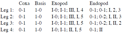
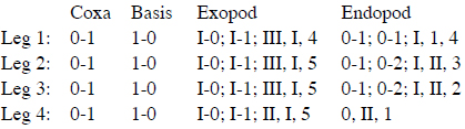
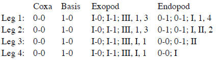
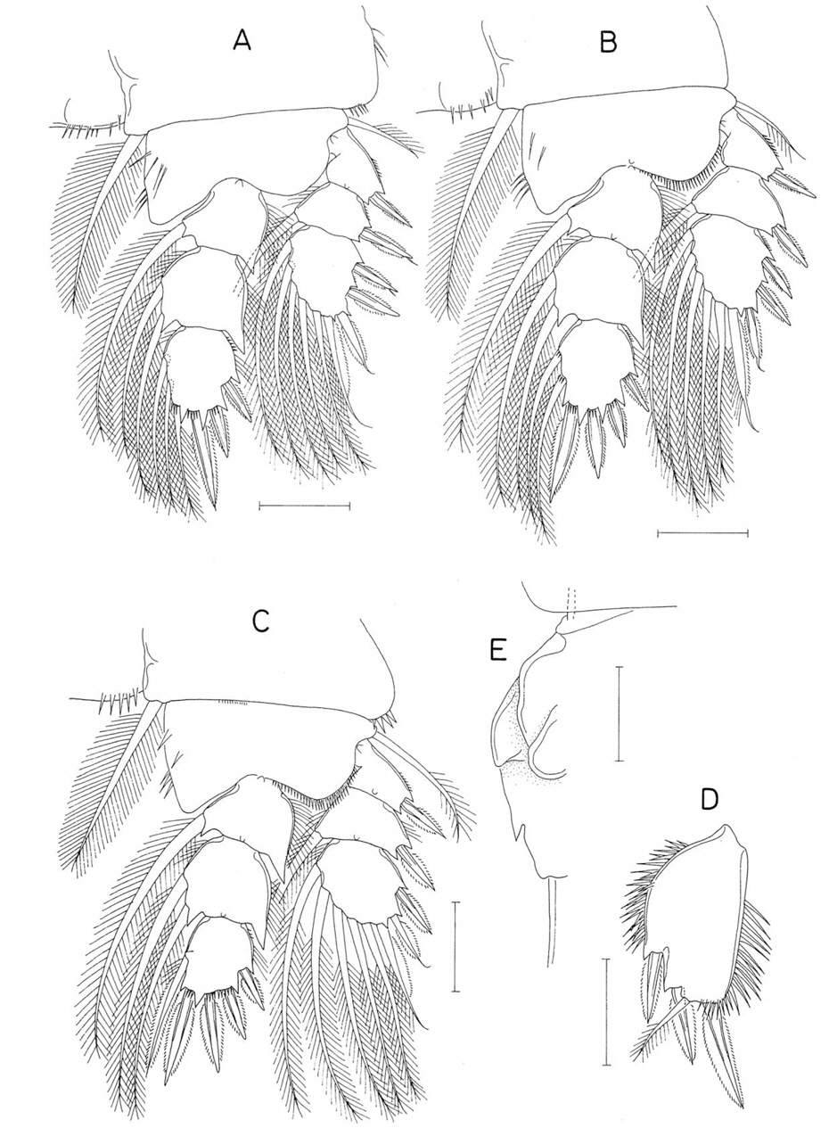
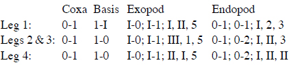
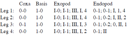
Legs 1-4 (Figs. 2F, 3A-C) with 3-segmented rami; endopods distinctly longer than exopods; coxa with setules on outer margin and patch of spinules at outer distal corner. Inner distal spine on basis of leg 1 large, spinulose, 78 μm long, and extending to distal border of second endopodal segment. Intercoxal plate with setules on posterior margin in leg 1, but with spinules in legs 2-4. Basis with row of spinules along distal margin between bases of rami and on inner side of anterior surface; inner side of distal margin with spinules in leg 1, but with setules in legs 2-4. Outer spines on leg 1 exopod tipped with setule. Outer distal corner of first and second endopodal segments of legs 1-4 projected, spiniform. Two inner spines on third endopodal segment of leg 4 elongate and pinnate in proximal 2/5 but spinulose in distal 3/5. Armature formula of legs 1-4 as follows:
Leg 5 (Fig. 2G) 2-segmented; basal segment with 1 unilaterally pinnate, dorsodistal seta and patch of spinules at outer distal corner; exopod (distal segment, Fig. 3D) narrow proximally but gradually broadened distally, 142×63 μm (ratio 2.25 : 1), ornamented with dense spinules on both lateral margins and armed with 3 spines and 1 seta on distal margin; lengths of spines 63, 56, and 69 μm, respectively, from outer to inner. Leg 6 not discernible.
Male. Unknown.
Etymology. The specific name
Remarks. Although only a single specimen is available to the present description of
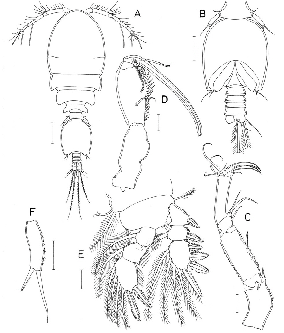
Material examined. 1♀ (holotype) from invertebrate burrows on the Ban Pa Khok seagrass bed, northeastern coast of Phuket Province, Thailand, 16 Dec 2012. Holotype (CR00 233152, dissected and mounted on a glass slide) has been deposited in the MABIK, Seocheon, Korea.
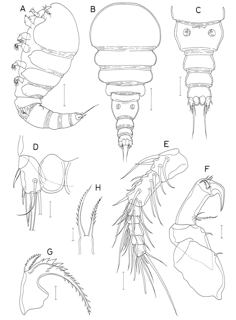
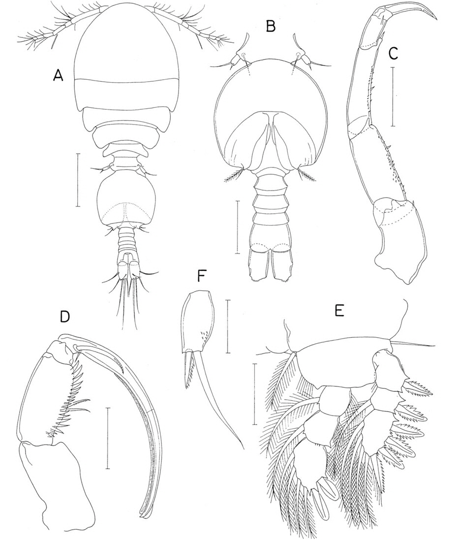
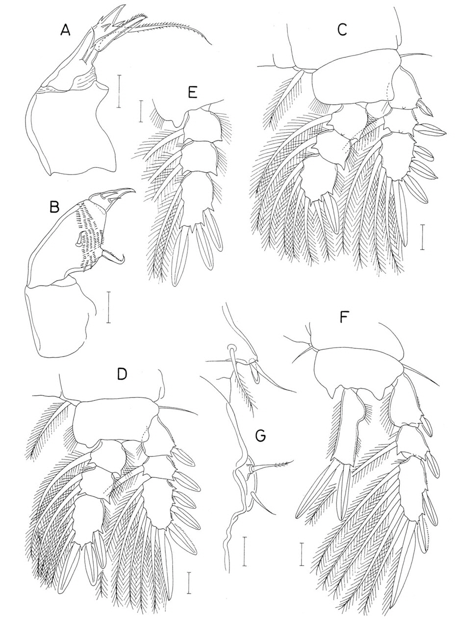
Female. Body (Fig. 4A) moderately narrow and 1.19 mm long. Prosome 655×465 μm and consisting of cephalothorax and second to fourth pedigerous somites. Posterolateral corners of prosomal somites not pointed. Cephalothorax 385 μm long. Second and third pedigerous somites fringed with membrane along posterior margin. Urosome (Fig. 4B) 5-segmented. First urosomal somite (fifth pedigerous somite) 185 μm wide, with smooth lateral and posterior margins. Genital double-somite (Figs. 4B, 6E) 227 μm long, comprising roundly expanded anterior 63% and narrower distal 37%; greatest width of double-somite 185 μm, and width of narrower distal part 115 μm; expanded anterior part with sharp point at subdistal region of its lateral margins; genital aperture locating at midlength of expanded anterior part. Three free abdominal somites 78×108, 60×100, and 50×103 μm, respectively. Anal somite with short, transverse row of fine spinules along lateral side of posterodorsal margin (Fig. 4C). Caudal rami (Fig. 4C) quadrate, 55×48 μm (ratio 1.15 : 1) measured in ventral view, with setules on inner margin, tapered, tube-like ventrodistal element bearing pore at tip (Fig. 4D), and finely spinulose posteroventral margin; outer lateral seta small, 35 μm long and pinnate along inner margin; outermost distal seta (seta III) spiniform proximally and tipped with pinnate seta.
Rostrum short, with round posterior apex. Antennule (Fig. 4E) 321 μm long; armature formula 4, 15, 6, 4, 4+aesthetasc, 2+aesthetasc, and 7+aesthetasc; pinnate setae: 1 on fourth, fifth, and sixth segments each, and 3 on terminal segment. Antenna (Fig. 4F) 4-segmented; first segment longest, with scattered setules and spinules and 1 long inner distal seta; second segment slightly shorter than first, with setules on outer margin, spinules on proximal part of inner margin and 1 seta (this seta spinulose on inner margin) at middle of inner margin; third segment with inflated inner distal corner, spinules on inner margin and 4 setae at inner distal region, 2 of these setae naked and remaining 2 spinulose; terminal segment 22×27 μm, slightly wider than long, and armed with 7 setae, 4 of them large and weakly geniculate.
Labrum (Fig. 5A) nearly hemicircular, with various spinules on distal and subdistal regions; posterior and lateral margins not defined. Mandible (Fig. 5B) with 2 massive elements and 2 setae distally. Paragnath (Fig. 5C) not articulated, slightly curved, with spinules and setules. Maxillule (Fig. 5D) distally bilobed, with 3 shorter setae (one of them broad) on narrower lobe and 5 longer seta on broader lobe. Maxilla (Fig. 5E) similar to that of preceding species; basal segment with 2 large setae and 1 small, weakly pinnate seta inserted to base of one of 2 larger setae. Maxilliped (Fig. 5F) with 2 large setae on inner margin and patch of spinules on outer side of first segment; second segment slightly inflated in middle, with 2 spinulose setae on inner margin and ornamented with row of large spinules on distal part of inner margin and row of several setules near inner distal corner; third segment small and unarmed; terminal segment terminating in long, smooth claw and proximally with 2 naked seta, 1 bifurcate seta, and 1 spinulose spine.
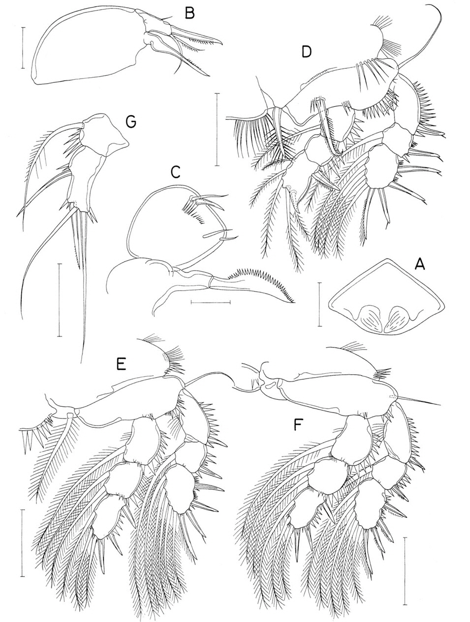
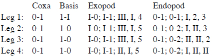
Leg 1 (Fig. 5G) with small tubercle on outer margin of coxa; inner distal spine on basis 54 μm long; distal process on third endopodal segment pronounced, with fine spinules on its outer margin; outermost terminal seta on third endopodal segment naked and smaller than nearby setae; all of outer spines on exopod tipped with setule. Posterior margin of coxa of leg 1 with setules (Fig. 5G), but those of legs 2-4 with sparse spinules (Fig. 6A-C). Inner proximal spine on third endopodal segment of leg 4 pinnate proximally. Armature formula of legs 1-4 as follows:
Leg 5 two-segmented (Fig. 4B); basal segment (protopod) with angular outer distal corner, setulose outer margin and armed with 1 dorsal, naked seta; exopod (Fig. 6D) 83×46 μm (ratio 1.80 : 1), with densely spinulose lateral margins, and armed with 3 spines and 1 pinnate seta; lengths of distal spines on exopod 35, 38, and 53 μm, respectively, from outer to inner. Leg 6 not discernible.
Male. Unknown.
Etymology. The specific name
Remarks.
As minor differences, the body length of the new species is 1.19 mm, which is compared to 1.36-1.50 mm in
It should be pointed out that
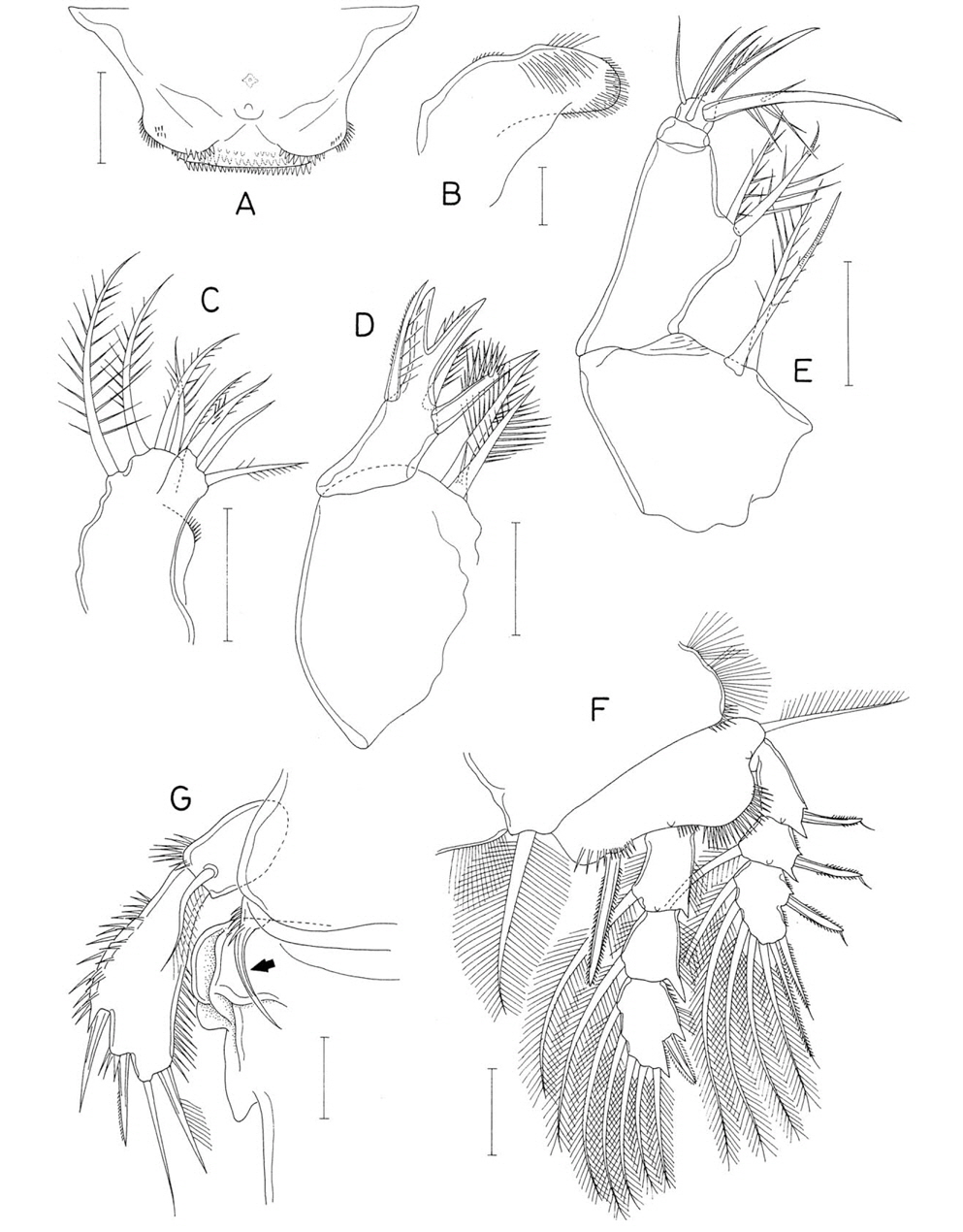
Family Synaptiphilidae Bocquet and Stock, 1957Genus Presynaptiphilus Bocquet and Stock, 1960
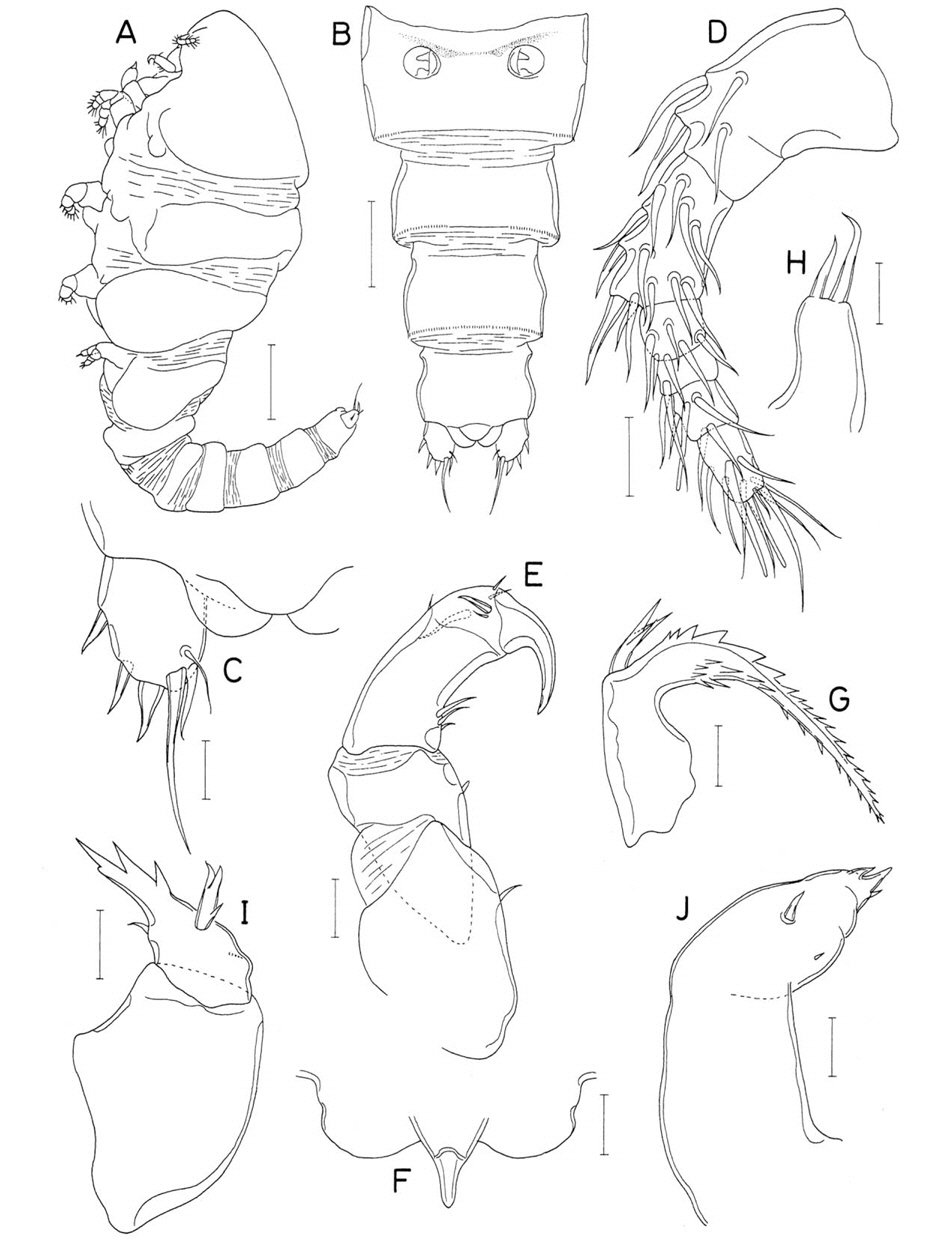
Material examined. 1♀, 1♂ from a brittle star (genus
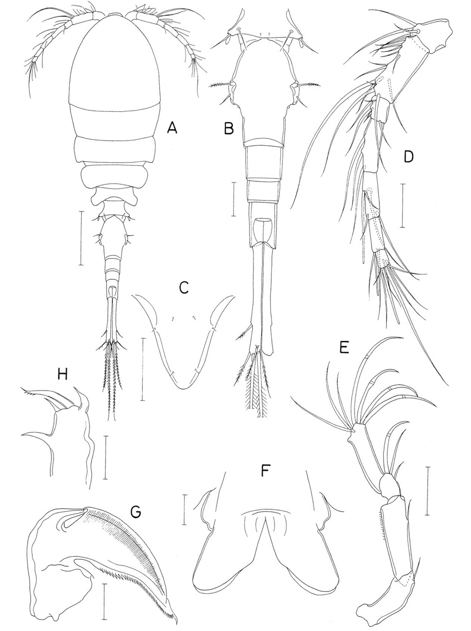
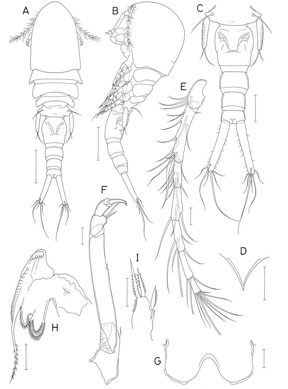
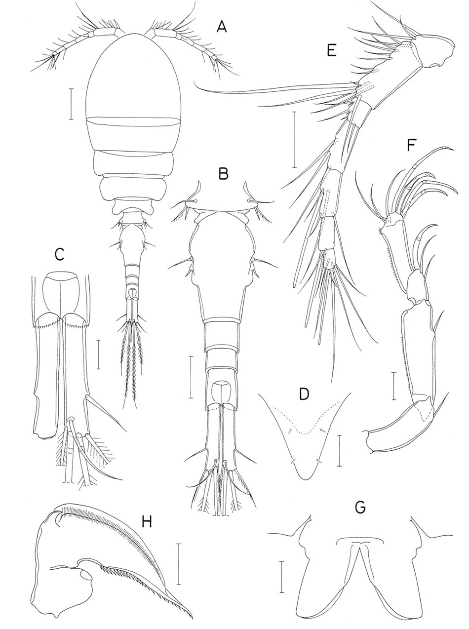
Female. Body (Fig. 7A) small, 692 μm long. Prosome 425×283 μm, consisting of cephalothorax and second to fourth pedigerous somites. Cephalothorax 238 μm long, wider than long, with blunt posterolateral corners. Second and third pedigerous somites with convex lateral margins, without corners. Fouth pedigerous somite reduced, much narrower than preceding somite. Urosome (Fig. 7B) 5-segmented. Fifth pedigerous somite (first urosomal somite) 90 μm wide, with smooth lateral and posterior margins. Genital double-somite (Fig. 7B) rectangular, 74×92 μm, wider than long, with weak anterolateral expansion, spinulose posteroventral margin, and 2 pairs of sensillae on posterodorsal region; genital aperture locating dorsally at region of anterolateral expansion. Three free abdominal somites 45×62, 58×53, and 21×39 μm, respectively. First and second free abdominal somites with spinules along posteroventral margin. Anal somite short, with 2 pair of denticles on posterior part of ventral surface (Fig. 7C); anal aperture large (Fig. 7D). Caudal ramus almost straightly directed backwards (Fig. 7C, D); each ramus 37×18 μm(measured in ventral view), 2.06 times as long as wide, armed with 7 naked setae, including setule-like outer proximal seta (seta I), and ornamented few spinules on posteroventral margin; one of caudal setae (seta II) located on proximal region of dorsal surface of ramus; outer one (seta IV) of 2 median terminal setae reduced, shorter than outermost terminal seta, but inner, nearby seta (seta V) large, more than 5 times as long as second longest seta (seta III).
Rostrum (Fig. 7E) nearly rectangular, much wider than long, with well-sclerotized posterior margin. Antennule (Fig. 7F) 265 μm long and 6-segmented; armature formula 4, 15, 9, 4+aesthetasc, 2+aethetasc, and 7+aesthetasc; all setae naked and all aesthetascs thin; first segment with additional patch of several setules at proximal region. Antenna (Fig. 7G) 4-segmented; first segment with 1 pinnate seta at inner distal corner and several rows of setules on distal half of surface; second segment nearly as long as first segment, with 1 naked seta on inner side; short third segment with produced inner distal corner and armed with 1 powerful claw and 3 setae, proximalmost one of latters small, setule-like; terminal segment wider than long and armed with 4 large, geniculate spiniform setae, 1 simple naked seta, and 2 pinnate setae.
Labrum (Fig. 8A) rhomboidal, wider than long, with tapering lateral margins and 1 pair of membranous lobes on posterior region of dorsal surface. Mandible (Fig. 7H) armed with 2 elements consisting of 1 subdistal seta and 1 distal spine bearing 1 subterminal cusp and 4 spinules. Paragnath not discernible. Maxillule (Fig. 7I) weakly bilobed distally and armed with 3 pinnate seta on outer lobe and 1 naked seta on inner lobe. Maxilla (Fig. 8B) 2-segmented; basal segment with elongate inner distal spine bearing minute spinules on distal part of outer margin; distal segment with with 1 spinulose spine, 2 slender seta and 1 small, setule-like, subdistal seta. Maxilliped (Fig. 8C) peculiar and 2-segmented; basal segment with projected inner distal corner tipped by large, saw-like spine bearing serrate distal margin; distal segment almost rectangular and plate-like, with 1 pair (one of them being spiniform) of distal and 1 pair of inner, subdistal setae and 1 transverse, subdistal row of setules.
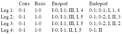
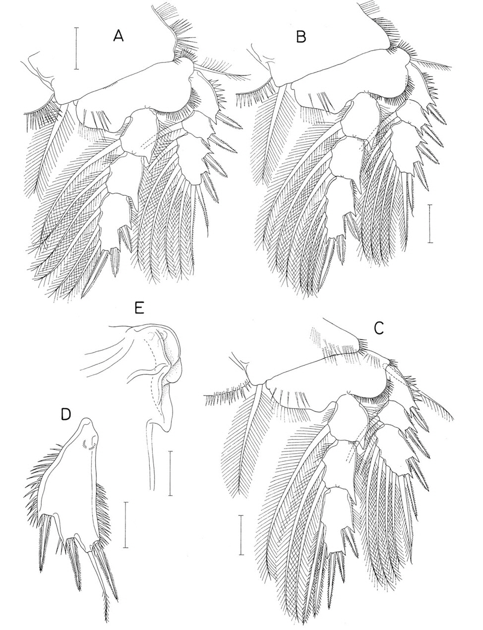
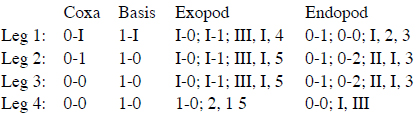
Legs 1-3 (Fig. 8D-F) with 3-segmented rami. Leg 4 (Fig. 9A) with 2-segmented rami. Posterior margin of coxa with large setules in leg 1, but with spinules in legs 2 and 3, and naked in leg 4. Inner element of coxa being setulose spine in leg 1, pinnate seta in leg 2, but absent in legs 3 and 4. Outer seta of legs 1-4 naked. Inner spine on basis of leg 1 setiform and spinulose. Third endopodal segment of leg 1 with pointed process at distal apex. Distal endopodal segment of leg 4 55×14 μm(ratio 3.93 : 1). Armature formula of legs 1-4 as follows:
Leg 5 (Fig. 8G) 2-segmented; basal segment (protopod) with 1 large seta and several spinules on outer margin; distal segment (exopod) 46×21 μm (ratio 2.19 : 1), with 2 spines (outer and outer subdistal) and 2 distal setae, all of these 4 elements naked. Spines and inner distal seta accompanied with 2 spinules near base of these elements; inner one of terminal setae longest, 107 μm long. Leg 6 (Fig. 9B) represented by 1 small, naked seta on genital operculum.
Male. Body (Fig. 9C) very similar to that of female, but smaller, 518 μm long. Greatest width 260 μm. Cephalothorax 190 μm long and distinctly wider than long. Urosome (Fig. 9D) 6-segmented. Fifth pedigerous somite 90 μm wide. Genital somite (Fig. 9B) rectangular, 38×87 μm, much wider than long, with indistinct genital opercula. Four abdominal somites 35×81, 31×69, 41×59, and 15×36 μm, respectively. First 3 abdominal somites with spinules along both sides of posteroventral margin. Anal somite ornamented as in female (Fig. 9E). Caudal ramus (Fig. 9E) 25×15 μm(ratio 1.67 : 1) and armed with 6 setae; seta I absent; outer one (seta IV) of 2 median terminal setae trifurcate at tip (Fig. 9E).
Rostrum, antennule and antenna as in female. Mouthparts, except for maxilliped, also as in female. Maxilliped (Fig. 9F) consisting of 3 segments and terminal claw; first segment unarmed; second segment greatly expanded proximally and strongly tapering distally, with inner hollow at proximal expansion receiving tip of terminal claw, 2 small setae and 4 broad, foliaceous spinules at hollow, and longitudinal row of spinules along inner margin distal to hollow; small third segment unarmed; terminal claw weakly curved and armed with 2 small setae prxoximally.
Leg 1 (Fig. 9G) with spinules (instead of setules as in female) on posterior margin of coxa; inner spine on coxa weakly pinnate distally; inner element on basis as spinulose seta. Leg 1 endopod 2-segmented; inner seta on proximal segment swollen proximally; distal segment armed with 1 outer spine and 4 inner setae (armature formula I, 4); distal process more pronounced than that of female.
Leg 5 (Fig. 9H) stout; exopod 31×22 μm(ratio 1.41 : 1), tapering, armed with 3 spines (distal element being a strong spine rather than a long seta as in female) and 1 naked seta, and ornamented with more number of spinules than in female. Leg 6 absent (Fig. 9D).
Etymology. The specific name
Remarks. All of four known species of the genus
The new species can be differentiated from its congeners on the basis of the female morphology. The endopod of female leg 1 is 3-segmented in all known species of the genus. The endopod of female leg 1 of the new species has 1, 0, and 6 elements, respectively, on the first to third segments (formula 0-1; 0-0; I, 5). This armature state is shared only with
Family Anchimolgidae Humes and Boxshall, 1996Genus Anchimolgus Humes and Stock, 1972
Material examined. 5♀♀, 8♂♂ from a scleractinian coral, on coral reef in Kantary Bay, southern coast of Phuket Province, Thailand, 7˚48′14′′N, 98˚24′20′′E, 15 Dec 2012. Holotype (♀, CR00233155), allotype (♂, CR00233156), and paratypes (3♀♀, 6♂♂, CR00233157) have been deposited in the MABIK, Seocheon, Korea. Dissected paratypes (1♀, 1♂) are retained in the collection of the senior author.
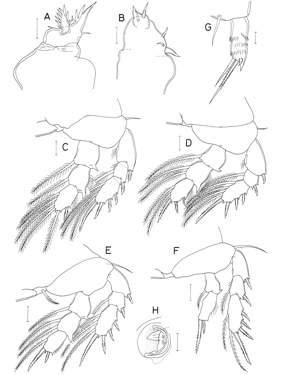
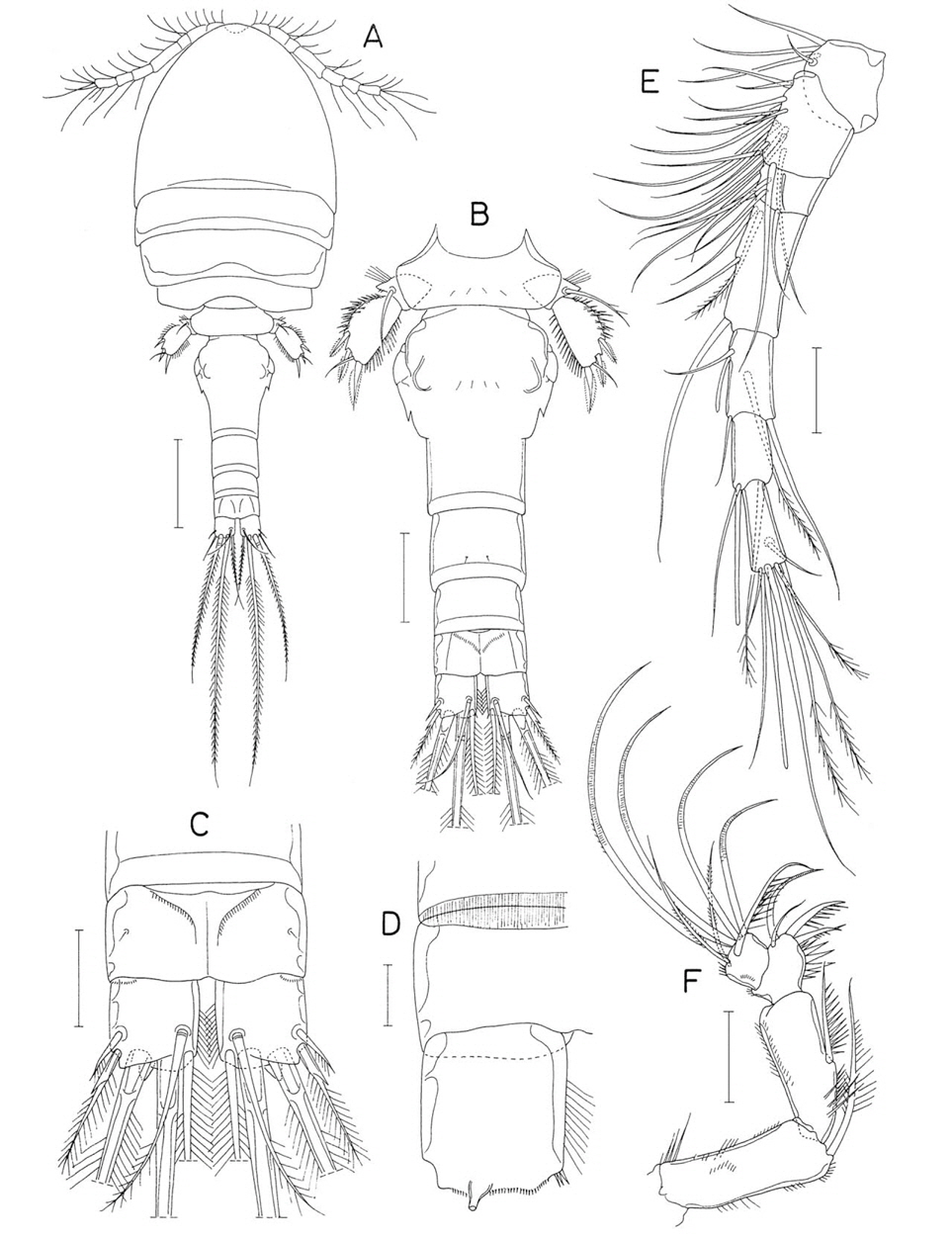
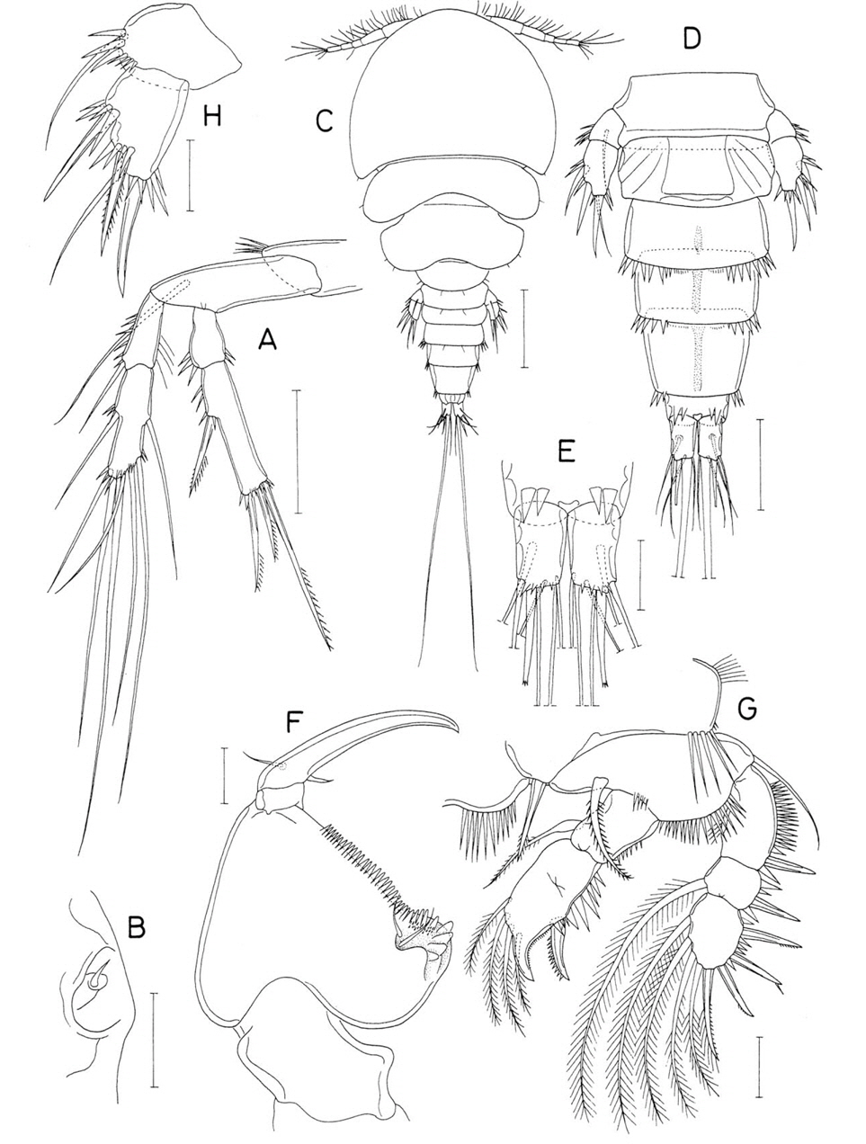
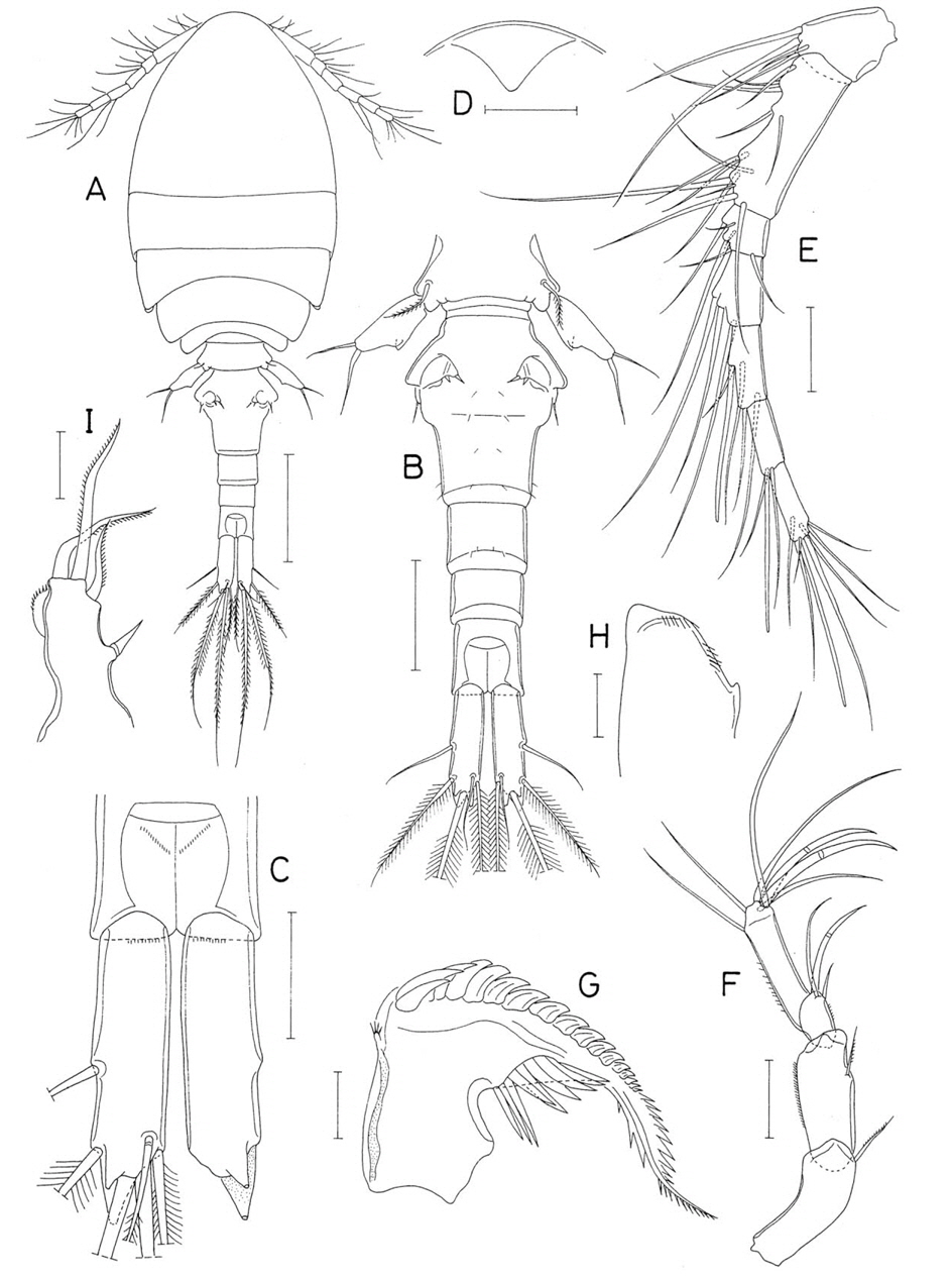
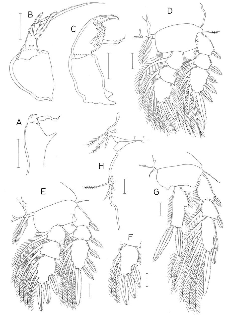
Female. Body (Fig. 10A) moderately broad, 1.35 mm long. Prosome 769×523 μm, tapering posteriorly. Cephalothorax 548 μm long, with dorsal suture line delimiting cephalosome and first pedigerous somite; posterolateral corners round. Fourth pedigerous somite small, only slightly wider than next somite. Urosome (Fig. 10B) 5-segmented. Fifth pedigerous somite 178 μm wide. Genital double-somite (Figs. 10B, 11H) 193 μm long and 178 μm in greatest width (measured at anterior 0.35 region); middle half of double-somite inflated laterally and invertedly trapezoidal; genital aperture large and locating in middle of double-somite. Three free abdominal somites 52×95, 40×80, and 78×100 μm, respectively. Anal somite with row of fine spinules along posteroventral margin. Caudal ramus (Fig. 10C) 82×43 μm (ratio 1.91 : 1) with 6 setae and with fine spinules on posteroventral margin; outer lateral seta naked and locating at distal third of outer margin; inner dorsal seta (seta VII) naked and articulated at base; 4 distal setae pinnate.
Rostrum (Fig. 10D) with posterior margin confluent with ventral surface of cephalothorax. Antennule (Fig. 10E) slender, 425 μm long, and 7-segmented; armature formula 4, 13, 6, 3, 4+aesthetasc, 2+aesthetasc, and 7+aesthetasc; one of 4 setae on first segment much larger than other 3. Antenna (Fig. 10F) slender and 4-segmented; segments gradually narrowing from proximal to distal; first segment unarmed; second segment 77 μm long, longer than first, with 1 small seta at distal fourth of inner margin; third segment 94 μm long, elongate, 5 times as long as wide, and armed with 2 setae, one of which at proximal third of inner margin and the other at inner distal corner; last segment short, 32 μm long, about 1/3 as long as third segment, and armed with 1 claw (58 μm long) and 1 small seta.
Labrum (Fig. 10G) unornamented, with broad posterior lobes. Mandible (Fig. 10H) with distinct proximal notch; inner margin bilobed and spinulose; distal lash thin and elongate; proximal region of outer margin protruding, with 5 slender digitiform processes. Maxillule (Fig. 10I) with 3 distal and 1 subdistal setae. Maxilla (Fig. 11A) with unarmed basal segment. Distal segment with large distal lash and 3 setae; distal lash perpendicular to axis of segment, with 1 proximal spine followed by row of spinules along distal margin and with smooth inner margin; inner seta spinulose, not longer than anterior seta; anterior seta flame-shaped distally; outer proximal seta minute. Maxilliped (Fig. 11B) 3-segmented; first segment unarmed; middle segment with 2 small inner setae of similar size; terminal segment with 1 spine and 1 seta, and terminating in sharp process.
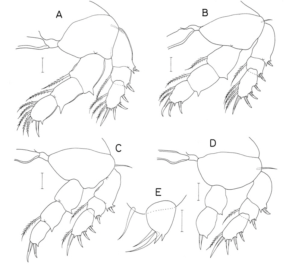
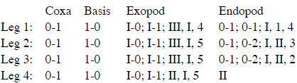
Legs 1-3 with 3-segmented rami (Fig. 11C, D). Outer spines on leg 1 exopod with serrate margins (Fig. 11C); other spines of legs with smooth or finely serrate margins. Outer seta on basis of legs 1-4 naked. Leg 4 (Fig. 11F) with 3-segmented exopod and 2-segmented endopod; inner coxal seta small and naked; distal endopodal segment 56×22 μm, its 2 distal spines 53 μm(inner) and 34 μm(outer). Armature formula of legs 1-4 as follows:

Leg 5 consisting of 1 pinnate dorsolateral seta on fifth pedigerous somite and free exopod (Fig. 10B); exopod (Fig. 11G) slender, 126×28 μm(ratio 4.50 : 1), with distinct inner proximal swelling, fine spinules on outer margin and 2 naked distal setae; slender distal region of exopod 16 μm wide; outer distal seta 74 μm long and inner one 104 μm long. Leg 6 represented by 2 small setae on genital operculum (Fig. 11H).
Male. Body (Fig. 12A) smaller than that of female, 1.05mm long. Prosome 585 μm long. Cephalothorax 404×400 μm. Urosome (Fig. 12B) 6-segmented. Fifth pedigerous somite 131 μm wide. Genital somite (Fig. 12B) large, globular, 212×217 μm. Abdomen gradually broadening from proximal to distal. Four abdominal somites small, 31×63, 34×67, 21×67, and 48×79 μm, respectively. Caudal ramus 67×38 μm (ratio 1.76 : 1).
Rostrum as in female. Antennule with 3 additional aesthetascs: 2 on second and 1 one fourth segments as indicated by dark dots in Fig. 10E. Antenna (Fig. 12C) shaped and armed as that of female, but with numerous, minute, additional spinules on inner margin of second and third segments.
Labrum, mandible, maxillule, and maxilla as in female. Maxilliped (Fig. 12D) consisting of 3 segments and terminal claw; first and third segments unarmed; second segment with 2 setae of similar length near middle of inner margin and longitudinal row of spinules along inner margin; terminal claw arched, as long as 3 combined proximal segments, with 1 minute and 1 large setae proximally and membrane along concave margin.
Leg 1 (Fig. 12E) with 2 spines and 4 setae on third endopodal segment. Legs 2-4 as in female. Exopod of leg 5 (Fig. 12F) small, nearly oval, 23×12 μm(ratio 1.92 : 1), with few minute spinules subdistally and armed distally with 1 serrate spine (15 μm long) and 1 naked seta; seta about 3 times as long as spine. Leg 6 represented by 1 pinnate seta and 1 minute, naked seta on genital operculum (Fig. 12B).
Etymology. The specific name
Remarks. In the genus
Material examined. 3♀♀ from a scleractinian coral, on coral reef in Kantary Bay, southern coast of Phuket Province, Thailand, 7˚48′14′′N, 98˚24′20′′E, 15 Dec 2012. Holotype (♀, CR00233158) and paratype (1♀, CR00233159) have been deposited in the MABIK, Seochon, Korea. Dissected paratype (1♀) is retained in the collection of the senior author.
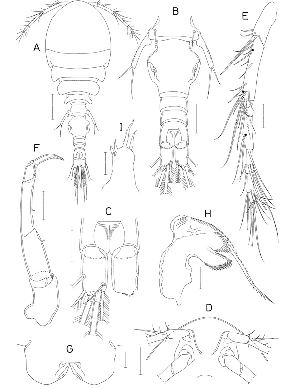
Female. Body (Fig. 13A, B) narrow, 1.23 mm long. Prosome 635 μm long and consisting of cephalothorax and second to fourth pedigerous somites. Greatest width of prosome 323 μm measured across second pedigerous somite. Cephalothorax 419×304 μm, distinctly longer than wide, dorsally elevated, 365 μm in dorsoventral depth, and dorsoventrally deeper than laterally wider, with nearly parallel lateral margins. Second pedigerous somite trapezoidal, with produced posterolateral corners. Urosome (Fig. 13C) 5-segmented. Fifth pedigerous somite 210 μm wide, much wider than next somite, with tapering lateral margins. Genital double-somite 177×144 μm, longer than wide, with slightly broadened anterior half and narrower posterior half; genital apertures locating dorsally at about anterior 0.3 of somite length. Three free abdominal somites 77×106, 67×90, and 48×90 μm, respectively; all of these somites smooth without any ornamentation. Caudal rami divergent (Fig. 13C); each ramus elongate, 205×33 μm (ratio 6.21 : 1), armed with 6 distal and subdistal, naked setae and ornamented with fine, sparse setules on outer and inner margins.
Rostrum (Fig. 13D) beak-like, as long as wide, with sharply pointed posterior apex. Antennule (Fig. 13E) slender, 263 μm long, and 7-segmented; armature formula 4, 13, 6, 3, 4+aesthetasc, 2+aesthetasc, and 7+aesthetasc; all of setae naked. Antenna (Fig. 13F) slender and 4-segmented; first segment with 1 inner distal seta; second segment elongate, about 127 μm long, with 1 small seta at distal third of inner margin; third segment short, 22 μm long with 3 setae, proximal one of them very small; terminal segment also short, 23 μm long, and armed with 2 minute setae and terminal claw; terminal claw 23 μm long, as long as segment and weakly curved.
Labrum (Fig. 13G) with broad median incision and broad posterior lobes fringed with membrane along posterior margin. Mandible (Fig. 13H) with deep proximal notch, strongly projected and bilobed inner margin (proximal lobe with 2 rows of spinules, and distal lobe with single row), and thin, spinulose distal lash; outer proximal region of blade strongly produced, with palm-like element bearing 5 or 6 blunt, digitiform processes. Maxillule (Fig. 13I) armed with 4 setae (2 distal and 2 lateral) and ornamented with spinules on distal region of outer margin; lateral proximal seta naked and other 3 setae spinulose. Maxilla (Fig. 14A) 2-segmented; basal segment unarmed but ornamented with several setules on outer subdistal region; distal segment armed with 2 setae: inner seta broad, foliaceous, with fine spinules and anterior seta naked and slender; outer proximal seta (seta III) absent; distal lash perpendicular to axis of segment, with 1 proximal spine followed by spinules along distal margin. Maxilliped (Fig. 14B) stout, curved and 3-segmented; first segment broadest and unarmed; middle segment with short inner margin and long, curved outer margin, and armed with 1 small and 1 longer, naked setae (longer one about 3 times as long as shorter one); terminal segment armed with 1 spinulose spine and 2 unequal, naked setae.
Legs 1 and 2 (Fig. 14C, D) and leg 3 with 3-segmented rami. Leg 4 (Fig. 14F) with 3-segmented exopod and 2-segmented endopod. Outer seta on basis of legs 1-3 small and naked; that of leg 4 also naked but large. All spines on leg 1 exopod spinulose and tipped with setule. Leg 3 similar to leg 2 but third endopodal segment (Fig. 14E) armed with 3 spines and 2 setae. Distal segment of leg 4 endopod 44×18 μm (ratio 2.44 : 1) and narrowing distally; its 2 terminal spines setiform, spinulose, and 26 μm(outer) and 45 μm long (inner). Armature formula of legs 1-4 as in preceding species.
Leg 5 consisting of 1 dorsolateral seta on fifth pedigerous somite and free exopod (Fig. 13C); exopod 120×35 μm(ratio 3.43 : 1), nearly rectangular, and spinulose on all surfaces, with small inner tubercle proximally and 2 naked distal setae (46 and 72 μm long, respectively). Leg 6 not discernible.
Male. Unknown.
Etymology. The specific name of the new species
Remarks. About one-third of described species of
Family Kelleriidae Humes and Boxshall, 1996Genus Kelleria Gurney, 1927
Material examined. 2♀♀ from invertebrate burrows on sands of coral reef in Kantary Bay, southern coast of Phuket Province, Thailand, 7˚48′14′′N, 98˚24′20′′E, 15 Dec 2012. Holotype (♀, CR00233160) has been deposited in the MABIK, Seocheon, Korea. Dissected paratype (1♀) is retained in the collection of the senior author.
Female. Body (Fig. 15A) narrow, 1.08 mm long. Prosome elliptical and 615×385 μm. Cephalothorax 438 μm long, with dorsal suture line delimiting cephalosome and first pedigerous somite. Second pedigerous somite with posterolateral corners tipped with short membranous fringe. Urosome (Fig. 15B) 5-segmented. Fifth pedigerous somite 137 μm wide. Genital double-somite 178×150 μm, with expanded anterior half and narrower posterior half; expanded anterior half with angular lateral apex, narrower posterior half gradually narrowing distally. Genital apertures located dorsally. Three free abdominal somites 61×75, 44×67, and 67×65 μm, respectively. Anal somite with minute spinules along posteroventral margin; anal aperture large. Caudal rami (Fig. 15C) directed backwards; each ramus 103×32 μm(ratio 3.22 : 1), with 6 setae and 2 large, conical ducts bearing terminal pore; outer lateral seta (seta II) naked and locating at 0.6 of outer margin; other 5 setae pinnate; inner dorsal seta articulated at base.
Rostrum (Fig. 15D) strongly tapering and wider than long. Antennule (Fig. 15E) 347 μm long, gradually narrowing distally, and 7-segmented; armature formula 4, 13, 6, 3, 4+aesthetasc, 2+aesthetasc, and 7+aesthetasc; setae generally long and all of them naked. Antenna (Fig. 15F) 4-segmented; first segment with 1 inner distal seta; second segment similar in length to first segment, with fine spinules along outer margin and 1 subdistal seta on inner margin; third segment short and armed with 2 setae and 1 setiform claw; terminal segment 80×22 μm(ratio 3.64 : 1), armed with 2 slender claws and 5 long setae, and ornamented with fine spinules on outer margin.
Labrum (Fig. 16A) with large posterior lobes and deep median incision; each lobe slightly broadened distally, with broad membrane on proximal half of inner margin. Mandible (Fig. 15G) with shallow proximal notch; inner margin oblique, not distinctly demarcated from distal lash, with about 9 large spines, proximal 3 of them being slender; convex margin with small knob tipped with 4 or 5 minute spinules, and followed by about 17 thick teeth (these teeth becoming gradually smaller from proximal to distal); distal lash spinulose on both margins. Paragnath (Fig. 15H) tapering in distal half, with 1 small, dentiform process laterally and setules subdistally. Maxillule (Fig. 15I) with serrate membrane on distal region of outer margin and armed with 1 lateral, 1 subdistal, and 2 large distal setae; lateral seta naked and articulated at base; other 3 setae weakly pinnate unilaterally. Maxilla (Fig. 16B) 2-segmented; basal segment with 1 small, tooth-like tubercle on outer side; distal segment armed with large inner spine, slender, naked anterior seta, and minute outer, proximal seta, and terminating in straight, rigid distal lash; distal lash almost perpendicular to proximal part of segment, articulated near middle, and armed with large teeth along distal margin (proximal third and fourth teeth smaller than other teeth and distal 3 teeth locating distal to articulation); inner spine extending slightly over distal lash, with 7 spinules on distal margin and about 10 spinules on proximal margin; spinules on both margins of inner spine becoming larger from proximal to distal. Maxilliped (Fig. 16C) 3-segmented. First segment longest but unramed. Second segment with protruded inner margin bearing 2 large setae; proximal seta with about 10 spinules a proximal part of proximal margin and row of setules along distal margin; distal seta setulose along both margins. Terminal segment terminating in elongate setiform process and armed with 1 pinnate seta and 2 smaller naked setae.
Legs 1 and 2 (Fig. 16D, E) and leg 3 with 3-segmented exopod and endopod. Leg 4 (Fig. 16F) with 3-segmented exopod and 1-segmented endopod. Leg 3 similar to leg 2, but its third endopodal segment armed with 3 spines and 2 setae (formula I, II, 2). Outer seta on basis of legs 2 and 3 small and naked; those of legs 1 and 4 larger and pinnate. Inner distal corner of basis of leg 2 weakly bilobed. Leg 4 endopod 67×21 μm (ratio 3.19 : 1), with small dentiform process at proximal 0.4 of inner margin; inner seta not extending to distal end of segment; 2 distal spines large, 65 μm (inner) and 46 μm long (outer); both outer and inner distal corners pointed. Armature formula of legs 1-4 as follows:

Leg 5 consisting of 1 pinnate dorsolateral seta on fifth pedigerous somite and free exopod; exopod (Fig. 16G) approximately 82×47 μm, with strongly projected inner margin tipped with digitiform process and 2 naked distal setae. Leg 6 (Fig. 16H) probably represented by 1 seta, 1 conical process, and laterally positioned, seta-tipped knob in genital region.
Male. Unknown.Etymology. The specific name
Remarks. The exopod of leg 5 in
In
In
Family Pseudanthessiidae Humes and Stock, 1972Genus Pseudanthessius Claus, 1889
Pseudanthessius stenosus n. sp. (Figs. 17, 18)
Material examined. 2♀♀ from invertebrate burrows on sands of coral reef in Kantary Bay, southern coast of Phuket Province, Thailand, 7˚48′14′′N, 98˚24′20′′E, 15 Dec 2012. Holotype (♀, CR00233161) has been deposited in the MABIK, Seocheon, Korea. Dissected paratype (♀) is retained in the collection of the senior author.
Female. Body (Fig. 17A) slender, 1.25 mm long. Prosome 704 μm long. Cephalothorax 473×342 μm, distinctly longer than wide and divided by dorsal suture line into cephalosome and first pedigerous somite. Urosome (Fig. 17B) 5-segmented. Fifth pedigerous somite 112 μm wide and laterally tapering. Genital double-somite 162×106 μm(ratio 1.53 : 1), consisting of moderately expanded anterior and narrower posterior parts; genital aperture locating laterally near halfway of lateral margin. Three free abdominal somites 53×57, 32×49, and 66×42 μm, respectively. Anal somite distinctly longer than wide and unornamented, without spinules or setules. Caudal rami close to each other and elongate; each ramus 170×17 μm, 10 times as long as wide, slightly broadened at distal region, and armed with 6 setae; outer lateral seta located at 0.73 of ramus length; all of setae pinnate.
Rostrum (Fig. 17C) tapering, as long as its basal width, with round posterior apex. Antennule (Fig. 17D) 320 μm long and 7-segmented; armature formula 4, 13, 6, 3, 4+aesthetasc, 2+aesthetasc, and 7+aesthetasc; all of setae naked and one of subdistal setae on second segment distinctly larger than others; terminal segment short, half as long as penultimate segment. Antenna (Fig. 17E) 4-segmented; first segment with 1 inner distal seta; second segment distinctly longer than first, with fine spinules on distal half of outer margin and 1 subdistal seta on inner margin; third segment with 1 slender claw and 2 setae; terminal segment 3.1 times as long as wide and armed with 4 slender claws (2 middle ones shorter than other 2) and 3 setae.
Labrum (Fig. 17F) with elongate, divergent, tapering posterior lobes fringed with narrow membrane along posterior margin and with deep median incision. Mandible (Fig. 17G) consisting of short proximal part and tapering blade, with deep but narrow proximal notch; inner margin not differentiated; convex outer side with large, well-sclerotized, distally-directed process; inner margin of blade serrate; outer convex margin of blade smooth. Maxillule (Fig. 17H) armed with 1 lateral, setiform process and 3 distal setae, median one of latters larger than other 2, with several minute spinules distally; outermost one of distal setae broad, process-like, with articulation at base. Maxilla (Fig. 18A) 2-segmented; basal segment unarmed; distal segment with large inner spine bearing membranous fringe along inner (proximal) margin and spinules along distal half of outer (distal) margin, spiniform anterior seta tipped with 1 seta and 1 small dentiform process, and terminating in long, thin, spinulose distal lash; distal region of second segment near base of distal lash with 1 large, tooth-like process and 3 small denticles; outer proximal seta (seta III) absent. Maxilliped (Fig. 18B) 3-segmented; first segment unarmed; second segment with slightly inflated inner margin, 2 unequal setae, and several oblique rows of minute spinules; terminal segment tapering, distally forming pointed claw, with 2 setae proximally.
Legs 1 and 2 (Fig. 18C, D) and leg 3 with 3-segmented rami. Leg 4 (Fig. 18F) with 3-segmented exopod and 1-segmented endopod. Leg 3 similar to leg 2, but its third endopodal segment armed with 3 spines and 2 setae (Fig. 18E). Outer seta on basis of legs 1-4 thin and naked. Inner coxal seta of legs 1-3 large and pinnate, but that of leg 4 small and naked. Leg 4 with conical process at inner distal corner of basis; endopod 69×23 μm(ratio 3.0 : 1), with inflated proximal third of outer margin; both lateral margins setulose; 2 distal setae large, 64 μm(inner) and 51 μm(outer). Armature formula of legs 1-4 as follows:
Leg 5 (Fig. 18G) represented by produced lateral region of fifth pedigerous somite bearing pinnate proximal seta and 2 distal elements (short spine of 18 μm long and longer, naked seta). Leg 6 represented by 2 setae on genital operculum (Fig. 18G).
Male. Unknown.
Etymology. The specific name
Remarks. One of diagnostic features of
Pseudanthessius phuketensis n. sp. (Figs. 19, 20)
Material examined. 1♀ (holotype) from invertebrate burrows on sands of coral reef in Kantary Bay, southern coast of Phuket Province, Thailand, 7˚48′14′′N, 98˚24′20′′E, 15 Dec 2012. Holotype (CR00233162, dissected and mounted on a glass slide) has been deposited in the MABIK, Seocheon, Korea.
Female. Body (Fig. 19A) slender, 942 μm long. Prosome 581×308 μm. Cephalothorax 385 μm long, with dorsal suture line delimiting cephalosome and first pedigerous somite. Urosome (Fig. 19B) 5-segmented. Fifth pedigerous somite 88 μm wide and laterally tapering. Genital double-somite 134×81 μm (ratio 1.65 : 1), with slightly expanded anterior 0.6 and narrower, weakly tapering posterior 0.4. Genital aperture locating laterally near midlength of somite. Three free abdominal somites 35×43, 25×40, and 46×38 μm, respectively. Anal somite (Fig. 19B) with fine spinules along posteroventral margin. Caudal rami (Fig. 19C) straight, close to each other; each ramus 88×17 μm, 5.18 times as long as wide; outer lateral seta naked and locating at 0.69 of ramus length; inner dorsal seta (seta VII) naked and lacking basal articulation; other 4 setae pinnate.
Rostrum (Fig. 19D) strongly tapering, as long as its basal width, with round posterior apex. Antennule (Fig. 19F) 248 μm long and 7-segmented; armature formula 4, 13, 6, 3, 4+ aesthetasc, 2+aesthetasc, and 7+aesthetasc; all of setae naked; one of distal setae on second segment very large. Antenna (Fig. 19F) 4-segmented; first segment with 1 inner distal seta; second segment distinctly longer than first, with fine spinules on outer margin and 1 subdistal seta on inner margin; third segment with 1 claw and 2 setae; terminal segment 3.0 times as long as wide and armed with 4 claws (second outer one broader and stronger than other 3 spines) and 3 setae.
Labrum (Fig. 19G) very similar to that of preceding species. Mandible (Fig. 19H) consisting of short proximal part and tapering blade, with deep proximal notch; inner margin not differentiated; convex outer side with claw-like process; inner margin of blade serrate and outer convex margin of balde smooth; distal lash short. Maxillule (Fig. 20A) with 1 digitiform process on lateral margin and 3 naked, unequal, distal setae. Maxilla (Fig. 20B) similar to that of preceding species; inner spine of distal segment serrate on distal part of distal margin; anterior seta bifurcate; distal part of segment near base of distal lash with 2 tooth-like processes. Maxilliped (Fig. 20C) 3-segmented; first segment longest but unarmed; second segment with 2 very unequal setae and scattered spinules on inner side; terminal segment narrow and tapering, with 1 small proximal seta and several spinules on distal region of distal margin.
Legs 1 and 2 (Fig. 20D, E) and leg 3 segmented and armed as in preceding species. Outer seta on basis of leg 1 weakly pinnate, and those of legs 2-4 naked. Terminal segment (Fig. 20F) of leg 3 endopod with 3 spines and 2 setae as in preceding species. Leg 4 (Fig. 20G) endopod 63×25 μm (ratio 2.52 : 1), with blunt process on outer margin; 2 distal spines 62 μm (inner) and 46 μm long (outer). Armature formula of legs 1-4 as in preceding species.
Leg 5 (Fig. 20H) represented by produced lateral region of fifth pedigerous somite, bearing pinnate proximal seta and 2 distal elements (short, naked spiniform seta and pinnate seta). Leg 6 (Fig. 20H) represented by 2 setae (pinnate anterior seta and smaller, naked, undulated posterior seta) on genital operculum.
Male. Unknown.
Etymology. The specific name is derived from the type locality of the new species, Phuket.
Remarks. In a key to species of
Family Rhynchomolgidae Humes and Stock, 1972Genus Critomolgus Humes and Stock, 1983
Critomolgus gemmatus (Humes, 1964)
Material examined. 2♀♀, 1♂ from 1
Remarks. In the female the body length is 1.27 mm. The caudal ramus is 58×42 μm(ratio 1.38 : 1). Leg 5 is exopod 131×49 μm(ratio 2.67 : 1) and bears with numerous minute scales on the outer surface. The specimens from Phuket showed no other significant differences from the original description of this species.
Genus Doridicola Leydig, 1853
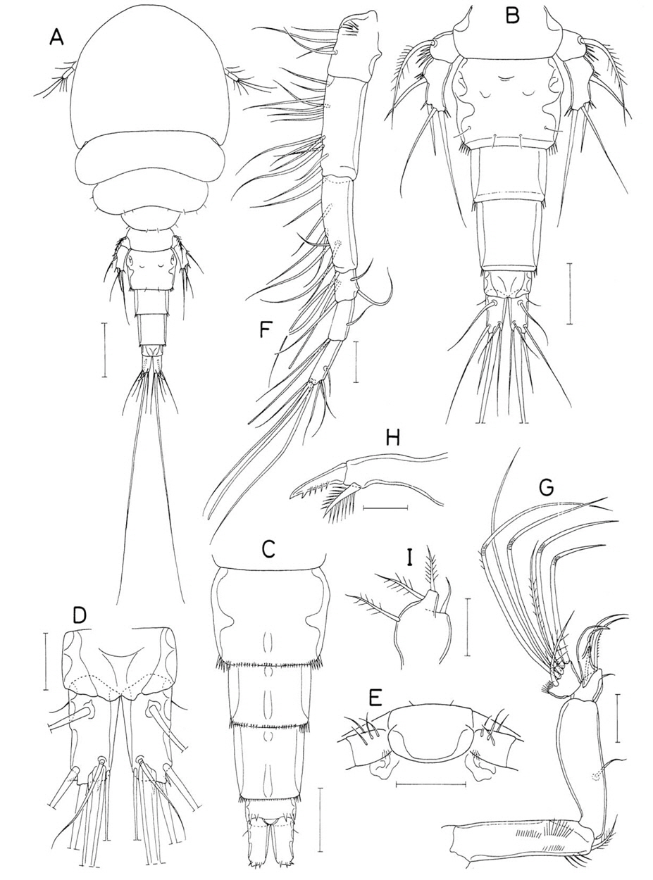
Doridicola pattayensis n. sp. (Figs. 21-23)
Material examined. 4♀♀, 3♂♂ from washings of the zoanthid coral
Female. Body (Fig. 21A) narrow, 1.00 mm long (other 2 measured females 0.83 and 0.84 mm, respectively). Prosome 737×458 μm. Cephalothorax 455 μm long and divided dorsally by suture line into cephalosome and first pedigerous somite. Second pedigerous somite with slightly projected, angular posterolateral corners. Urosome (Fig. 21B) 5-segmented, short and stout, about 0.36 times as long as prosome. Fifth pedigerous somite 112 μm wide. Genital double-somite 115×115 μm, as long as wide, and consisting of expanded anterior 0.75 and narrower posterior 0.35; genital aperture locating at 0.55 of somite length. Three free abdominal somites 19×66, 16×62, and 26×59 μm, respectively. Anal somite unornamented. Caudal ramus (Fig. 21C) very short, 27×25 μm(ratio 1.08 : 1) with 6 setae and with fine spinules on posteroventral margin; outer lateral seta naked; outermost distal seta (seta III) pinnate only along inner margin; other 4 setae bilaterally pinnate.
Rostrum hemicircular, with round posterior apex. Antennule (Fig. 21D) slender, 336 μm long, and 7-segmented; armature formula 4, 13, 6, 3, 4+aesthetasc, 2+aesthetasc, and 7+aesthetasc; all of setae naked. Antenna (Fig. 21E) 4- segmented; first segment slightly curved, with 1 seta at inner distal corner; second segment as long as first, with 1 seta on inner margin; short third segment with 1 slender claw and 2 setae; terminal segment 66×17.5 μm, about 3.8 times as long as wide, with 2 claws of similar length (slender inner one 45 μm long and outer one slightly broader and 44 μm long) and 5 setae, one of latters inserting to base of inner spine.
Labrum (Fig. 21F) with broad posterior lobes and deep median incision; inner distal corner of lobes pointed. Mandible (Fig. 21G) with broad proximal notch; inner margin distinct, perpendicular to distal lash, and densely spinulose; outer proximal region of blade projected, with row of small denticle; cutting edge finely serrate; distal lash elongate and spinulose on both margins. Paragnath (Fig. 21H) lobate, bearing setules distally. Maxillule (Fig. 21I) armed with 1 small, setiform lateral element and 3 distal, naked setae. Maxilla (Fig. 22A) with unarmed basal segment; distal segment with minute outer proximal seta, weakly spinulose anterior seta, 5-spinules-bearing inner spine, and elongate distal lash; distal lash with 6 proximal teeth followed by fine spinules on outer margin. Maxilliped (Fig. 22B) 3-segmented; first segment with several, scattered spinules; second segment slightly swollen, with 1 small and 1 extremely large setae, latter 104 μm long, about 20 times as long as shorter seta, and unilaterally spinulose; terminal segment (Fig. 22C) armed with 1 naked seta and 1 spinulose spine and terminating in spinulose, spiniform process.
Legs 1 and 2 (Fig. 22D, E) and leg 3 with 3-segmented rami. Leg 4 (Fig. 22G) with 3-segmented exopod and 2-segmented endopod. Leg 3 similar to leg 2, but its third endopodal segment (Fig. 22F) with 3 spines and 2 setae. Inner coxal seta of leg 4 small and naked. Distal segment of leg 4 endopod 59×21 μm, about 2.8 times as long as wide, distally with 3 pointed processes and 2 unequal spines; outer spine 27 μm long and inner one 55 μm long. Armature formula of legs 1-4 as follows:

Leg 5 consisting of 1 pinnate dorslateral seta on fifth pedigerous somite and free exopod; exopod (Fig. 22H) 81×29 μm, 2.79 times as long as its greatest width, with prominent inner proximal swelling, scattered spinules on outer surface and 2 distal setae; outer seta 41 μm long, and inner seta 66 μm long, with thin membranous fringe along outer margin. Leg 6 represented by 1 small seta, 1 small spine and 1 dentiform process on genital operculum (Fig. 22I).
Male. Body (Fig. 23A) narrower than that of female and 775 μm long (other 2 examined specimens 744 and 697 μm, respectively). Prosome 495×277 μm. Cephalothorax 313 μm long; dorsal suture line between cephalosome and first pedigerous somite very faint and limited to lateral sides. Urosome (Fig. 23B) 6-segmented. Fifth pedigerous somite 71 μm wide. Genital somite large, 146×137 μm. Four abdominal somites small, 17×43, 14×45, 11×43, and 15×47 μm, respectively. Caudal ramus (Fig. 23B) 18×20 μm, slightly wider than long.
Rostrum as in female. Antennule with 3 additional aesthetascs, 2 on second and 1 on fourth segments, as indicated by dark dots in Fig. 21D. Antenna (Fig. 23C) with spinules on inner margin of 2 proximal segments and on outer margin of second segment; setae on 2 proximal segments finely spinulose along their inner margin.
Labrum, mandible, paragnath, maxillule, and maxilla as in female. Maxilliped (Fig. 23D) consisting of 3 segments and terminal claw; first segment unarmed; second segment slightly expanded around 0.6 region of segment length, with 2 small setae near 0.4 region and 2 longitudinal rows of spinules, one of latters limited to dital half of segment; small third segment umarmed; terminal claw elongate, proximally with 1 seta and 1 setule.
Leg 1 (Fig. 23E) armed with 2 spines and 4 setae on third endopodal segment, otherwise same as that of female. Legs 2-4 as in female.
Leg 5 exopod (Fig. 23F) small, 36×11 μm, 3.27 times as long as wide, slightly curved, and armed distally with 1 spinulose spine and 1 naked seta. Leg 6 (Fig. 23B) represented by 2 thin, unequal setae on genital operculum.
Etymology. The specific name is derived from Pattaya, the type locality of the new species.
Remarks. Ten species of the genus
The possession by the new species of the two greatly unequal setae on the second segment of the female maxilliped is remarkable; one of these setae is extremely enlarged, about 20 times as long as the other. Similar form of the maxilliped is possessed by a few species, such as
Species of
Doridicola inaequalis (Humes and Ho, 1966)
Material examined. 26♀♀, 7♂♂ from washings of the zoanthid coral
Female. The mean body length is 938 μm (815-1,023 μm), based on 10 specimens. The body length of dissected specimen is 1.01 mm. The cepahlothorax is 446×525 μm. The genital double-somite is 131×120 μm. The caudal ramus is 48×32 μm(ratio 1.50 : 1). Two setae on the second segment of maxilliped are equal in size; the terminal segment of the maxilliped bears 1 spiniform process, 1 small spine and 2 setae. The outer seta on the basis of legs 1-4 are weakly pinnate. Leg 5 exopod is 97×27 μm(ratio 3.57 : 1).
Male. The mean body length is 815 μm(777-857 μm), based on 6 specimens. Leg 5 exopod is 28×13 μm (ratio 2.15 : 1) and armed distally with 1 spine (18 μm) and 1 seta (40 μm).
Remarks. This species was originally recorded as an associate of species of
Doridicola cuspis (Humes, 1964)
Material examined. 3♀♀, 2♂♂ from 1
Remarks. The body length of a dissected female is 1.52mm, not significantly different from those of type specimens of Humes (1964). This species is characteristic in having greatly expanded cephalothorax which is much wider than the second pedigerous somite.
Genus Indomolgus Humes and Ho, 1966
Indomolgus brevisetosus Humes and Ho, 1966
Material examined. 1♂ from washings of the zoanthid coral
Remarks. The body length of the observed single male is 2.33 mm. The caudal ramus is 246×81 μm (ratio 3.04 : 1). Leg 5 exopod is 60×33 μm (ratio 1.82 : 1), a measurement quite different from that of type specimens of Humes and Ho (1966), probably due to the difference of viewing angles. This is the first record on this species outside of Madagascar.
Genus Lambanetes Humes, 1982
Lambanetes stichodactylae Humes, 1982 (Figs. 24, 25)
Material examined. 3♀♀, 10♂♂ from
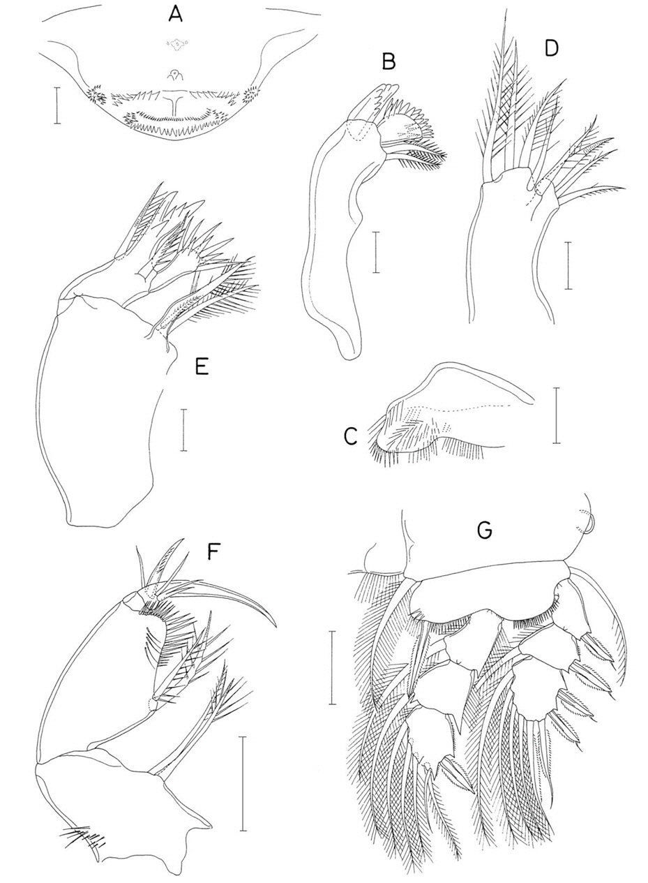
Female. Body tapering (Fig. 24B), with dorsally directed urosome (Fig. 24A). Body length 1.35 mm. Greatest width 656 μm. Exoskeleton moderately hard. Prosome 531 μm long. All prosomal somites with rounded lateral margins. Cephalothorax 451 μm long, 1.45 times as wide as long. Second to fourth pedigerous somites 574, 503, and 365 μm wide, respectively. Fourth pedigerous somite with posterolateral protrusions. Urosome (Fig. 24C) 5-segmented. Fifth pedigerous somite 267 μm wide. Genital double-somite (Fig. 24C) 172×263 μm, with convex lateral margins; genital aperture circular and located dorsally, slightly anterior to midway. Three free abdominal somites 96×171, 77×147, and 77×131 μm, respectively. Anal region with 2 baloonlike swellings (Fig. 24C). Caudal ramus (Fig. 24D) 47×38 μm(ratio 1.24 : 1), strongly tapering, with 6 setae; setae II-IV spiniform and 27, 23, and 51 μm, respectively; seta V largest, 181 μm long, 3.85 times as long as caudal ramus; all setae naked.
Rostrum absent. Antennule (Fig. 24E) 7-segmented, with thick, crowded setae; armature formula 4, 13, 6, 2, 4+aesthetasc, 2+aesthetasc, and 7+aesthetasc; all setae naked. Antenna (Fig. 24F) 3-segmented; first and second segments broad, each with 1 seta on inner margin; third segment much narrower than 2 proximal segments, about 2.3 times as long as wide, proximally with 3 setae on inner margin and distally with 4 small setae and 2 strongly curved claws, proximal one of latters much smaller than terminal one and directed laterally.
Labrum with tapering pasteromedian process. Mandible (Fig. 24G) with narrow blade; convex side with large, serrate spine bearing 2 rows of spinules; convex margin distal to spine with 5 denticles followed by row of spinules; concave margin also spinulose. Maxillule (Fig. 24H) lobate, with 2 distal spinulose setae. Maxilla (Fig. 25A) 2-segmented; basal segment very broad and unarmed; distal segment terminating in acute process bearing 3 broad spinules along proximal part of convex margin, armed with 2 setae (small setae I and III) and large anterior spine (originally seta II) bearing about 10 broad spinules along margins. Maxilliped (Fig. 25B) incompletely 2-segmented; basal segment leaf-like, with 3 unequal setae on inner side; small distal segment armed with 1 spine and 1 seta, and terminating in spiniform process.
Legs 1-3 (Fig. 25C-E) with 3-segmented rami; leg 4 (Fig. 25F) with 3-segmented exopod and 2-segmented endopod. Inner seta on coxa present only on leg 3. Distal segment of leg 4 endopod 38×17 μm; its 2 distal spines 42 μm (inner) and 17 μm(outer). Armature formula of legs 1-4 as follows:
Leg 5 (Fig. 25G) locating ventrally on somite and consisting of 1 outer lateral seta on somite and free exopod; exopod 65×22 μm (length measured to tip of distal process), about 3 times as long as wide, terminating in 2 unequal, spiniform processes, ornamented with 3 transverse rows of spinules on outer surface, and armed distally with 2 weakly spinulose setae; outer seta 45 μm long and inner seta 65 μm long. Leg 6 represented by 2 minute setae on genital operculum (Fig. 25H).
Male. Generally as in original description by Humes (1982).
Remarks. Humes (1982) originally described this species, based only on the male. Therefore, the above description is the new record on the female of
Lambanetes mollis n. sp. (Figs. 26-28)
Material examined. 1♀, 7♂♂ from
Female. Body (Fig. 26A) inflated and flexed, with soft exoskeleton. Length 1.82 mm. Prosome 1.10 mm long. Cephalothorax wider than long. Urosome directed dorsally and 5- segmented. Genital double-somite 170×269 μm, 1.58 times as wide as long, with slightly convex lateral margins and row of minute spinules along posteroventral and posterolateral margins; genital aperture circular and locating dorsally at anterior third of somite length (Fig. 26B). Three free abdominal somites 115×195, 103×167, and 103×141 μm, respectively. First and second free abdominal somites with row of minute spinules along posterior margin. Anal somite (Fig. 26B) with well-developed anal operculum and posterior inflations. Caudal ramus (Fig. 26C) 46×38 μm, 1.21 times as long as wide, with 6 setae; setae II-VII 17, 19, 21, 66, 28, and 25 μm long, respectively; setae II-IV spiniform; seta V less than twice as long as ramus; all setae naked.
Rostrum absent. Antennule (Fig. 26D) 6-segmented; armature formula 4, 13, 6, 2, 4+aesthetasc, and 9+2 aesthetascs; all setae naked. Antenna (Fig. 26E) 3-segmented; first and second segments each with 1 small seta on inner margin; third segment about 1.75 times as long as wide and armed proximally with 3 setae on inner margin and distally with 5 setae and 2 claws; proximal one of claws small and directed laterally; terminal claw much larger and strongly curved.
Labrum (Fig. 26F) weak but with well-sclerotized, tapering posteromedian process. Mandible (Fig. 26G) narrow; spine on outer, convex side trifurcate; convex and concave margins serrate; several spinules present on dorsal surface near proximal region of distal lash. Maxillule (Fig. 26H) with 2 naked distal setae. Maxilla (Fig. 26I) 2-segmented; basal segment unarmed; distal segment terminating in process bearing 3 spines, and proximally with 2 small setae and 1 spine, the latter with 3 spinules (1 proximal and 2 distal). Maxilliped (Fig. 26J) unsegmented, with trace of segmentation near middle, and armed with 2 very unequal setae (one of them minute and transparent) near distal third and distally with 2 spiniform processes and 1 small seta.
Legs 1-3 (Fig. 27A-C) with 3-segmented rami. Leg 4 (Fig. 27D) with 3-segmented exopod and 2-segmented endopod. All of these legs without inner seta on coxa. First endopodal segment of legs 3 and 4 unarmed. Distal segment of leg 4 endopod 35×23 μm. Armature formula of legs 1-4 as follows:

Leg 5 (Fig. 27E) consisting of 1 outer seta on fifth pedigerous somite and free exopod; exopod 41×27 μm, 1.52 times as long as wide, terminating in 2 dentiform processes, with 2 naked distal setae. Leg 6 not seen,
Male. Body (Fig. 28A) straight and not inflated. Mean body length 0.95 mm (0.85-1.01 mm), based on 8 specimens. Length of dissected specimen 1.00 mm. Prosome 654 μm long. Cephalothorax 388×462 μm, with angular posterolateral corners. Second to fourth pedigerous somites 381, 343, and 300 μm wide, respectively. Third and fourth pedigerous somites with rounded, posteriorly protruded posterolateral corners. Urosome (Fig. 28B) 6-segmented. Fifth pedigerous somite 238 μm wide. Genital somite (Fig. 28B) 117×250 μm, about twice as wide as long. Four abdominal somites 52× 122, 54×106, 38×90, and 52×81 μm, respectively. First to third abdominal somites with row of minute spinules along posterior border. Anal somite (Fig. 28B) with row of minute spinules near base of caudal rami. Caudal ramus (Fig. 28C) 35×25 μm, 1.40 times as long as wide; setae II-VII 18, 19, 38, 86, 38, and 27 μm, respectively; longest seta V about 2.5 times as long as ramus.
Rostrum absent. Antennule, antenna, labrum, mandible, and maxillule as in female. Maxilla (Fig. 28D) similar to that of female, but inner seta (seta I) spiniform, with several spinules; distal process with 3 or 4 spines (probability about 1 : 1); anterior spine with 3 or 4 (usually 3) spinules. Maxilliped (Fig. 28E) consisting of 3 segments and terminal claw; first segment longest but unarmed; second segment with projected inner proximal corner, 2 inner setae slightly distal to midlength of segment, and small scales along inner margin; small third segment unarmed; terminal claw extending to proximal border of second segment, strongly curved in distal part, with 2 proximal setae.
Leg 1 (Fig. 28F) with third endopodal segment armed with 2 spines and 4 setae (armature formula I, I, 4), otherwise same as that of female. Legs 2-4 as in female.
Leg 5 (Fig. 28B) exopod 25×17 μm, 1.47 times as long as wide. Three setae on leg 5 feebly pinnate and longer than exopod. Leg 6 (Fig. 28B) represented by 1 dentiform process and 2 weakly pinnate setae on genital operculum.
Etymology. The specific name
Remarks. Three known species of
Besides the difference in leg armature, other appendages of
Family Synapticolidae Humes and Boxshall, 1996Genus Scambicornus Heegaard, 1944
Scambicornus affinis Ho, 1982
Material examined. 70♀♀, 35♂♂ from sea cucumbers on coral reef in Kantary Bay, southern coast of Phuket Province, Thailand, 7˚48′14′′N, 98˚24′20′′E, 15 Dec 2012.
Remarks.