검색 전체 메뉴
PDF
맨 위로
OA 학술지
Akashiwo sanguinea (Dinophyceae) extruding mucous from pores on the cell surface
  • 비영리 CC BY-NC
  • 비영리 CC BY-NC
ABSTRACT
Akashiwo sanguinea (Dinophyceae) extruding mucous from pores on the cell surface
KEYWORD
Akashiwo sanguinea , cyst , dinoflagellate , HAB , mucocyst , reproduction , trichocyst
  • INTRODUCTION

    Akashiwo sanguinea (Hirasaka) G. Hansen et Moestrup was first described in 1922 from water samples collected in Kozusa-ura Gokasho Bay, Japan (Hirasaka 1922). A. sanguinea is cosmopolitan in its distribution and has been observed to form blooms in coastal ecosystems around the world (Kim et al. 1993, Voltolina 1993, Horner et al. 1997, Lee et al. 2001, Lu and Hodgkiss 2004, Feyzioğlu and Öğüt 2006), including the Gulf of Mexico and the Atlantic coast of North America (Robichaux et al. 1998, Steidinger et al. 1998, Badylak and Phlips 2004, Badylak et al. 2007, Phlips et al. 2011, 2012). The cosmopolitan distribution of A. sanguinea is in part attributable to its eurythermal and euryhaline character (Matsubara et al. 2007, Badylak et al. 2014).

    Blooms of A. sanguinea have been associated with mass mortalities of invertebrates and fish in various regions of the world (Cardwell et al. 1979, Harper and Guillen 1989, Schumway 1990, Kahru et al. 2004). In the Gulf of Mexico, blooms of A. sanguinea were the suspected cause of fish kills and marine mammal stranding in the mid- 1990’s (Robichaux et al. 1998, Steidinger et al. 1998). One ecologically important feature of dinoflagellate blooms which can contribute to harmful effects is the production of mucous. For example, the smothering of fish by Cochlodinium polykrikoides blooms containing dense concentrations of mucous has been observed in Korea (Kim et al. 2000). Thin horizontal layers of concentrated A. sanguinea densities have been observed in blooms in Monterey Bay, California, USA (Rines et al. 2010). Zooplanktons were found just above and below the thin layer but not within the layer, suggesting that high concentrations of A. sanguinea can negatively influence the distribution of zooplankton.

    While A. sanguinea has been observed in association with mucous (Voltolina 1993, Robichaux et al. 1998), there have not been clear observations of this phenomenon at the detailed cellular level. In this study we present previously undocumented observations of A. sanguinea extruding mucous substances from pores on the cell surface. The A. sanguinea cells were observed in water samples collected in the Caloosahatchee Estuary, Florida during a bloom event.

    MATERIALS AND METHODS

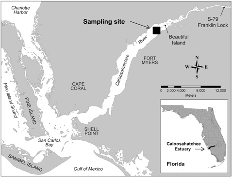
    A. sanguinea cells observed in this study were from water samples collected in the Caloosahatchee Estuary, Florida during a bloom event. Caloosahatchee Estuary is on the subtropical southwest Florida coastline adjacent to the Gulf of Mexico. The Caloosahatchee Estuary is subject to wide salinity variations due to fresh water releases from Lake Okeechobee, connected to the estuary through a flow-regulated canal.

    Salinity measurements were taken with a salinity refractometer (Fisher Scientific, Pittsburgh, PA, USA). Water was collected with a sample pole that allowed the even collection of water from the surface to 0.1 m from the bottom of the water column. Aliquots of well-mixed sample water were preserved with Lugol fixative (i.e., 1 mL concentrate added per 100 mL to yield strong tea color) and stored in an amber bottle for light microscopy and another aliquot was preserved with buffered glutaraldehyde (2% final concentration) for scanning electron microscopy (SEM) observation.

    Lugol preserved samples were used to enumerate A. sanguinea cells using the Utermöhl settling method (Utermöhl 1958). In a separate treatment, aliquots of glutaraldehyde preserved water were filtered onto 8 μm pore size nylon membrane filters for SEM analysis, rinsed three times with deionized water, dehydrated in a graded ethanol series and critical point dried (Baltec CPD; Leica Microsystems, Bannockburn, IL, USA). Each filter was placed on aluminum specimen mounts and sputter coated with Au/Pd (Deskll; Denton Vacuum, Moorestown, NJ, USA). High resolution digital micrographs were acquired with a field emission scanning electron microscope (S-4000; Hitachi High Technologies America Inc., Schaumberg, IL, USA).

    RESULTS AND DISCUSSION

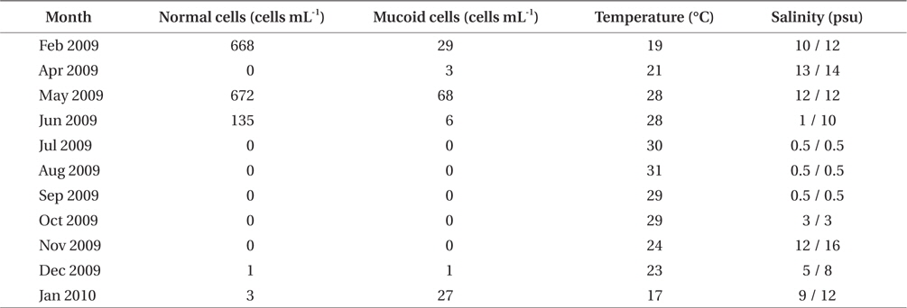
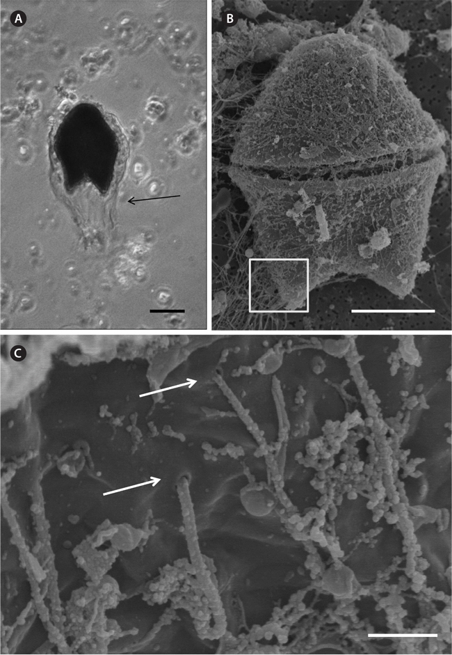
    A bloom of A. sanguinea was observed in water samples collected in the upper Caloosahatchee Estuary in February and May of 2009 (Table 1). Salinities during the sample collection were less than as 14 psu. In May A. sanguinea cell densities were 672 cells mL-1. A. sanguinea represented over 90% of total phytoplankton biovolume during the bloom event (Badylak et al. 2014). The sample also contained 68 cells mL-1 of A. sanguinea cells with mucoid substances surrounding the cells (Fig. 1). SEM investigations provided a closer view of A. sanguinea and revealed granular strands of mucous covering the cell surface (Fig. 2A). The SEM images also revealed that the mucous strands were secreted from thecal pores on the cell surface (Fig. 2B). The presence of thecal pores has been observed in other dinoflagellate species and has been linked to the excretion of mucilage (Honsell et al. 2013). It is possible to hypothesize concerning the origin of the mucous strands. One cellular component of dinoflagellates associated with the excretion of extracellular material is extrusomes (Dodge 1973). Another feature is the mucocyst, a type of ejectile organelle found in dinoflagellates that contains fine granular material that can be secreted onto the outer surface of cells (Dodge 1987). There has been no previous description of A. sanguinea possessing mucocysts, but the granular strands extruding from A. sanguinea pores observed in this study could be associated with mucocysts. Transmission electron microscopy is needed to confirm this hypothesis, and to identify the type of mucocyst present, since more than one type of mucocyst exists within dinoflagellates (Zhou and Fritz 1993). It is also possible that the mucous comes from a complex of multiple extrusomes in the cells (Hoppenrath and Leander 2008).

    [Table 1.] Cell densities of normal and mucous covered Akashiwo sanguinea cells at the sampling site in the Caloosahatchee Estuary, along with surface water temperature and salinity (surface / bottom)

    label

    Cell densities of normal and mucous covered Akashiwo sanguinea cells at the sampling site in the Caloosahatchee Estuary, along with surface water temperature and salinity (surface / bottom)

    From a functional perspective, excretion of mucilage has been associated with a number of potential benefits, such as dampening turbulence, attachment to benthic substrates, reduction in rates of sedimentation, sequestration of nutrients and resistance to grazing (Smayda 2002, Reynolds 2006). The excretion of mucous has also been linked to the formation of cysts (Reynolds 2006). Resting cysts or hypnozygotes are associated with life cycles and can require a mandatory dormancy period, whereas pellicle or thin walled cysts are a response to adverse conditions (Anderson and Wall 1978, Bravo et al. 2010). The ability to form cysts provides a mechanism for expanding geographical distribution and can act as a defense mechanism to deal with adverse conditions (Smayda 2002).

    The highest density of mucous covered A. sanguinea cells was observed in the upper Caloosahatchee, during blooms associated with salinities (i.e., <14 psu) lower than observed in the rest of the estuary during the same time period (Badylak et al. 2014). Salinities during the bloom were on the low end of the range previously reported for A. sanguinea blooms, i.e., 5-30 psu (Matsubara et al. 2007, Badylak et al. 2014). It is possible that the presence of mucoid cells is a response to suboptimal environmental conditions, perhaps a precursor to the formation of resting cysts.

    CONCLUSION

    The results of this study demonstrate the excretion of mucilage from thecal pores on the surface of A. sanguinea cells. The observation that A. sanguinea produces mucous provides new insights into the character of the process and possible functional attributes, which may relate to a range of potential benefits, such as protection of cells from adverse conditions and formation of resting cysts.

참고문헌
  • 1. Anderson D. M., Wall D. 1978 Potential importance of benthic cysts of Gonyaulax tamarensis and G. excavata in initiating toxic dinoflagellate blooms [J. Phycol.] Vol.14 P.224-234 google cross ref
  • 2. Badylak S., Phlips E. J. 2004 Spatial and temporal patterns of phytoplankton composition in a subtropical coastal lagoon the Indian River Lagoon Florida USA [J. Plankton Res.] Vol.26 P.1229-1247 google cross ref
  • 3. Badylak S., Phlips E. J., Baker P., Fajans J., Boler R. 2007 Distributions of phytoplankton in Tampa Bay estuary USA 2002-2003 [Bull. Mar. Sci.] Vol.80 P.295-317 google
  • 4. Badylak S., Phlips E. J., Mathews A. L. 2014 Akashiwo sanguinea (Dinophyceae) blooms in a sub-tropical estuary: an alga for all seasons [Plankton Benthos Res.] Vol.9 P.147-155 google cross ref
  • 5. Bravo I., Figueroa R. I., Graces E., Fraga S., Massanet A. 2010 The intricacies of dinoflagellate pellicle cysts: the example of Alexandrium minutum cysts from a bloom-recurrent area (Bay of Baiona NW Spain) [Deep-Sea Res. Part II Top. Stud. Oceanogr.] Vol.57 P.166-174 google cross ref
  • 6. Cardwell R. D., Olsen S., Carr M. I., Sanborn E. W. 1979 Causes of oyster mortality in South Puget Sound. NOAA Tech. Mem. ERL MESA-39 P.73 google
  • 7. Dodge J. D., Dodge J. D. 1973 Ejectile organelles P.167-175 google
  • 8. Dodge J. D., Taylor F. J. R. 1987 General ultrastructure P.92-119 google
  • 9. Feyzio?lu A. M., O?ut H. 2006 Red tide observations along the Eastern Black Sea coast of Turkey [Turk. J. Bot.] Vol.30 P.375-379 google
  • 10. Harper D. E., Guillen G. 1989 Occurrence of a dinoflagellate bloom associated with an influx of low salinity water at Galveston Texas and coincident mortalities of demersal fish and benthic invertebrates [Contrib. Mar. Sci.] Vol.31 P.147-161 google
  • 11. Hirasaka K. 1922 Honnen Shunki ni Okoreru Akashiwo [Red tides appearing in the spring] [Zool. Mag.] Vol.34 P.740-748 google
  • 12. Honsell G., Bonifacio A., De Bortoli M., Penna A., Battocchi C., Ciminiello P., Dell’Aversano C., Fattorusso E., Sosa S., Yasumoto T., Tubaro A. 2013 New insights on cytological and metabolic features of Ostreopsis cf ovata Fukuyo (Dinophyceae): a multidisciplinary approach [PLoS One] Vol.8 P.e57291 google cross ref
  • 13. Hoppenrath M., Leander B. S. 2008 Morphology and molecular phylogeny of a new marine sand-dwelling Prorocentrum species P tsawwassenense (Dinophyceae Prorocentrales) from British Columbia Canada [J. Phycol.] Vol.44 P.451-466 google cross ref
  • 14. Horner R. A., Garrison D. L., Plumley F. G. 1997 Harmful algal blooms and red tide problems on the US west coast [Limnol. Oceanogr.] Vol.42 P.1076-1088 google cross ref
  • 15. Kahru M., Michell B. G., Diaz A., Miura M. 2004 MODIS detects a devastating algal bloom in Paracas Bay Peru [Eos Trans. AGU] Vol.85 P.465-472 google
  • 16. Kim C. S., Lee S. G., Kim H. G. 2000 Biochemical responses of fish exposed to a harmful dinoflagellate Cochlodinium polykrikoides [J. Exp. Mar. Biol. Ecol.] Vol.254 P.131-141 google cross ref
  • 17. Kim H. G., Park J. S., Lee S. G., An K. H. 1993 Illustrations of plankton responsible for the blooms in Korean coastal waters P.97 google
  • 18. Lee J. -B., Shynn B., Lee M. -H 2001 Seasonal dynamics of microalgal assemblages at tidepools in the southern intertidal zone of Cheju Island, Korea [Algae] Vol.16 P.197-207 google
  • 19. Lu S., Hodgkiss I. J 2004 Harmful algal bloom causative collected from Hong Kong waters [Hydrobiologia] Vol.512 P.231-238 google cross ref
  • 20. Matsubara T., Nagasoe S., Yamasaki Y., Shikata T., Shimasaki Y., Oshima Y., Honjo T. 2007 Effects of temperature salinity and irradiance on the growth of the dinoflagellate Akashiwo sanguinea [J. Exp. Mar. Biol. Ecol.] Vol.342 P.226-230 google cross ref
  • 21. Phlips E. J., Badylak S., Christman M., Wolny J., Brame J., Garland J., Hall L., Hart J., Landsberg J., Lasi M., Lockwood J., Paperno R., Scheidt D., Staples A., Steidinger K. 2011 Scales of temporal and spatial variability in the distribution of harmful algae species in the Indian River Lagoon Florida USA [Harmful Algae] Vol.10 P.277-290 google cross ref
  • 22. Phlips E. J., Badylak S., Hart J., Haunert D., Lockwood J., O’Donnell K., Sun D., Viveros P., Yilmaz M. 2012 Climatic influences on autochthonous and allochthonous phytoplankton blooms in a subtropical estuary St Lucie Estuary Florida USA [Estuar. Coasts] Vol.35 P.335-352 google cross ref
  • 23. Reynolds C. S. 2006 Ecology of phytoplankton P.507 google
  • 24. Rines J. E. B., McFarland M. N., Donaghay P. L., Sullivan J. M. 2010 Thin layers and species-specific characterization of the phytoplankton community in Monterey Bay California USA [Cont. Shelf Sci.] Vol.30 P.66-80 google cross ref
  • 25. Robichaux R. J., Dortch Q., Wrenn J. H. 1998 Occurence of Gymnodinium sanguineum in Louisiana and Texas coastal waters 1989-1994 [NOAA Tech. Rep. NMFS] Vol.143 P.19-25 google
  • 26. Schumway S. E. 1990 A review of the effects of algal blooms on shellfish and aquaculture [J. World Aquac. Soc.] Vol.21 P.65-104 google cross ref
  • 27. Smayda T. J. 2002 Turbulence watermass stratification and harmful algal blooms: an alternative view and frontal zones as pelagic seed banks [Harmful Algae] Vol.1 P.95-112 google cross ref
  • 28. Steidinger K. A., Stockwell D. A., Truby E. W., Wardle W. J., Dortch Q., Van Dolah F. M. 1998 Phytoplankton blooms off Louisiana and Texas May-June 1994 [NOAA Tech. Rep. NMFS] Vol.143 P.13-17 google
  • 29. Utermohl H. 1958 Zur vervollkommnung der quantitativen phytoplankton-methodik [Miteilungen der Internationalen Vereinigung fur Theoretische und Angewandte Limnologie] Vol.9 P.1-38 google
  • 30. Voltolina D. 1993 The origin of recurrent blooms of Gymnodinium sanguineum Hirasaka in a shallow lagoon [J. Exp. Mar. Biol. Ecol.] Vol.168 P.217-222 google cross ref
  • 31. Zhou J., Fritz I. 1993 Ultrastructure of two toxic marine dinoflagellates Prorocentrum lima and Prorocentrum maculosum [Phycologia] Vol.32 P.444-450 google cross ref
OAK XML 통계
이미지 / 테이블
  • [ Table 1. ]  Cell densities of normal and mucous covered Akashiwo sanguinea cells at the sampling site in the Caloosahatchee Estuary, along with surface water temperature and salinity (surface / bottom)
    Cell densities of normal and mucous covered Akashiwo sanguinea cells at the sampling site in the Caloosahatchee Estuary, along with surface water temperature and salinity (surface / bottom)
  • [ Fig. 1. ]  Location of sampling site in the Caloosahatchee Estuary.
    Location of sampling site in the Caloosahatchee Estuary.
  • [ Fig. 2. ]  (A) Light microscopy observation of Akashiwo sanguinea cells. (B) Scanning electron microscopy observation of A. sanguinea and mucous strands. (C) Scanning electron microscopy observation of mucous strands extruding from A. sanguinea pores (e.g., locations shown by arrows), in the area outlined in (A). Scale bars represent: A & B, 20 μm; C, 1 μm.
    (A) Light microscopy observation of Akashiwo sanguinea cells. (B) Scanning electron microscopy observation of A. sanguinea and mucous strands. (C) Scanning electron microscopy observation of mucous strands extruding from A. sanguinea pores (e.g., locations shown by arrows), in the area outlined in (A). Scale bars represent: A & B, 20 μm; C, 1 μm.
(우)06579 서울시 서초구 반포대로 201(반포동)
Tel. 02-537-6389 | Fax. 02-590-0571 | 문의 : oak2014@korea.kr
Copyright(c) National Library of Korea. All rights reserved.