Little is known about the sponge fauna from Kosrae, one of four Federated States of Micronesia (FSM). The sponge fauna of FSM are summarized by Kelly-Borges and Valentine (1995), based upon the work of de Laubenfels (1954) and Bergquist (1965). De Laubenfels (1954) report one species,
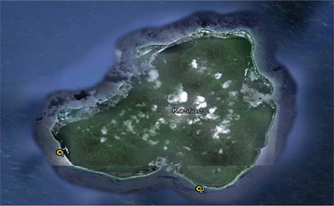
Sponge collections were made from the region of Kosrae, the most eastern of the Caroline Islands. The island is located approximately 370 miles (600 km) north of the equator (5°19′ N, 162°59′E) between Guam and the Hawaiian Islands. They were taken from depths of 10-50 m using scuba diving, 23- 30 Jan 2011, 8-15 Jan 2012, 18-28 Oct 2012 and 20-24 Nov 2013. The GPS coordinates of each site were recorded (Table 1, Fig. 1). Collected specimens were frozen and some preserved in 95% ethyl alcohol and were identified based on their morphological characters. The external feature of sponges was observed with a stereo microscope (Stemi SV 6, Carl Zeiss, Jana, Germany). The skeletal arrangements and spicules were studied under a light microscope (Axioscope II, Carl Zeiss) and SEM (HITACHI S-3500; Hitachi, Tokyo, Japan).
[Table 1.] Geological information for collection sites
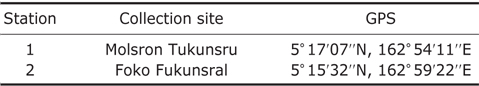
Geological information for collection sites
Order Agelasida Hartman, 1980 Family Agelasidae Verrill, 1907 Genus Agelas Duchassaing & Michelotti, 1864
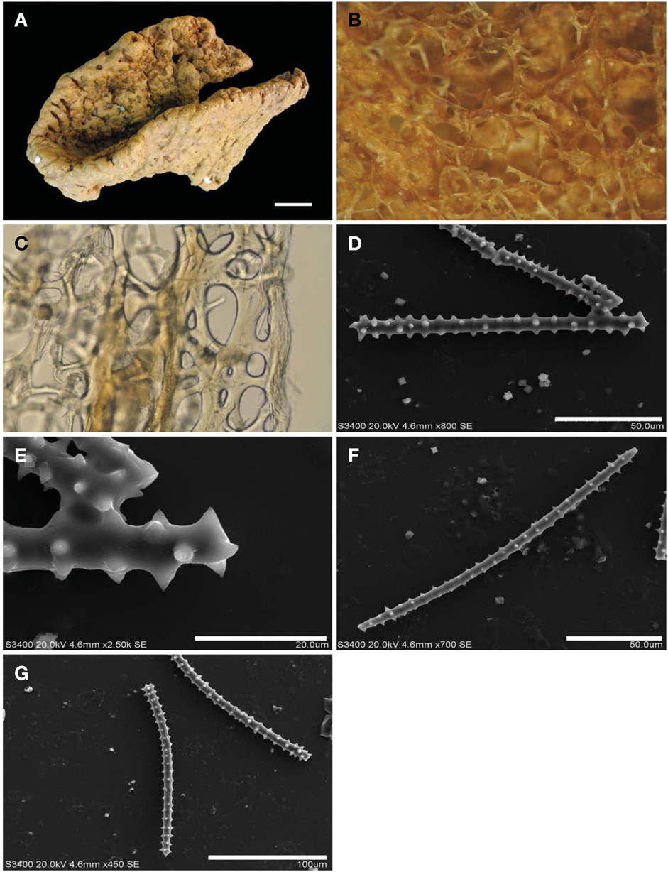
Type specimen. Holotype (MABIK IV00151590), Molsron Tukunsru Village, Kosrae, Micronesia, 22 Oct 2012, Rho HS, by scuba, depth 10 m, deposited in the MABIK, Seocheon, Korea.
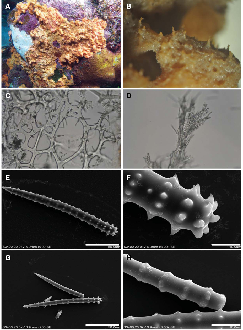
Description. Thin encrusting small pieces, size up to 9×6×0.1-0.3 cm, more or less tuberculated on the surface, attached tightly to the broad rocky substratum. Microscopically the tubercles are rugose with openings and with spicule clusters that protrude from the surface. Surface of many round projecting conules (1-2 mm height) with oscules. Texture hard at surface, soft at bottom. Color dark red.
Skeleton. Primary fibres cored and echinated, 65-78-232 μm in diameter, the end of primary fibres have densely echinated spicules. Secondary fibres 35 μm in diameter. Secondary fibres make large meshes, less echinated at the sponge base. The top of the surface, tubercle has many echinated brush-like bundles of spicules, mixed with thin smooth Acanthostyles. Tertiary fibres 20-30 μm in diameter, no echinating spicules.
Spicules. Acanthostyles 110-200×10-15 μm, number of spine whorls 11-17.
Etymology. This species named after the strawberry-like surface characteristics of the specimen.
Remarks. This sponge is similar to
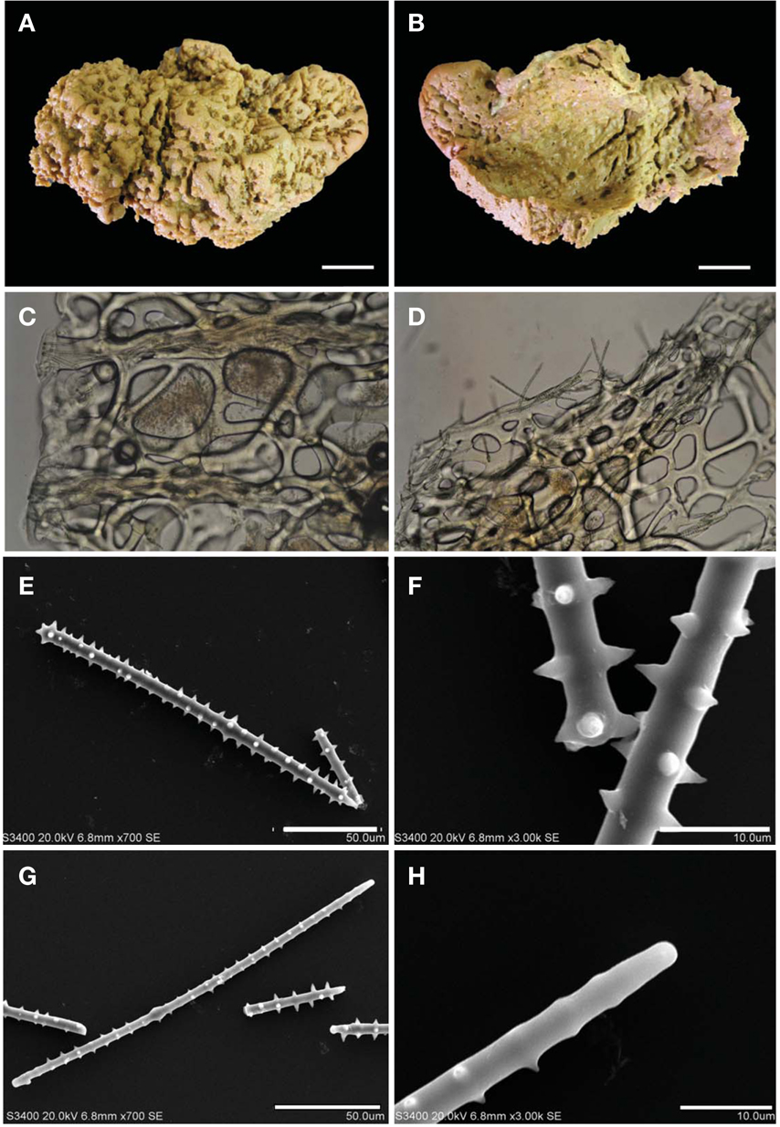
Type specimen. Holotype (MABIK IV00151591), Molsron Tukunsru Village, Kosrae, Micronesia, 22 Oct 2012, Rho HS, by scuba depth 15 m, deposited in the MABIK, Seocheon, Korea.
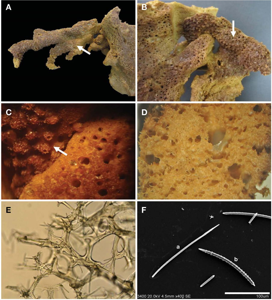
Description. Irregular elongated repent sponge with several branches, size up to 20×6×1.5-2 cm thick. Some wide parts of sponge attached tightly to the substrate. Surface smooth. Texture firm, compressible and hard to tear. Oscules sparse on the surface. Color live, purple on the surface and beige in the choanosome.
Skeleton. Primary fibres rarely cored and rarely echinated, 100-200 μm in diameter. Secondary fibres echinated, 30-60 μm in diameter. Secondary fibres meshes 83-163-232 μm in diameter. Tertiary fibres 10-20 μm in diameter are very rarely echinated.
Spicules. Acanthostyles 110-140×6-8 μm, number of spine whorls 19-22. Acanthoxeas 150-170×6-8 μm, number of spine whorls 18-22.
Etymology. This species is named after the type locality, Kosrae, Micronesia.
Remarks. This new sponge is similar to
Type specimen. Holotype (MABIK IV00151592), Molsron Tukunsru Village, Kosrae, Micronesia, 22 Oct 2012, Rho HS, by scuba, depth 15 m, deposited in the MABIK, Seocheon, Korea.
Description. Subcylindrical, irregular branched, size up to 13×2×1.8 cm, attached to the sponge
Skeleton. Primary fibres, 50-100 μm in diameter, heavily cored; secondary fibres, 30-40 μm diameter, cored; tertiary fibres 10 μm in diameter, cored. All fibres rarely echinated; free spicules occur in the choanosome.
Spicules. Acanthoxeas 150-180×6 μm, number of spine whorls 20-21. Acanthostyles 150-220×5-8 μm, number of spine whorls 19-21.
Etymology. This species named after the live color purple.
Remarks. This new species is similar to
Type specimen. Holotype (MABIK IV00151593), Foko Fukunsral, Kosrae, Micronesia, 19 Jan 2011, Rho HS, by scuba, depth 17 m, deposited in the MABIK, Seocheon, Korea.
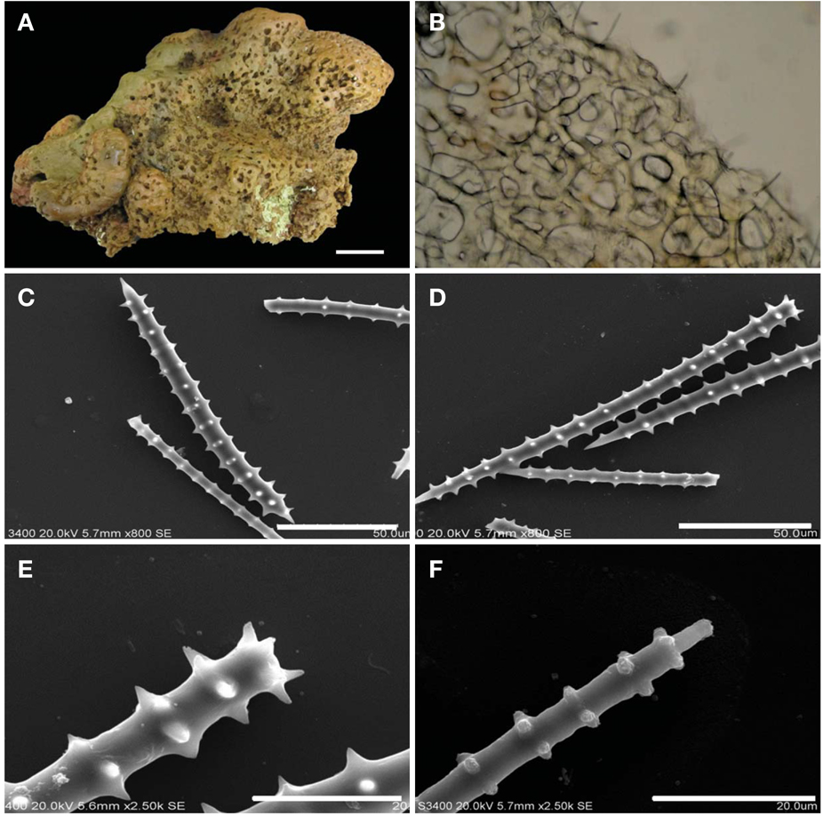
Description. Thickly encrusting, size up to 22×20×2-3 cm. Surface smooth with many hole-like pits. Consistency firm. Color orange in life. Cavernous interior.
Skeleton. Ectosome consists of a thick membrane with free spicules (mostly acanthoxeas) and mixed fibres. Choanosome consists of a dense network of primary and secondary fibres, not distinct. Primary fibres 50-130 μm in diameter, lightly cored with spicules, echinated and reticulated. Secondary fibres 40-70 μm in diameter, rarely echinated; meshes 50- 230 μm in diameter. No tertiary fibres. Choanosome weak cavernous, many spicules.
Spicules. Acanthostyles 150-200×8-10 μm, acanthoxeas 140-250×6-8 μm. Number of spine whorls, acanthostyles 10-18, acanthoxeas 14.
Etymology. This species name
Remarks. This species is similar to
Type specimen. Holotype (MABIK IV00151594), Foko Fukunsral, Kosrae, Micronesia, 19 Jan 2011, Rho HS, by scuba, depth 17 m, deposited in the MABIK, Seocheon, Korea.
Description. Flabellate, cup-shaped, size up to 23×10×1.5 cm. Surface very rough inside but outside smooth with slight protuberances. No oscules. Consistency firm and difficult to tear. Color yellowish brown in life.
Skeleton. Ectosome a thin membrane, very resilient with cored fibres and free spicules. Choanosome a irregular reticulation with loose primary fibres 150 μm in diameter, heavily cored and echinated, primary fibres form a simple fascicle; secondary fibres 60-70 μm in diameter, not cored and rarely echinated; tertiary fibres 20-30 μm in diameter, not cored and rarely echinated, The secondary fibre mesh is 30 -350 μm in diameter.
Spicules. Acanthostyles 180-200×8-10 μm, number of spine whorls, 24. Acanthostrongyles 160-200 μm, number of spine whorls 19-24. Acanthoxeas 200-220 μm, number of spine whorls 22-24 (very rare).
Etymology. This species name
Remarks. The new species is similar to
Type specimen. Holotype (MABIK IV00151595), Molsron Tukunsru Village, Kosrae, Micronesia, 22 Oct 2012, Rho HS by scuba, depth 17 m, deposited in the MABIK, Seocheon, Korea.
Description. Thick encrusting sponge, size up to 18×10×2 cm. Surface with many protuberances but smooth. Texture hard. Color light orange in life.
Skeleton regularly arranged, interior not cavernous; many fasciculate, primary fibres with numerous spicule; echinating spicules rare. Mesh variable. Primary fibres 140-150 μm in diameter and make a fascicle complex, spaced at intervals of 400 μm in diameter. Secondary fibres 30-40 μm. Mesh 31- 179-184-238 μm with rare echinating spicules.
Spicule. Acanthostyles 170-210×5-10 μm, number of spine whorls 21-23.
Etymology. This species named after the encrusting habit.
Remarks. This species is similar to
The genus
These three group show different spicule types. Rough surface with brushed spicules have very distinct verticilliate shape and thick diameter. In repent sponge, spicules have irregular spine of whorls and thinner than brush-like spicules. Thick encrusting or funnel shape sponge has also irregular spine of whorls but thicker than repent sponge.
Although Kosrae is geographically close to Truk, Kosrae has a high diversity of species of
Thirteen species of Indo pacific