The genus
Samples were collected by washing of algae using a sieve in the intertidal zone. Specimens were initially fixed with 5% formaldehyde-seawater solution, and preserved with 85% ethyl alcohol after sorting in the laboratory. The appendages of specimens were dissected in a petri dish filled with glycerol using a dissection pincer and a needle under the stereomicroscope (SZH10; Olympus, Japan), and mounted on temporary or permanent slides. Drawings and measurements were performed by the light microscope (LABOPHOT-2; Nikon, Japan) with the aid of a drawing tube.
For the scanning electron microscopic study, examined specimens were treated as follows. Intact specimens or dissected parts were preserved in 70% ethanol and cleaned ultrasonically with distilled water. To obtain the dried specimens, a freeze drier was used after freezing at −40℃. The dried specimens were attached to aluminum stubs and examined with scanning electron microscopy (TM-1000; HITACHI, Japan).
Order Amphipoda Latreille, 1816 Suborder Gammaridea Latreille, 1803 Family Ampithoidae Stebbing, 1899 Genus Ampithoe Leach, 1814
Key to Korean species of the genus
1. Pleonal epimeron 3 with pointed posteroventral corner ································2 -Pleonal epimeron 3 with rounded posteroventral corner································3 2. Gnathopod 1, propodus short, less than 0.7 times as long as carpus ································A. tarasovi -Gnathopod 1, propodus moderate, more than 0.7 times as long as carpus ································A. lacertosa 3. Gnathopod 2, palm not defined ································A. youngsanensis -Gnathopod 2, palm defined ································4 4. Gnathopod 2, palm of propodus with posterior process ································5 -Gnathopod 2, palm of propodus without posterior process································6 5. Gnathopod 2, posterior process acute································A. akuolaka -Gnathopod 2, posterior process blunt································A. ramondi 6. Antenna 2 with dense plumose setae posteriorly; gnatho- pod 2, propodus widening distally································A. koreana -Antenna 2 without plumose setae posteriorly; gnathopod 2, propodus uniform in width································7 7. Gnathopod 2, palm rather short, concave································A. brevipalma - Gnathopod 2, palm rather long, not concave································8 8. Antenna 2, peduncle 4 rather expanded; gnathopod 1, carpus transverse posterodistally································A. shimizuensis - Antenna 2, peduncle 4 linear and ordinary; gnathopod 1, carpus produced posterodistally································A. valida
Material examined. Korea: 4♂♂ 3♀♀, Jeollanam-do: Wando-gun, Bogil-myeon, Yesong-ri, 10 Feb 2010, Kim JG; 1♂, Wando-gun, Geumil-eup, Sadong-ri, Sorang Bridge, 30 Mar 2010, Jung TW.
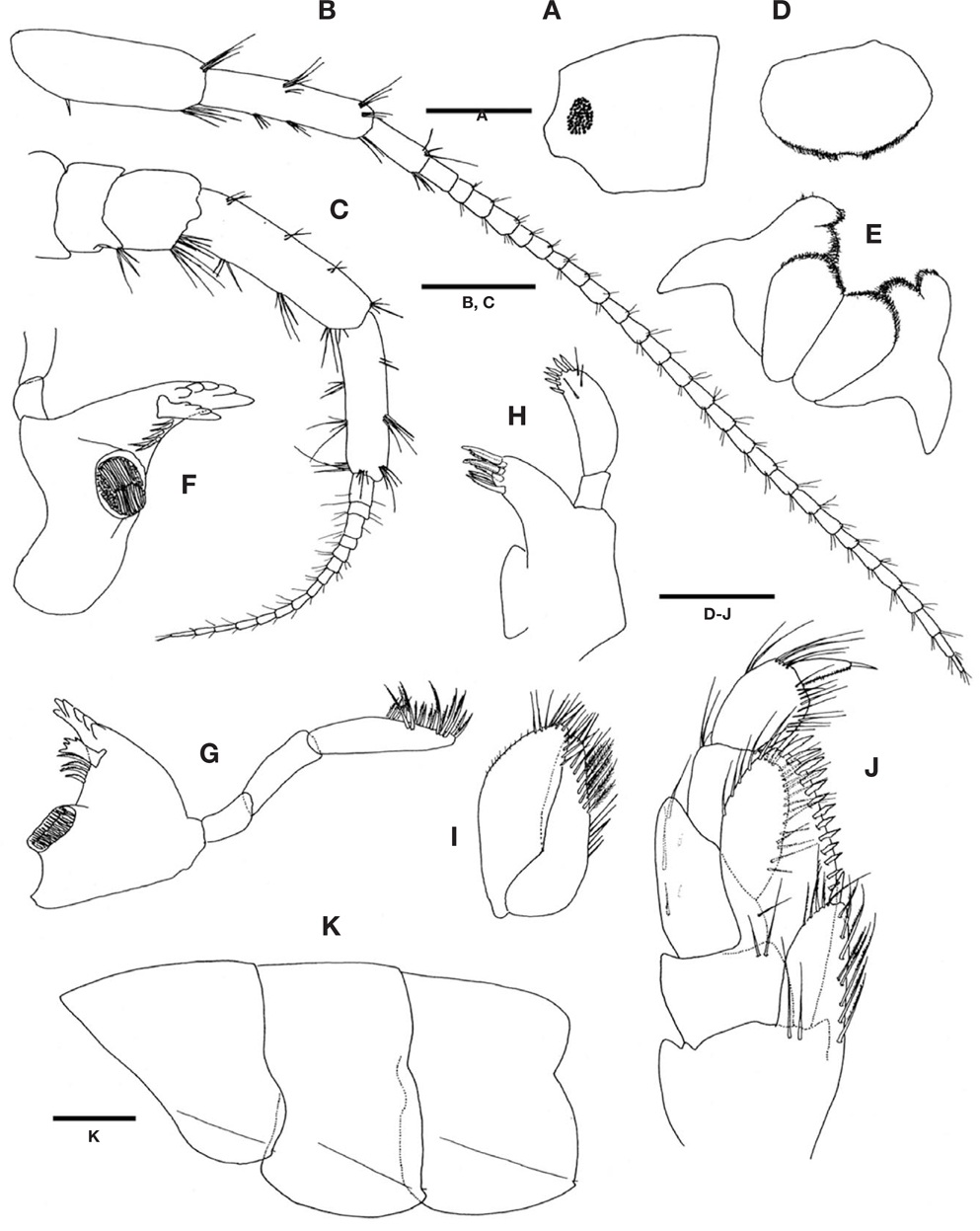
Description. Male: Body 9.8 mm long.
Head (Fig. 1A), rostrum indistinct; anterior cephalic lobe blunt, truncated obliquely; eyes medium, oval.
Pleonal epimera (Fig. 1K), each posterovental corner with small notch bearing minute setule and lateral surface with faint ridges obliquely.
Antenna 1 (Fig. 1B) weakly setose, longer than antenna 2; length ratio of peduncular articles 1-3 1.00 : 0.83 : 0.36; peduncular article 1 stouter than article 2 with posteroproximal spine, antero- and posterodistal corners with 1 group of several setae; peduncular article 2 slender, anterior margin with 1 group of mesial and 2 groups of distal setae, posterior margin with 2 groups of mesial and 1 group of distal setae; flagellum elongate, 27-articulate, each with short setae distally.
Antenna 2 (Fig. 1C), gland cone produced; peduncular articles 4, 5 stout, anterior and posterior margins with several groups of setae; peduncular article 4 longer than article 5; flagellum short, 15-articulate, each with short setae distally.
Upper lip (Fig. 1D), ventral margin rounded and densely pubescent.
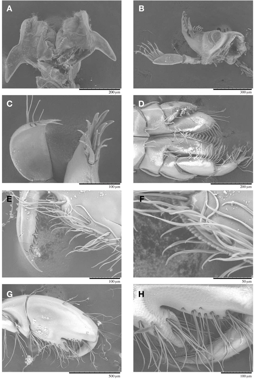
Lower lip (Figs. 1E, 5A), apical lobules of outer lobe separated, but short and tumid; apexs of inner and outer lobes blunt and densely pubescent; mandibular process well developed.
Right mandible (Fig. 1G), incisor 6-toothed; lacinia mobilis 4-toothed; accessory setal row with 4 dentate setae; molar process triturative, well developed; palp triarticulate, length ratio of articles 1-3 1.00 : 1.58 : 2.35, apex of article 3 obliquely truncated with many pectinate setae subapically.
Left mandible (Figs. 1F, 5B), incisor 5-toothed; lacinia mobilis 4-toothed; accessory setal row with 5 dentate setae.
Maxilla 1 (Figs. 1H, 5C), inner plate short; outer plate with 9 stout denticulate spines apically; palp biarticulate, article 1 short, article 2 stout with 6 denticulate apical spines and 3 subapical setae.
Maxilla 2 (Fig. 1I), surface of inner plate with oblique row of pinnate setae, inner and apical margin lined with simple setae; outer plate broader than inner, outer margin swollen mesially.
Maxilliped (Figs. 1J, 5D), inner plate with stout spine apically and several pinnate setae on inner and subapical margins; outer plate enlarged, reaching distal end of palp article 2, subinner margin lined with denticulate spines; palp 4-articulate, articles 1 and 2 stout, inner margins of article 2 and 3 lined with several long setae, article 4 produced apically and inner margin pectinate with elongate apical spine.
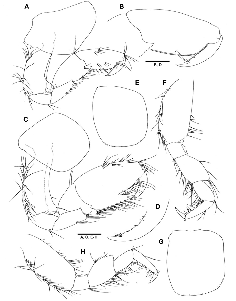
Gnathopod 1 (Figs. 2A, B, 5E, F), coxa produced anteroventrally, ventral margin lined with short setules; basis trapezoidal with large rounded anterodistal lobe, posterior margin swollen with long setae; ischium short, anterior margin with small lobe, posterior margin with pair of setae distally; merus tapering and truncated distally, distal half of posterior margin with several setae; carpus gradually widening, anterior margin bare, anterodistal corner with pair of unequal setae, posterior margin expanded with triangular extension posterodistally and several setae posteriorly; propodus subrectangular, as long as carpus, slightly tapering toward apex; palm short with large defining spine, edge of palm with row of scabrous clusters composed of numerous fine setae (Fig. 5E, F); dactylus slightly curved, exceeding palm, inner margin serrulate with accessory tooth.
Gnathopod 2 (Figs. 2C, D, 5G, H), coxa quadrate with rounded anteroventral corner, ventral margin lined with short setules; basis stout, slightly curved, anterior margin lobate, anterodistal lobe well developed, posterior margin with several clusters of long setae; ischium short, anterior margin with small lobe, posterior margin with pair of setae distally; merus, distal margin concave, fitting carpal lobe, posterior margin with 2 groups of mesial and distal setae; carpus, anterior margin swollen with 2 distal setae, carpal lobe well developed, apex blunt with group of setae; propodus, anterior margin convex and distal half with 4 groups of long setae, posterior margin with 5 clusters of setae; palm deeply bifid, apex of produced lobe acute; dactylus large, exceeding end of produced lobe of palm, with serrulation at inner margin.
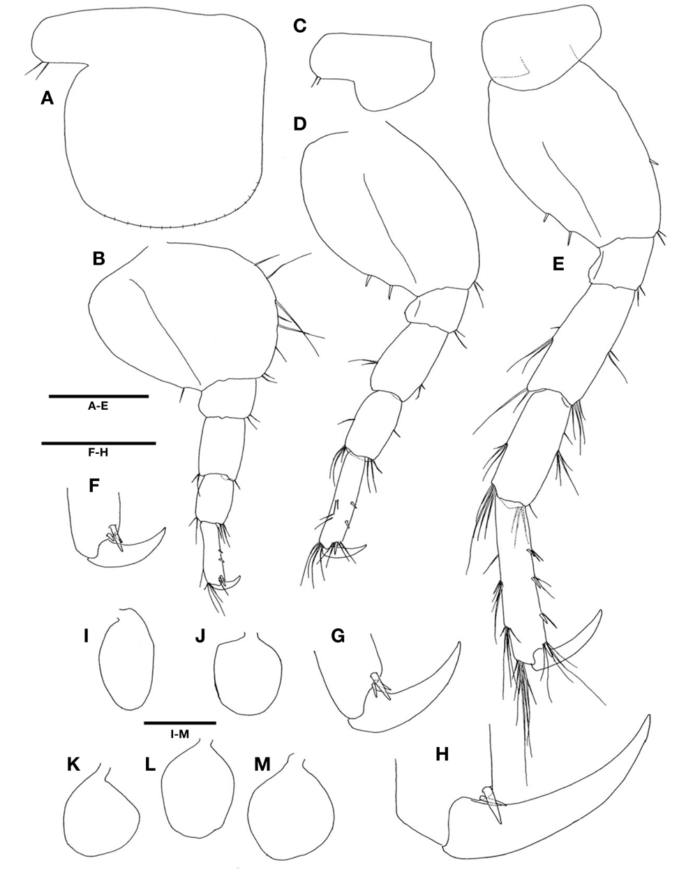
Pereopod 3 (Fig. 2E, F), coxa quadrate, anterior and posterior margins slightly convex, ventral margin lined with short setules; basis, anterior margin straighten, anterodistal half with setae, posterior margin convex with several groups of setae; ischium quadrate, anterior margin with small lobe, posterior margin with group of 3 setae; length ratio of merus, carpus, and propodus 1.00 : 0.79 : 1.07; merus about half of basis, anterior margin lobate with 2 mesial setae, anterodistal corner produced with group of 4 setae, posterior margin with long setae; carpus, anterior and posterior margins swollen, anterior margin bare, anterodistal corner with pair of setae; posterior margin with long setae; propodus gradually slender, anterior margin with pair of medial setae; posterior margin with several setae; dactylus falcate, slightly shorter than half of propodus; inner margin with weakly swollen protuberance.
Pereopod 4 (Fig. 2G, H), nearly similar to pereopod 3 in shape, but slightly shorter and wider; anterior margin of coxa straighter.
Pereopod 5 (Fig. 3A, B, F), coxa bilobate, anterior lobe enlarged and rounded-quadrate, posterior lobe small with 2 setae; basis suboval, anterior margin convex with several setae; posterior margin strongly expanded and dilated proximally, with short seta subdistally; ischium quadrate, posterior margin with small lobe, anterodistal corner with pair of short setae; merus and carpus rectangular, length ratio 1.00 : 0.84; propodus slender, anterior margin with 2 mesial spines, anterodistal corner with group of 3 spines and several setae, posterodistal corner with group of long setae; dactylus falcate, simple.
Pereopod 6 (Fig. 3C, D, G) longer than pereopod 5; coxa bilobate, anterior lobe expanded ventrally and larger than posterior lobe, posterior lobe with 2 setae; basis suboval, width 0.75 times length, anterior margin with pair of short setae distally; posterior margin expanded and slightly dilated proximally, with 2 spines subdistally; ischium, posterior margin with small lobe, anterodistal corner with pair of setae; merus and carpus rectangular, length ratio 1.00 : 0.92; merus slightly swollen distally, each distal 2/3 of anterior and posterior margins with pair of setae; carpus slender than merus, anterior and posterior margins with one mesial and one group of long distal setae; propodus slender, shorter than merus and carpus combined in length, anterior margin with 2 mesial spines, anterodistal corner with group of one large, one pair of small spines and several setae, posterior margin with 2 groups of one mesial and one group of distal setae; dactylus falcate, 0.44 times as long as propodus.
Pereopod 7 (Fig. 3E, H) longer than pereopod 6; coxa unilobate, swollen posteroventrally; basis elongate oval, width 0.62 times length, anterior margin convex with mesial spine and pair of distal setae, posterior margin with 2 spines subdistally, posteroproximal corner angulate and weakly produced; ischium, posterior margin with small lobe, anterodistal corner with pair of setae; merus and carpus rectangular, length ratio 1.00 : 0.87; merus, anterior margin with pair of mesial and group of 4 distal setae, posterior margin with one short and one pair of long mesial setae, posterodistal corner with pair of distal setae; carpus slightly slender than merus, anterior margin with one and one group of mesial setae, anterodistal corner with 4 setae; posterior margin with group of one short and 2 long setae, posterodistal corner with 4 long setae; propodus slender, shorter than merus and carpus combined in length, anterior margin with 3 groups of mesial spines and setae, anterodistal corner with group of 2 unequal spines and several setae, posterior margin with seta, 2 groups of mesial and one group of distal setae; dactylus falcate, 0.51 times as long as propodus.
All coxal gills oval in shape (Fig. 3I-M).
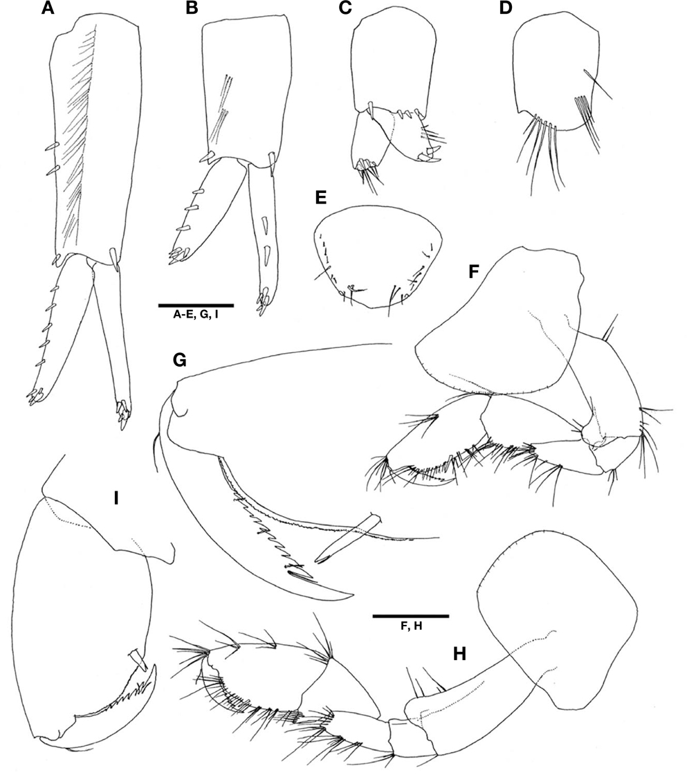
Uropod 1 (Fig. 4A), peduncle longer than rami, gradually narrowing distally, dorsolateral margin with 2 mesial and one distal spines, ventrolateral margin with row of setae, dorsomedial surface with stout spine distally; outer ramus slightly shorter and stouter than inner, outer-lateral margin with 5 small spines, apex with 2 apicolateral and 2 unequal apical spines; inner ramus without lateral spine; apex with 2 apicolateral and 2 unequal apical spines.
Uropod 2 (Fig. 4B) 0.72 times as long as uropod 1; peduncle subequal to inner ramus, dorsal surface with one lateral and one medial spines distally; lateral surface with 2 groups of setae; outer ramus shorter than inner, outer-lateral margin with 3 small spines, apex with 2 apicolateral and 2 unequal apical spines; inner ramus slender than outer, dorsolateral margin with 2 mesial spines; apex with 2 apicolateral and 2 unequal apical spines.
Uropod 3 (Fig. 4C, D), peduncle 0.58 times as long as uropod 2, dorsodistal margin with 3 small spines, lateral corner with spine distally, ventrodistal margin convex with row of 6 long setae, ventral surface with row of 4 setae and 1 seta; inner ramus with 4 spines and 6 setae subapically; outer ramus subequal to inner with 2 curved large spines subapically.
Telson (Fig. 4E). broadly subtriangular, 1.2 times wider than long, each lateral margin with row of several setae.
Female: Body 9.2 mm long.
Gnathopod 1 (Fig. 4F, G) similar to that of male, except for lacking posterodistal extention of carpus.
Gnathopod 2 (Fig. 4H, I) smaller than that of male in size. Basis more slender than that of male; posterior margin bare. Propodus not bifid, similar to that of gnathopod 1.
Remarks. Barnard (1970) mentioned that
But, the Korean specimens of A.
The crustacean cuticle has numerous kinds of projections, and some of these, such as setae, have important mechanical as well as sensory functions (Hindley and Alexander, 1978; Garm, 2004). Some workers such as Watling (1989) and Calazans and Ingle (1998) suggested that the fine structures of the cuticle, mainly the setae, are important tools for the study of crustacean taxonomy, and a previous study on the genus
Distribution. Korea, Hawaii.