검색 전체 메뉴
PDF
맨 위로
OA 학술지
Effect of Residual Lithium Ions on the Structure and Cytotoxicity of Silk Fibroin Film
  • 비영리 CC BY-NC
  • 비영리 CC BY-NC
ABSTRACT
Effect of Residual Lithium Ions on the Structure and Cytotoxicity of Silk Fibroin Film
KEYWORD
silk fibroin , dialysis , lithium ion , secondary structure , cytotoxicity
  • Introduction

    Silk fibroin (SF) is a biocompatible material with various biomedical applications, for instance as a drug delivery vehicle or a scaffold in tissue engineering (Ki et al., 2009). SF also possesses the advantages of tunable mechanical properties and biodegradability; for instance, a more flexible SF film can be made by increasing water content (Hu et al., 2008), while the time required for SF degradation can be altered by controlling molecular weight distribution (Cao et al., 2009) and crystallinity (Hu et al., 2012). It is also possible to prepare a transparent film from SF (Lawrence et al., 2009), making it a highly versatile substance for optoelectronics (Capelli et al., 2011).

    SF is initially prepared as an aqueous solution before being manufactured into various forms including fiber, film, sponges, nano- and microparticles, and hydrogel. The multi-step process includes the removal of sericin by degumming using sodium carbonate solution with or without surfactant. The SF is then dissolved in 9.3 M LiBr solution, which is accelerated by higher temperatures: complete dissolution requires more than 12 h at room temperature, but at 60℃ or higher the time is reduced to less than an hour. The removal of excess LiBr by dialysis is the rate-limiting step during the preparation of SF solution, with a recommended time of more than 48 h (Rockwood et al., 2011).

    Despite being a time-consuming process, dialysis is the only applicable method for complete LiBr removal. Other techniques, such as membrane separation or electrodialysis, each have their limitations. Although LiBr can be extracted using a membrane within a few hours, membrane fouling by SF necessitates high pressures to maintain adequate flux, which induces SF crystallization and leads to the formation of insoluble fleeces. Electrodialysis is also a well-established technique for removing salt from a solution, but in the case of LiBr, hazardous Br2 gas is generated at the anode. LiBr is harmful if swallowed and causes skin and eye irritation; thus, the removal of LiBr is important for biomedical applications of SF. However, there is little information available regarding the effects of residual LiBr on the structure or cytotoxicity of SF-based materials; these were investigated in the present study using SF films with variable amounts of LiBr prepared by varying the dialysis time. The secondary structure of SF films was determined by Fourier transform infrared (FT-IR) analysis, while cytotoxicity was assessed by treating cultured cells with SF film extract or by growing cells directly on SF film and evaluating their viability using the MTT assay.

    Materials and Methods

      >  Materials

    Silk cocoons were obtained from the National Academy of Agricultural Sciences (Seoul, Korea). Sodium oleate was purchased from Junsei Chemical Co., Ltd. (Tokyo, Japan), and LiBr was from Thermo Scientific (Waltham, MA, USA). All other chemicals were purchased from Sigma-Aldrich (Seoul, Korea).

      >  Preparation of SF aqueous solution

    Cocoons were cut into 8 equal-sized pieces, which were immersed in degumming solution consisting of 0.3 wt% sodium oleate and 0.2 wt% sodium carbonate. Degumming was performed at 100℃ for 1 h at a final rate of 28.1%. SF fibers were dried in an oven at 50℃ for 2 d, and 40 g were dissolved in 200 mL 9.3 M LiBr solution at 50℃ for 4 h.

      >  Dialysis conditions

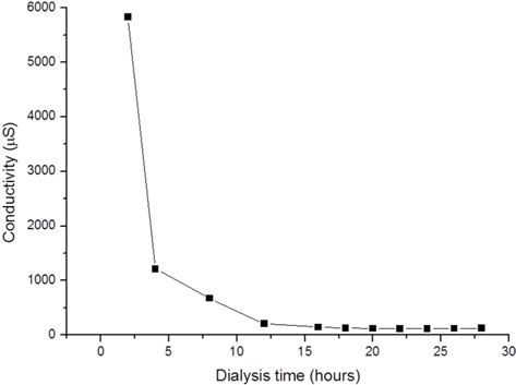
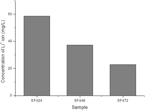
    After dissolving the SF fibers, the solution was dialyzed using a dialysis tube (Spectrum Laboratories, Inc., Rancho Dominguez, CA, USA) with a diameter and length of 25.5 and 300 mm, respectively, and a molecular weight cut-off (MWCO) of 6–8 kDa; each tube containing 50 mL solution was hermetically sealed and immersed in 5 L distilled water. The dialysate was changed every 2 h five times, then every 4 h until the end of the dialysis. To obtain solutions with different amounts of residual LiBr, the dialysis was terminated at 8, 24, 48, and 72 h. The conductivity of the dialysate during the procedure was measured using a conductivity meter (Model 115; ATI Orion, Boston, MA, USA). The amount of residual lithium ion in the dialyzed SF solution was measured with an inductively coupled plasma optical emission spectrometer (ICP-OES) (ICP-730-ES; Varian, Inc., Belrose, Australia).

      >  Preparation and characterization of SF films

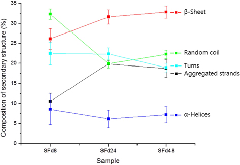
    To obtain SF films from solutions corresponding to different dialysis times—which were designated as SFd8, SFd24, SFd48, and SFd72—Petri dishes were coated with solution and dried at 50℃ for 24 h. With the exception of SFd8, the films were insoluble after casting. The secondary structure of the films was determined using an attenuated total reflectance (ATR) FT-IR spectrometer (ATR-FTIR, Nicolet 6700; Thermo Scientific) as in previous studies, while composition was determined at the amide I band between 1600 and 1700 cm-1 using the peakfitting method. The second-derivative spectra for calculating the number and position of overlapped single bands and the deconvolution spectra for fitting were obtained using Origin 8.0 software (Northampton, MA, USA). Curve-fitting proofs were performed until the χ2 distribution reached the minimum value with multi-peak Gaussian fitting. The relative area of the overlapped single bands was used to estimate the proportion of each type of secondary structure, and the assignment of single bands was as described in previous studies: 1605–1615 cm-1, aggregated strands; 1616–1637 cm-1, β-sheet; 1638–1655 cm-1, random coil; 1656–1662 cm-1, α-helices; and 1663–1695 cm-1, turns (Zhang et al., 2011).

      >  Cytotoxicity tests

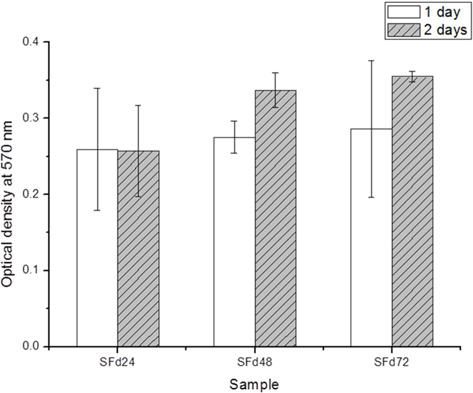
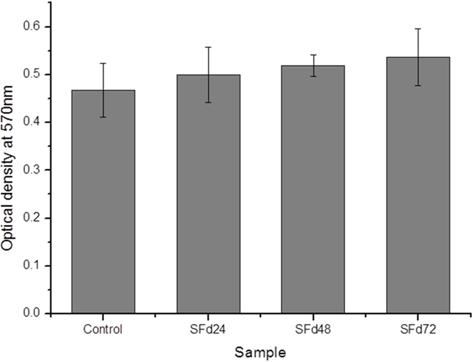
    The cytotoxicity of the SF film was evaluated directly and indirectly using the MTT cell viability assay. For the indirect assessment, cultured NIH3T3 fibroblasts (SPL Life Sciences Co., Ltd., Pocheon, Korea) were treated with SF film extracts, obtained by immersing SF film prepared from solutions with different dialysis times in serum-free medium for 24 h and collecting the medium. Cells were seeded in a 24-well plate (SPL Life Sciences Co., Ltd.) at a density of 1.0 × 10⁴ cells/well and incubated at 37℃ and 5% CO2/95% air for 24 h to ensure complete attachment. The culture medium was then replaced with extract-containing medium and incubated for 24 h before cytotoxicity was assessed using a standard MTT assay: 200 μL of 0.5 mg/mL MTT was added to each sample, and after 4 h, the supernatant containing formazan crystals produced from the MTT substrate by mitochondrial activity was removed and solubilized in 200 μL dimethylsulfoxide (DMSO; Sigma-Aldrich, St. Louis, MO, USA). UV absorbance was read at 570 nm using a microplate reader (Synergy H1; BioTek, Winooski, VT, USA).

    For the direct contact test, cells were cultured on SF film sterilized by immersion in 70% ethanol for 30 min followed by three 20-min phosphate-buffered saline (PBS) washes. The film was transferred to 24-well plates, and cells were seeded at a density of 1.0 × 10⁴ cells/well and maintained at 37℃ and 5% CO2. Cell culture was performed at 37℃ and 5% CO2. After the incubation period, 200 μl of 0.5 mg/mL MTT was added to each sample for 24 h. The MTT solution was removed, and samples were washed twice with Dulbecco’s PBS (DPBS), and 200 μL DMSO was added to solubilize the formazan crystals. After 30 min, UV absorbance at 570 nm was measured. Each experiment was performed in triplicate and data are shown as the mean ± standard error of three measurements.

    Results and Discussion

      >  LiBr concentration during dialysis

    Dialysis removes excess LiBr used to dissolve SF, and requires several dialysate changes; according to one report, at least 6 changes within 48 h are needed (Rockwood et al., 2011). However, there is little information on how LiBr can be effectively removed and on the biological consequences of incomplete lithium removal. In this study, LiBr content was monitored during dialysis by measuring the conductivity of the dialysate over time (Fig. 1). Conductivity decreased rapidly over the first 12 h; after 24 h, the rate of decrease was negligible. Since the volume of dialysate was 100-fold higher than the SF solution, the concentration of LiBr would in theory be reduced by a factor of 1.0 × 10⁸ after 4 dialysate changes (i.e., after 12 h of dialysis) if equilibrium was attained. Therefore, the predicted concentration of LiBr in the solution would be 9.3 × 10⁻² µM or 8.077 µg/L LiBr after 12 h. However, in practice this calculation does not hold for the following reasons. First, the volume inside the dialysis tube increases with time. During this process, an equilibrium is achieved between solutions inside (SF solution) and outside (dialysate) the tube, corresponding to the inflow of water and outflow of LiBr; this leads to an increase in volume inside the tube and a concomitant decrease in the dilution factor as the dialysis proceeds. This volumetric change is minimized if the SF solution completely fills the tube; however, osmotic pressure will cause it to expand, rupturing the tube or changing the MWCO. Second, ionic interactions would necessitate a higher concentration gradient in order for the LiBr to be separated from the SF. The conductivity of the dialysate did not decrease after 24 h (Fig. 1); however, this may have been due to limitations of the conductivity meter used in this study. Therefore, ICP-OES measurements were performed to quantify the residual lithium ion in the solution (Fig. 2). The results indicated that the ion cannot be completely removed: even after 72 h of dialysis, the concentration was 22.85 mg/L, compared to the theoretical value of 8.077 µg/L at 12 h. The pI and pH of SF are around 4 (Kang et al., 2000) and 6.0-7.0, respectively; thus, the SF has a net negative charge and can interact with the positively charged lithium ion. Since the presence of ions can alter various properties of a protein, the effect of residual LiBr on the secondary structure and cytotoxicity of SF film was investigated.

      >  Effect of residual LiBr on the secondary structure of SF films

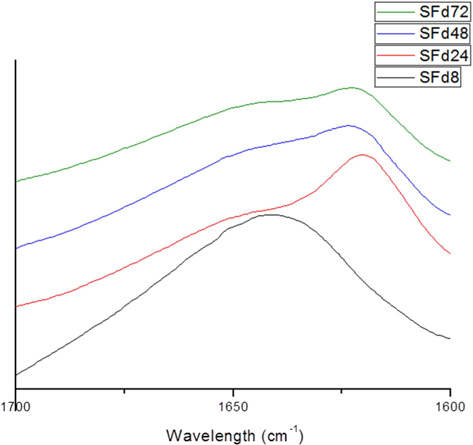
    The original ATR-FTIR spectra of SF films and secondary structure composition after deconvolution are shown in Fig. 3 and 4, respectively. Only SFd8 film was still soluble after casting; the insolubility of the other films was likely caused by a transition in secondary structure. Indeed, the secondary structure of the SFd8 film consisted mostly of random coils, whereas β-sheets were more prominent in SFd24, SFd48, and SFd72. The deconvolution could not be carried out on SFd72 due to low intensity in the range of 1700–1680 cm-1. However, the peak intensity at 1620 cm-1 was higher for SFd72 than SFd24 and SFd48, indicating a higher degree of β-sheet formation. These results suggest that a high residual lithium ion content hinders conformational changes, as in the case of SFd8. Conversely, the removal of lithium allows the structural transition of SF to a β-sheet structure. The role of various cations on structural transitions has been previously studied; for instance, it has been reported that divalent calcium ions can induce β-sheet formation by bringing SF molecules closer together and acting as an ion bridge (Foo et al., 2006). A similar effect is observed with copper, but in this case the β-sheet arrangement is mediated through the chelation of histidine residues (Zong et al., 2004). Other divalent cations such as magnesium and zinc can also promote the β-sheet structure (Zhou et al., 2005); however, monovalent ions such as sodium and potassium have the opposite effect on SF structural transitions, and can break down the protein network (Zhou et al., 2005), which could explain the effects of the monovalent lithium ion.

    Effect of residual LiBr on the cytotoxicity of SF films

    Residual LiBr was detected in SF films even after extensive dialysis (Fig. 2). LiBr has acute oral toxicity with an LD50 of 1800 mg/kg, which is low enough to exclude it from categorization as a toxic substance. Nonetheless, any biomaterials require rigorous testing in order to ensure that they are safe for use. In particular, SF has low biodegradability, and would therefore persist at the site of implantation over a long period. Even at a low level of toxicity, prolonged exposure can be harmful. The cytotoxicity of the SF film was verified indirectly by using SF film extract to treat cultured cells, and directly by seeding cells onto the film. When dialysis was performed for more than 48 h, the cytotoxicity of the SF film was negligible (Figs. 5 and 6). The results of the MTT assay revealed that the soluble SF extract was not toxic to cells for any dialysis time (Fig. 5). However, at least 48 h of dialysis were required to counter any adverse effects on cell viability, which was unaffected by SFd48 and SFd72, but was significantly reduced by SFd24 (Fig. 6). Lithium reportedly inhibits the glycogen synthase kinase-3β, a serine/threonine kinase that controls cell cycle progression; lithium treatment causes G2/M arrest while preserving cell viability (Mao et al., 2001), and inhibits growth in NIH3T3 cells (Candia et al., 2011). Therefore, the decreased viability associated with SFd24 is likely due to the high lithium content. In conclusion, the results indicate that residual LiBr can affect the secondary structure and cytotoxicity of SF films when dialysis time is insufficient, and provide justification for using the traditional dialysis time of 48 h during the preparation of aqueous SF solution.

참고문헌
  • 1. Candia PD, Minopoli G, Verga V, Gargiulo A, Vanoni M, Alberghina L 2011 Nutritional Limitation Sensitizes Mammalian Cells to GSK-3β Inhibitors and Leads to Growth Impairment [Am J Pathol] Vol.178 P.1814-1823 google
  • 2. Cao Y, Wang B 2009 Biodegradation of silk biomaterials [Int J Mol Sci] Vol.10 P.1514-1524 google
  • 3. Capelli R, Amsden JJ, Generali G, Toffanin S, Benfenati V, Muccini M, Kaplan DL 2011 Integration of silk protein in organic and lightemitting transistors [Org Electron] Vol.12 P.1146-1151 google
  • 4. Foo CWP, Bini E, Hensman J, Knight DP, Lewis RV, Kaplan DL 2006 Role of pH and charge on silk protein assembly in insects and spiders [Appl Phys A] Vol.82 P.223-233 google
  • 5. Hu X, Kaplan D, Cebe P 2008 Dynamic protein-water relationships during β-sheet formation [Macromolecules] Vol.41 P.3939-3948 google
  • 6. Hu Y, Zhang Q, You R, Wang L, Li M 2012 The relationship between secondary structure and biodegradation behavior of silk fibroin scaffolds [Adv Mater Sci Eng] google
  • 7. Kang GD, Nahm JH, Park JS, Moon JY, Cho CS, Yeo JH 2000 Effects of poloxamer on the gelation of silk fibroin [Marcromol Rapid Commun] Vol.21 P.788-791 google
  • 8. Ki CS, Park YH, Jin HJ 2009 Silk protein as a fascinating biomedical polymer: structural fundamentals and applications [Macromol Res] Vol.17 P.935-942 google
  • 9. Lawrence BD, Marchant JK, Pindrus MA, Omenetto FG, Kaplan DL 2009 Silk film biomaterials for cornea tissue engineering [Biomaterials] Vol.30 P.1299-1308 google
  • 10. Mao CD, Hoang P, DiCorleto PE 2001 Lithium inhibits cell cycle progression and induces stabilization of p53 in bovine aortic endothelial cells [J Biol Chem] Vol.276 P.26180-26188 google
  • 11. Rockwood DN, Preda RC, Yucel T, Wang X, Lovett ML, Kaplan DL 2011 Materials fabrication from Bombyx mori silk fibroin [Nat Protoc] Vol.6 P.1612-1631 google
  • 12. Zhang H, Deng L, Yang M, Min S, Yang L, Zhu L 2011 Enhancing Effect of Glycerol on the Tensile Properties of Bombyx mori Cocoon Sericin Films [Int J Mol Sci] Vol.12 P.3170-3181 google
  • 13. Zhou L, Chen X, Shao Z, Huang Y, Knight DP 2005 Effect of Metallic Ions on Silk Formation in the Mulberry Silkworm, Bombyx mori [J Phys Chem B] Vol.109 P.16937-16945 google
  • 14. Zong XH, Zhou P, Shao ZZ, Chen SM, Chen X, Ju BW, Deng F, Yao WH 2004 Effect of pH and Copper(II) on the conformation transitions of silk fibroin based on EPR, NMR, and raman spectroscopy [Biochemistry] Vol.43 P.11932-11941 google
OAK XML 통계
이미지 / 테이블
  • [ Fig. 1. ]  Change in dialysate conductivity over time during the dialysis of LiBr from silk fibroin solution. Data represent mean ± standard error of three independent experiments.
    Change in dialysate conductivity over time during the dialysis of LiBr from silk fibroin solution. Data represent mean ± standard error of three independent experiments.
  • [ Fig. 2. ]  Lithium ion concentration in silk fibroin (SF) solution after dialysis for 24 (SFd24), 48 (SFd48), and 72 (SFd72) h. Data represent mean ± standard error of three independent experiments.
    Lithium ion concentration in silk fibroin (SF) solution after dialysis for 24 (SFd24), 48 (SFd48), and 72 (SFd72) h. Data represent mean ± standard error of three independent experiments.
  • [ Fig. 3. ]  ATR-FTIR of the amide I peak of silk fibroin (SF) films prepared from solutions with dialysis times of 8 (SFd8), 24 (SFd24), 48 (SFd48), and 72 (SFd72) h.
    ATR-FTIR of the amide I peak of silk fibroin (SF) films prepared from solutions with dialysis times of 8 (SFd8), 24 (SFd24), 48 (SFd48), and 72 (SFd72) h.
  • [ Fig. 4. ]  Secondary structure composition of silk fibroin (SF) films prepared from solutions with dialysis times of 8 (SFd8), 24 (SFd24), and 48 (SFd48) h, as determined by deconvolution of the amide I peak in the ATR-FTIR spectra. Data represent mean ± standard error of three independent experiments.
    Secondary structure composition of silk fibroin (SF) films prepared from solutions with dialysis times of 8 (SFd8), 24 (SFd24), and 48 (SFd48) h, as determined by deconvolution of the amide I peak in the ATR-FTIR spectra. Data represent mean ± standard error of three independent experiments.
  • [ Fig. 5. ]  Cytotoxicity of soluble part of SF film prepared from the solutions with dialysis times of 24 (SFd24), 48 (SFd48), and 72 (SFd72) h in NIH3T3 cells. Data represent mean ± standard error of three independent experiments.
    Cytotoxicity of soluble part of SF film prepared from the solutions with dialysis times of 24 (SFd24), 48 (SFd48), and 72 (SFd72) h in NIH3T3 cells. Data represent mean ± standard error of three independent experiments.
  • [ Fig. 6. ]  Attachment and proliferation of NIH3T3 cells seeded onto SF film prepared from the solutions with dialysis times of 24 (SFd24), 48 (SFd48), and 72 (SFd72) h. Data represent mean ± standard error of three independent experiments.
    Attachment and proliferation of NIH3T3 cells seeded onto SF film prepared from the solutions with dialysis times of 24 (SFd24), 48 (SFd48), and 72 (SFd72) h. Data represent mean ± standard error of three independent experiments.
(우)06579 서울시 서초구 반포대로 201(반포동)
Tel. 02-537-6389 | Fax. 02-590-0571 | 문의 : oak2014@korea.kr
Copyright(c) National Library of Korea. All rights reserved.