검색 전체 메뉴
PDF
맨 위로
OA 학술지
A Study of Nine Newly Reported Species of the Order Chlorococcales (Chlorophyta) in Hongcheon River, Korea
  • 비영리 CC BY-NC
  • 비영리 CC BY-NC
ABSTRACT
A Study of Nine Newly Reported Species of the Order Chlorococcales (Chlorophyta) in Hongcheon River, Korea
KEYWORD
Chlorococcales , epilithic algae , Hongcheon river , new recorded species , phytoplankton
  • INTRODUCTION

    Order Chlorococcales is taxa belong to phylum Chlorophyta and class Chlorophyceae. Chlorococcales is considered a main taxa researches about rivers, lakes, and dams (Kim and Chung 1993). These plants, along with cyanophytes and diatoms, are biological indicators of the water environment (Thunmark 1945, Reynolds 1984, Kim 1998). Chlorococcales exist as unicells or colonies with diverse sizes and shapes, and do not have flagella, eye spots, contractile vacuoles, or basal bodies (Wilcox et al. 1992).

    The order Chlorococcales was classified by Pascher in 1915 based on electron microscopic microstructure and molecular biology evidence; however, due to insufficient classification standards and descriptions, the synonyms have been confused, resulting in difficulties identifying the order Chlorococcales.

    The order Chlorococcales is one of the largest taxa of green algae in freshwater worldwide. Komárek and Fott (1983) recorded 15 families 248 genera and 1,025 species worldwide, whereas Hindák (1977, 1980, 1984, 1988) recorded 12 families 96 genera, and 168 species excluding the genus Scenedesmus in Western Slovakia. The Algaebase includes 20 families 109 genera, and 292 species (Guiry 2013).

    In Korea, order Chlorococcales contains 246 taxa classified into 12 families, 56 genera, 181 species, 64 varieties, and 20 forms (Lee 1996). Studies including Ann and

    Chang (1985), An and Chang (1990), Kim et al. (1991) and Kim and Chang (1997) have been conducted on the order Chlorococcales in Korea but they are far behind international studies.

    In this study, we collected phytoplankton and epilithic algae in Hongcheon River, and newly recorded species to contribute the Korean flora in the order Chlorococcales.

    MATERIALS AND METHODS

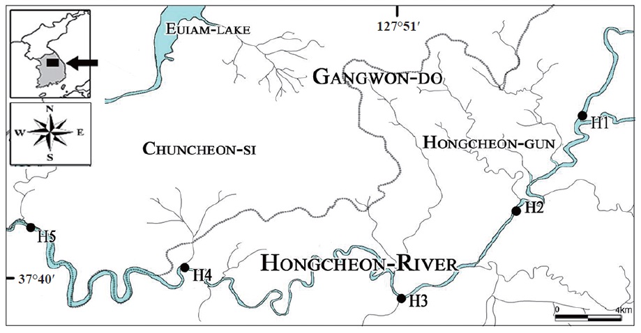
    Order Chlorococcales specimens were collected in Hongcheon River from December 2011 to September 2012 (Fig. 1). The Hongcheon River starts in Saengok-ri Seosuk-myun Hongcheon-gun and is a second tributary of the Han River, which is 120 km in length (Yang et al. 1991). The vicinity of Hongcheon River is mostly mountains and farmlands, but parts go through city and factory or leisure areas (Choi and Kim 2004).

    H1 is Cheoljeong-kyo, Cheoljeong-ri Duchon-myeon Hongcheon-gun Gangwon-do, H2 is Gurun-kyo Gurun-ri Hwachon-myeon H3 is Dunji-kyo Hahwagye-ri Bukbangmyeon, H4 is Palbong-kyo Eoyupo-ri Seo-myeon, H5 is Chungui-bridge Balsan-ri Nam-myeon Chuncheon-si (Fig 1).

    Aquatic plants, submerged land plants, and rocks were scrubbed off to collect the attached algae, and surface water was sampled with a 20 μm mesh, 30 cm diameter phytoplankton net (Wildco, Yulee, FL, USA) to collect phytoplankton.

    The collected specimens were separated by species using a Pasteur pipette under a light microscope and cul-tured in solid media. When a colony formed as a single species, it was transferred to liquid media.

    All samples were examined under ×1000 magnification, and pictures were taken of the cell’s outer shape using an Olympus DP70 camera (Olympus, Tokyo, Japan). Then, using Adobe Photoshop CS4 (Adobe Systems, Mountain View, USA) and WACOM bamboo MTE-450K (WACOM, Seoul, Korea), the outer shapes of the cells and chlorophyll were depicted, and size is shown with a scale bar. All of specimens were deposited in the algal culture collection of Kyonggi University (ACKU) and the National Institute of Biological Resources (NIBR) in Korea.

    The collected taxa were identified using the Komárek and Fott (1983) classification system, and Chung (1993), Prescott (1973), Prescott et al. (1972, 1977, 1981, 1982), Hindák (1977, 1980, 1984, 1988), Hirose et al. (1977), John et al. (2002), John and Robert (2003) were used as references. The species distribution was performed referring to Algaebase (Guiry 2013).

    RESULTS AND DISCUSSION

    The morphological and ecological characteristics of the nine Korean unrecorded species including the unrecorded genera Oocystidium and Heleococcus from Hongcheon River are as follows.

      >  Order Chlorococcales

      >  Family Oocystaceae

      >  Subfamily Oocystoideae

      >  Genus Oocystidium

    Genus Oocystidium was named by Korshikov in 1953. The Cell is broad oval, unicell or in 2-4 cells in mucilage. The Cell wall is smooth and thick. Chloroplasts 1-2 or parietal with pyrenoid (Komárek and Fott 1983, Guiry 2013). Three species such as O. ovale, O. ovale var. polymammilatum, and O. polymammilatum, have been recorded; however, only O. ovale is currently accepted (Guiry 2013).

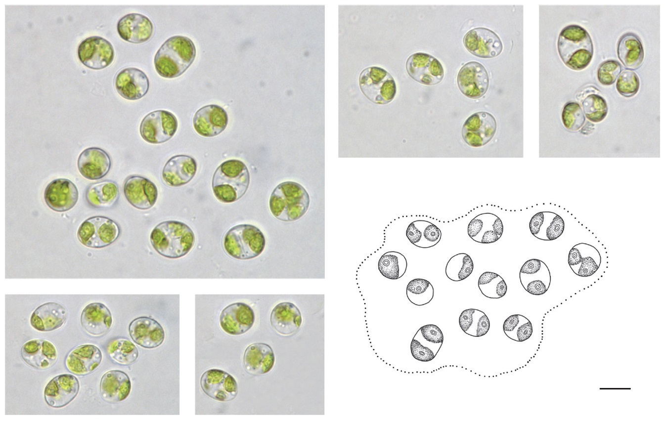
    Oocystidium ovale Korshikov 1953 (Fig. 2)

    Description: Cells spherical or ellipsoidal; usually 1.3 times as long as wide, sometimes twice. Cells individually or combined into 2–4 in colonies (Komárek and Fott 1983). The apical tips of the cells have chloroplasts and are divided in two, each with a pyrenoid. Cell size is 10–19μm long, 8–14 μm wide.

    Ecology and distribution: Ponds in Romania, Spain, Argentina, Czechoslovakia, and Hungary (Komárek and Fott 1983). We found it as phytoplankton.

    Site of collection: Chungui-bridge Balsan-ri Nam-myeon Chuncheon-si (H5).

    Specimen: ACKU 9-296.

      >  Order Chlorococcales

      >  Family Palmellaceae

      >  Subfamily Hormotiloideae

      >  Genus Heleococcus

    Genus Hormotila was first named by Bourrelly in 1966; however, some species in this genus were transferred to Heleococcus by Korshikov in 1953. The cell combines amorphous colonies with mucilage. Sometimes layered. The cell wall is smooth and smaller vacuoles. Chloroplast single or parietal with pyrenoid (Komárek and Fott 1983, Guiry 2013). Three species have been recorded such as H. mucicola, H. polyvacuolatus, H. ramosissimus; however, only H. mucicola is currently accepted taxonomically (Guiry 2013).

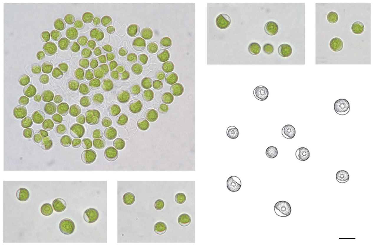
    Heleococcus mucicola Korshikov 1953 (Fig. 3)

    Synonym: Hormotila mucicola Bourr. 1966.

    Description: Cell wall is thin and smooth. Cells individual or 2–4-celled colonies with end of branch mucilage. Cells spherical or ellipsoidal. Cells 10–20 μm in diameter. Chloroplast thin, parietal. The center of the cell has a vacuole (Komárek and Fott 1983).

    Ecology and distribution: Ireland and Sweden in overgrown swamps and sphagnum ponds (Komárek and Fott 1983). We found this species as epilithic algae in a river.

    Site of collection: Dunji-kyo Hahwagye-ri Bukbangmyeon (H3).

    Specimen: NIBRCL0000104603, ACKU 9-302.

      >  Order Chlorococcales

      >  Family Chlorellaceae

      >  Subfamily Chlorelloideae

    Chlorella mirabilis V. M. Andreyeva 1975 (Fig. 4)

    Description: Cells individual, most spherical; young cells spherical and slight ellipsoidal. Cells 2–8 μm in diameter. Chloroplast parietal, usually filling two-thirds of the cell. Pyrenoid has small fragment.

    Ecology and distribution: The Black Sea, Russia and at the bottoms of rivers in the tundra (Komárek and Fott 1983). It is also found in terrestrial habitats (Guiry 2013). We found it as phytoplankton.

    Site of collection: Dunji-kyo Hahwagye-ri Bukbang-myeon (H3).

    Specimen: ACKU 9-301.

      >  Order Chlorococcales

      >  Family Chlorellaceae

      >  Subfamily Ankistrodesmoideae

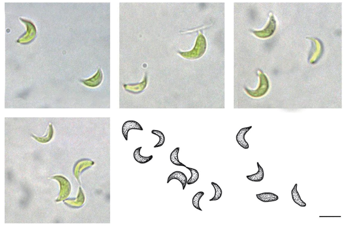
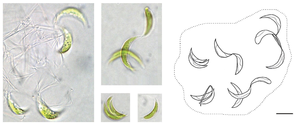
    Monoraphidium convolutum var. pseudosabulosum Hindak 1970 (Fig. 5)

    Description: Cells individual or in a colony, Cell size is 4–6 μm wide, 12–16 μm long. Cells irregular or crescent; side of cell is convex and other side of cell is concave or spindle shape. Cell is blunt rather than acuminated, often associated with the end (Komárek and Fott 1983).

    Ecology and distribution: Eutrophic water in Britain and the Hawaiian Islands, scattered as plankton and metaphyton mesotrophic, but widespread, probably cosmopolitan (Komárek and Fott 1983).

    Site of collection: Gurun-kyo Gurun-ri Hwachon-myeon (H2).

    Specimen: NIBRCL0000104605, ACKU 9-299.

      >  Order Chlorococcales

      >  Family Chlorellaceae

      >  Subfamily Ankistrodesmoideae

    Monoraphidium minutum Komarkova-Legnerova 1969 (Fig. 6)

    Synonyms: Raphidium minutum Nág. 1849, Selenastrum minutum Collins 1907, Ankistrodemus convolutes var. obtusus Printz 1916, A. minutissimus Kors 1953, and A. lunulatus Belcher & Swale 1962.

    Description: Cells very small and individually crescent shaped, end part of cell is spiral. Chloroplast parietal, filling most of the cell, without a pyrenoid. Cells connected with a cell wall (Komárek and Fott 1983). Cell size is 1–3 μm wide, 3–10 μm long.

    This species is smaller than the recorded description (3.5–20 μm long) by Komárková and Legnerová (1969).

    Ecology and distribution: Ontario, China, and New South Wales, scattered in plankton and metaphyton in mesotrophic water; probably cosmopolitan but more in tropical and subtropical waters (Komárek and Fott 1983).

    Site of collection: Gurun-kyo Gurun-ri Hwachon-myeon (H2).

    Specimen: ACKU 9-300.

      >  Order Chlorococcales

      >  Family Chlorellaceae

      >  Subfamily Ankistrodesmoideae

    Raphidocelis mucosa Komarek 1979 (Fig. 7)

    Synonyms: Ankistrodesmus mucosus Korschikov 1953, Kirchneriella mucosa Hind 1977.

    Description: Cells individual or in colonies, with irregular arrangement of cells in mucilage. Cells narrowly fusiform, straight or slightly curved; crescent shape at a blunt end. Chloroplast parietal without a pyrenoid. Cells size is 2–5 μm wide, 4–12 μm long.

    This species was wider than the recorded description (1.5–2.5 μm wide) by Komárková (1979).

    Ecology and distribution: Planktonic in Romania; however, it has been specified up to now only in Czechoslovakia and Russia (Komárek and Fott 1983).

    Site of collection: Palbong-kyo Eoyupo-ri Seo-myeon (H4).

    Specimen: ACKU 9-298.

      >  Order Chlorococcales

      >  Family Radiococcaceae

      >  Subfamily Radiococcoideae

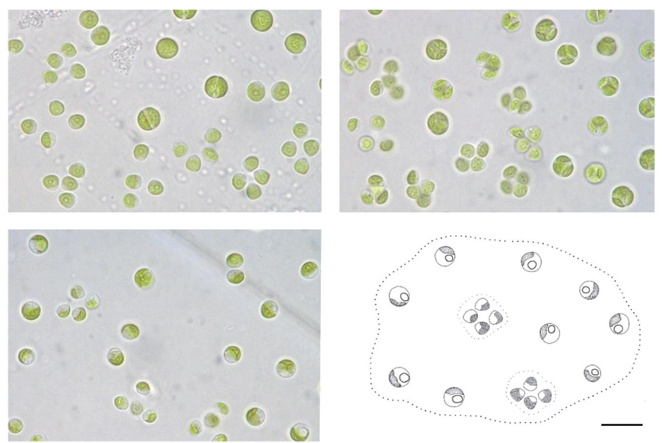
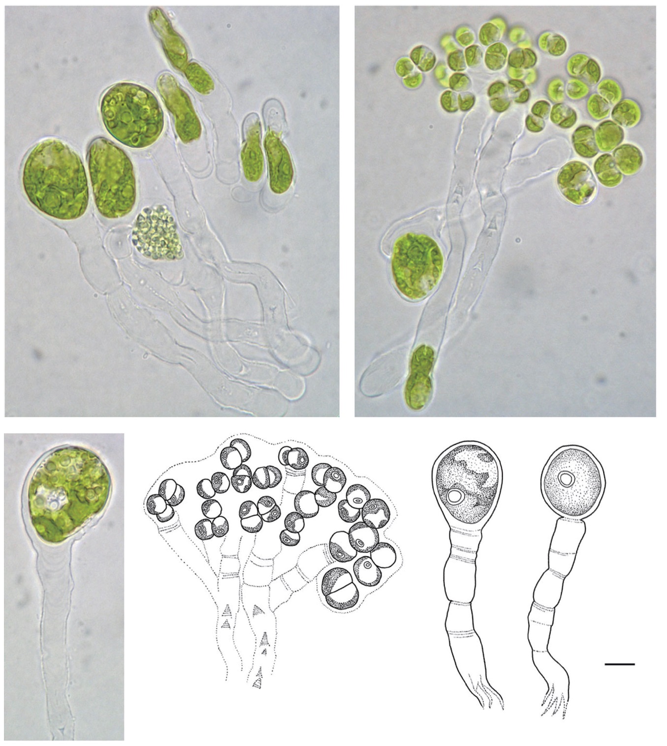
    Eutetramorus tetrasporus Komarek 1983 (Fig. 8)

    Description: Cell size is 3–5 μm in diameter. Colonies 4, 8, or 16-celled but mostly four-celled (Komárek and Fott 1983), Cells spherical, early young cells are triangular in shape with mother cell in mucilage. Mature cells mostly spherical. Chloroplast parietal, each with a pyrenoid.

    Ecology and distribution: Romania, Brazil, and Cuba in eutrophic water as plankton (Komárek and Fott 1983).

    Site of collection: Gurun-kyo Gurun-ri Hwachon-myeon (H2).

    Specimen: ACKU 9-297.

      >  Order Chlorococcales

      >  Family Scenedesmaceae

      >  Subfamily Desmodesmoideae

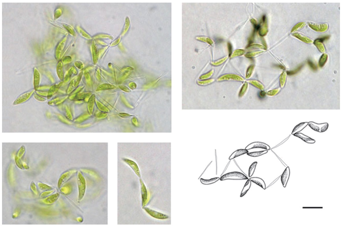
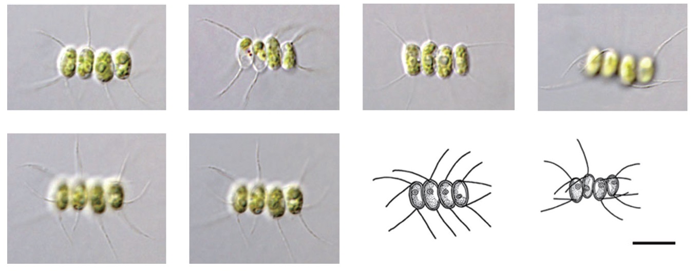
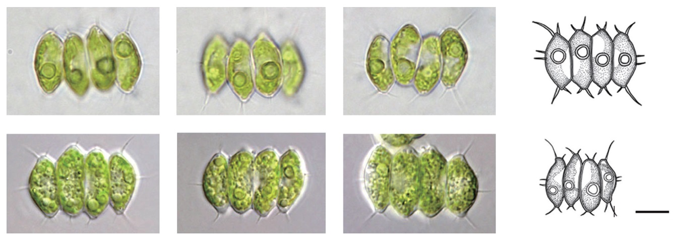
    Scenedesmus flavescens Chodat 1913 (Fig. 9)

    Description: Colonies 2, 4, or 8-celled, most are 4-celled colonies. Outer cell is ellipsoidal, spindle shape, the lateral margins are swollen. Inner cell is elongated oval, spindle shape sometimes rounded and pentagon shaped. Both ends of the outer cell have two spines with a variety of sizes (long and short); center of outer cells has 1–2 short spine. Both ends of the inner cells have 1–2 short spines. Cell size is 8–10 μm wide, 18–30 μm long.

    This species was larger than the recorded description (1.7–7 μm wide, 5.5–15 μm long) by Chodat (1913).

    Ecology and distribution: Britain and Portugal and is planktonic in rivers.

    Site of collection: Palbong-kyo Eoyupo-ri Seo-myeon (H4).

    Specimen: NIBRCL0000104608, ACKU 7-257.

      >  Order Chlorococcales

      >  Family Scenedesmaceae

      >  Subfamily Scenedesmoideae

    Scenedesmus multicauda Masyuk 1962 (Fig. 10)

    Description: Cells ellipsoidal, linear, slightly curved toward the outside. Four-celled colonies, sometimes found as two-celled colonies (Komárek and Fott 1983). Both inner and outer cells outside are long; 1–2 spines at the poles. Cell size is 3–10 μm wide, 7–13 μm long.

    Masyuk (1962) described the spines as long or slightly shorter yet similar, but this species has longer spines than the cell size.

    Ecology: Ponds and lakes as phytoplankton (Komárek and Fott 1983).

    Site of collection: Cheoljeong-kyo Cheoljeong-ri Duch on-myeon (H1). Site of collection: Cheoljeong-kyo Cheoljeong-ri Duch on-myeon (H1).

    Specimen: NIBRCL0000104609, ACKU 7-258.

    The morphological characteristics of the nine taxa

    identified in this study mostly corresponded to the reported characteristics; however, Monoraphidium minutum had a shorter cell length than the recorded description and Raphidocelis mucosa and S. flavescens were larger than their previous descriptions. S. multicauda had larger spines than the cell itself.

    Eutetramorus planctonicus and E. polycoccus were transferred to genus Coenococcus by Komárek and Fott due to a lack of taxonomic characters; however, E. tetrasporus is considered a species.

    The genus Scenedesmus was named by Meyen in 1829 and was classified into four subgenera in 1926 by Cho-dat (Clathrodesmus, Desmodesmus, Euscenedesmus and Rhynchodesmus). However, a recent taxonomic evaluation by Trainor et al.(1976) has divided it into the spiny genus Desmodesmus and the non-spiny genus Scenedesmus (Lürling 2003). S. flavescens and S. multicauda are also considered as genus Desmodesmus according to Trainor et al. (1976).

    The reported number of Korean species in the order Chlorococcales flora is very low compared to the internationally reported taxa. Therefore, more studies in various habitats are needed.

참고문헌
  • 1. An SS, Chang YK 1990 A taxonomic study on the genus Scenedesmus in Korea (1). [Algae] Vol.5 P.123-129 google
  • 2. Andreyeva VM 1975 Novye vidy Chlorella Beijer [New species of the Chlorella Beijer]. [Bot Zhurn (St. Petersburg)] Vol.58 P.1735 google
  • 3. Ann SS, Jang YK 1985 Morphological variation of Scenedesmus quadricauda (Turpin) de Brebisson and Scenedesmus armatu (Chodat) G. M. Smith in culture. [Kor J Bot] Vol.28 P.305-315 google
  • 4. Bourrelly P. 1966 Quelques algues d’eau douce du Canada. [Int. Rev ges Hydrobiol] Vol.51 P.45-126 google cross ref
  • 5. Chodat R 1913 Monographies d’algues en culture pure. Materiaux pour la flore cryptogamique Suisse. Vol 5 P.2 google
  • 6. Chodat R 1926 Scenedesmus Etude de genetique, de systematique experimentale et d’hydrobiologie. [Rev Hydrobiol] Vol.3 P.71-258 google
  • 7. Choi JS, Kim JK 2004 Ichthyofuna and fish community in Hongcheon river, Korea. [Kor J Environ Biol] Vol.18 P.446-455 google
  • 8. Chung J 1993 Illustration of the freshwater algae of Korea. google
  • 9. Guiry MD 2013 Algaebase google
  • 10. Hindak F 1970 A contribution to the systematics of the family Ankistrodesmaceae (Chlorophyceae). [Algol Stud] Vol.1 P.7-32 google
  • 11. Hindak F 1977 Studies on the chlorococcal algae (Chlorophyceae). Vol I. Biologicke prace 23. google
  • 12. Hindak F 1980 Studies on the chlorococcal algae (Chlorophyceae). Vol II. Biologicke prace 26. google
  • 13. Hindak F 1984 Studies on the chlorococcal algae (Chlorophyceae). Vol III. Biologicke prace 30. google
  • 14. Hindak F 1988 Studies on the chlorococcal algae (Chlorophyceae). Vol IV. Biologicke prace 34. google
  • 15. Hirose HM, Akiyama T, Imahori K, Kasaki H, Kumano S, Kobayasi H, Takahashi E, Tsumura T, Hirano M, Yamagishi T 1977 Illustrations of the Japanese freshwater algae. google
  • 16. John DM, Whitton BA, Brook AJ 2002 The freshwater algal flora of the British Isles: An Identification guide to freshwater and terrestrial algae. google
  • 17. John DW, Robert GS 2003 Freshwater algae of North America: Ecology and Classification. google
  • 18. Kim HS, Chung J 1993 Freshwater algal flora of natural swamps in Changnyong County. [Korean J Limnol] Vol.26 P.305-319 google
  • 19. Kim MR, An SS, Chang YK 1991 Morphological variations of Scenedesmus quadricauda var. biornatus Kiss. [Korean J Limnol] Vol.24 P.109-116 google
  • 20. Kim MR, Chang YK 1997 Taxonomic studies on some species of Pediastrum Meyen in Korea. [Algae] Vol.12 P.159-165 google
  • 21. Kim YJ 1998 Ecological characteristics of phytoplankton community in lake Paltang Dam. [Korean J Limnol] Vol.31 P.225-234 google
  • 22. Komarek J 1979 Anderungen in der taxonomie der chlorokokkalalgen. [Archiv Hydrobiol Suppl] Vol.56 P.239-263 google
  • 23. Komarek J 1983 Contribution to the chlorococcal algae of Cuba. [Nova Hedwigia] Vol.37 P.65-180 google
  • 24. Komarek J, Fott B 1983 Chlorophyceae (Grunalgen) ordnung: Chlorococcales. In: Das phytoplankton des susswassers, 7 Teil, 1 Halfte (Thienemann A, ed). E Schweizerbart’sche Verlagsbuchhandlung (Nagele u. Obermiller) google
  • 25. Komarkova ? Legnerova J 1969 The systematics and ontogenesis of the genera Ankistrodesmus Corda and Monoraphidium gen. nov. In: Studies in Phycology (Fott B, ed). E Schweizerbart’sche Verlagsbuchhandlung (Nagele u. Obermiller) P.75-144 google
  • 26. Korshikov O 1953 The Freshwater algae of the Ukrainian SSR. V. Sub-Class Protococcineae. Vacuolales and Protococcales. google
  • 27. Lee OM 1996 Freshwater chlorophyta. In: Species List in Korea (Lee IK, ed). P.405-422 google
  • 28. Lurling M 2003 Phenotypic plasticity in the green algae Desmodesmus and Scenedesmus with special reference to the induction of defensive morphology. [Ann Limnol Int J Lim] Vol.39 P.85-101 google cross ref
  • 29. Masyuk NP 1962 New and interesting species of the genus Scenedesmus MEYEN from basins of the Volynian forest zone. [U krain Bot ?hurn] Vol.19 P.73-83 google
  • 30. Meyen FJF 1829 Beobachtungen uber einige niedere Algenformen. [Nova Acta Physico-Medica Academiae Cae-sareae Leopoldino-Carolinae Naturae] Vol.14 P.768-778 google
  • 31. Pascher A 1915 Die Susswasser-Flora Deutschlands, Osterreichs und der Schweiz. Heft 5. Chlorophyceae II. google
  • 32. Prescott GW, Bicudo CEM, Vinyard WC 1982 A synopsis of North American Desmids. Part II. Section 4. google
  • 33. Prescott GW, Croasdale HT, Vinyard WC 1972 North American flora, Series II. Part 6. Desmidiales. google
  • 34. Prescott GW, Croasdale HT, Vinyard WC 1977 A synopsis of North American Desmids. Part II. Desmidiaceae: Placodermae. Section 2. google
  • 35. Prescott GW, Croasdale HT, Vinyard WC, Bicudo CEM 1981 A synopsis of North American Desmids. Part II, Section 3. google
  • 36. Prescott GW 1973 Algae of the western great lakes area. google
  • 37. Reynolds CS 1984 Phytoplankton periodicity: the interaction of form, function and environmental variability. [Freshw Biol] Vol.14 P.111-142 google cross ref
  • 38. Thunmark S 1945 Zur soziologie des subwasser planktons. Fine methodologisch- okologische Studie. [Folia Limnol Scand] Vol.3 P.1-6 google
  • 39. Trainor FR, Cain JR, Shubert EL 1976 Morphology and nutrition of the colonial green alga Scenedesmus : 80 years later. [Bot Rev] Vol.42 P.5-25 google cross ref
  • 40. Wilcox LW, Lewis LA, Fuerst PA, Floyd GL 1992 Assessing the relationships of autosporic and zoosporic chlorococcalean green algae with 18S rDNA sequence data. [J Phycol] Vol.28 P.381-386 google cross ref
  • 41. Yang HJ, Chae BS, Nam MM 1991 The ichthyofauna in autumn at upper reach of Hongchon River. [Korean J Limnol] Vol.24 P.37-44 google
OAK XML 통계
이미지 / 테이블
  • [ Fig. 1. ]  Sampling sites on Hongcheon River at Gangwon-do from December 2011 to September 2012. H1, Cheoljeong-kyo; H2, Gurun-kyo; H3, Dunji-kyo; H4, Palbong-kyo; H5, Chungui-bridge.
    Sampling sites on Hongcheon River at Gangwon-do from December 2011 to September 2012. H1, Cheoljeong-kyo; H2, Gurun-kyo; H3, Dunji-kyo;
H4, Palbong-kyo; H5, Chungui-bridge.
  • [ Fig. 2. ]  Microscopic photographs and illustration of a newly recorded species, Oocystidium ovale Korshikov, in Hongcheon River of Korea from December 2011 to September 2012. Scale bar, 10 μm.
    Microscopic photographs and illustration of a newly recorded species, Oocystidium ovale Korshikov, in Hongcheon River of Korea from December
2011 to September 2012. Scale bar, 10 μm.
  • [ Fig. 3. ]  Microscopic photographs and illustrations of a newly recorded species, Heleococcus mucicola Korshikov, in Hongcheon River of Korea from December 2011 to September 2012. Scale bar, 10 μm.
    Microscopic photographs and illustrations of a newly recorded species, Heleococcus mucicola Korshikov, in Hongcheon River of Korea from
December 2011 to September 2012. Scale bar, 10 μm.
  • [ Fig. 4. ]  Microscopic photographs and illustration of newly recorded species, Chlorella mirabilis V. M. Andreeva, in Hongcheon River of Korea from December 2011 to September 2012. Scale bar, 10 μm.
    Microscopic photographs and illustration of newly recorded species, Chlorella mirabilis V. M. Andreeva, in Hongcheon River of Korea from
December 2011 to September 2012. Scale bar, 10 μm.
  • [ Fig. 5. ]  Microscopic photographs and illustration of a newly recorded species, Monoraphidium convolutum var. pseudosabulosum Hindak, in Hongcheon River of Korea from December 2011 to September 2012. Scale bar, 10 μm.
    Microscopic photographs and illustration of a newly recorded species, Monoraphidium convolutum var. pseudosabulosum Hindak, in Hongcheon
River of Korea from December 2011 to September 2012. Scale bar, 10 μm.
  • [ Fig. 6. ]  Microscopic photographs and illustrations of newly recorded species, Monoraphidium minutum Komarkova-Legnerova, in Hongcheon River of Korea from December 2011 to September 2012. Scale bar, 10 μm.
    Microscopic photographs and illustrations of newly recorded species, Monoraphidium minutum Komarkova-Legnerova, in Hongcheon River of
Korea from December 2011 to September 2012. Scale bar, 10 μm.
  • [ Fig. 7. ]  Microscopic photographs and illustration of newly recorded species, Raphidocelis mucosa Komarek, in Hongcheon River of Korea from December 2011 to September 2012. Scale bar, 10 μm.
    Microscopic photographs and illustration of newly recorded species, Raphidocelis mucosa Komarek, in Hongcheon River of Korea from December
2011 to September 2012. Scale bar, 10 μm.
  • [ Fig. 8. ]  Microscopic photographs and Illustration of newly recorded species, Eutetramorus tetrasporus Komarek in Hongcheon River of Korea from December 2011 to September 2012. Scale bar, 10 μm.
    Microscopic photographs and Illustration of newly recorded species, Eutetramorus tetrasporus Komarek in Hongcheon River of Korea from
December 2011 to September 2012. Scale bar, 10 μm.
  • [ Fig. 9. ]  Microscopic photographs and illustrations of newly recorded species, Scenedesmus flavescens Chodat, in Hongcheon River of Korea from December 2011 to September 2012. Scale bar, 10 μm.
    Microscopic photographs and illustrations of newly recorded species, Scenedesmus flavescens Chodat, in Hongcheon River of Korea from
December 2011 to September 2012. Scale bar, 10 μm.
  • [ Fig. 10. ]  Microscopic photographs and illustrations of newly recorded species, Scenedesmus multicauda Masyuk, in Hongcheon River of Korea from December 2011 to September 2012. Scale bar, 10 μm.
    Microscopic photographs and illustrations of newly recorded species, Scenedesmus multicauda Masyuk, in Hongcheon River of Korea from
December 2011 to September 2012. Scale bar, 10 μm.
(우)06579 서울시 서초구 반포대로 201(반포동)
Tel. 02-537-6389 | Fax. 02-590-0571 | 문의 : oak2014@korea.kr
Copyright(c) National Library of Korea. All rights reserved.