검색 전체 메뉴
PDF
맨 위로
OA 학술지
Antimicrobial Resistance and Virulence Genes Presence in Escherichia coli Strains Isolated from Gomso Bay, Korea
  • 비영리 CC BY-NC
  • 비영리 CC BY-NC
ABSTRACT
Antimicrobial Resistance and Virulence Genes Presence in Escherichia coli Strains Isolated from Gomso Bay, Korea
KEYWORD
Antimicrobial resistance , Escherichia coli , Gomso Bay , Virulence genes
  • Introduction

    Escherichia coli is a facultative anaerobic bacterium found in the normal flora of the intestinal tract of humans and animals; in contrast, it has also been implicated in infectious diseases (Rosas et al., 2006). Currently, E. coli are widely used as a sanitation indicator of microbiological contamination in water and food (Lang et al., 1999). While, E. coli is harmless in general, certain virulent strains are common causes of infectious diarrhea and other enteric diseases (Clements et al., 2012). Currently, the following E. coli pathotypes have been recognized to cause diarrhea in humans: EPEC, enteropathogenic E. coli; EIEC, enteroinvasive E. coli; STEC, Shiga toxin-producing E. coli; EAEC, enteroaggregative E. coli; ETEC, enterotoxigenic E. coli; and DAEC, diffusely adhering E. coli (Turner et al., 2006).

    For livestock farming, antimicrobial agents are abundantly used not only for medical treatment and prophylaxis of bacterial infections but also in the animals’ food to promote growth, which has resulted in an increase in antimicrobial-resistant bacterial strains (Aarestrup, 2005; Sapkota et al., 2008). The critical factor in the development of antimicrobial resistance is the ability of the bacteria to acquire and disseminate exogenous genes via mobile genetic elements, such as plasmids, transposons, DNA insertion elements, and genomic islands (Rowe-Magnus et al., 2002). Horizontal gene transfer is one of the most common mechanisms, by which antimicrobial resistance traits are transferred from one organism to another (Martinez, 2009). Since the discovery of integrons, a unique mechanism for the dissemination of antibiotic resistance genes (Stokes and Hall, 1989), the increase in bacterial strains resistant to several antimicrobial agents has raised public health concerns about treatment options, heath care costs, and foodborne illness (Hammerum and Heuer, 2009). Food contamination of antimicrobial-resistant bacteria could be a major public health threat, due to the possibility that the responsible genes, which are carried on mobile genetic elements, may be transferred to other bacteria that confer high risks to human health (Rowe-Magnus et al., 2002). E. coli strains, in particular, are candidate vehicles for such genetic transfers, not only due to their diversity but also because they exist as common microflora of the gastrointestinal tracts of humans and animals (Clements et al., 2012).

    In Korea, numerous studies have focused on antimicrobial resistance patterns and genes in E. coli strains isolated from various sources, including humans, food and animals (You et al., 2006; Koo and Woo, 2011; Ryu et al., 2012a). However, very few of these studies provided adequate data regarding the prevalence of E. coli in seawater or seafood.

    In this study, E. coli strains isolated from surface seawater of Gomso Bay, on the west coast of Korea, were characterized for their prevalence, antimicrobial resistance, and virulence gene expression using the disk diffusion method and polymerase chain reaction (PCR).

    Materials and Methods

      >  E. coli isolation and identification

    In total, 131 environmental strains of E. coli were isolated from the surface seawater of Gomso Bay, Jeollabuk-do, Korea, between May and December of 2012. Seawater samples collected from sampling stations were transported within 4 h to the laboratory in ice-cold containers. The number of total coliform and fecal coliform bacteria was determined using the most probable number (MPN) method (American Public Health Association, 1970). Seawater was inoculated with lauryl tryptose broth (Difco; Becton, Dickinson and Co., Franklin Lakes, NJ, USA) and incubated for 48 ± 2 h at 35℃. One loop from each gas-positive tube was inoculated with Brilliant Green Bile Broth (Difco) and EC medium (Difco), and incubated for 48 ± 2 h at 35℃ or 24 ± 2 h at 44.5 ± 0.2℃. Each gas-positive EC tube was plated onto eosin methylene blue agar (Difco) and incubated for 24 h at 35℃. One E. coli isolate per each seawater sample was randomly selected and validated using the API 20E Kit (bioMerieux, Marcy-l’Etoile, France).

      >  Antimicrobial susceptibility testing

    Antimicrobial susceptibility testing against 22 antimicrobials was performed on 131 E. coli isolates using the disk diffusion method on Mueller Hinton agar plates (Difco), according to the standards and interpretive criteria of the Clinical and Laboratory Standards Institute (CLSI) (2010). The following 22 antibiotics were used: amikacin (30 µg), amoxicillin (20 µg), ampicillin (10 µg), cefepime (30 µg), cefotetan (30 µg), cefoxitin (30 µg), ceftriaxone (30 µg), cephalothin (30 µg), chloramphenicol (30 µg), ciprofloxacin (5 µg), erythromycin (15 µg), gentamicin (10 µg), imipenem (10 µg), kanamycin (30 µg), nalidixic acid (30 µg), norfloxacin (10 µg), rifampin (5 µg), streptomycin (10 µg), sulfamethoxazole/trimethoprim (23.75/1.25 µg), tetracycline (30 µg), ticarcillin (75 µg), and trimethoprim (5 µg). Antimicrobial susceptibility test discs were purchased from Oxoid Ltd. (Basingstoke, Hampshire, UK), and the results were classified as susceptible, intermediate, or resistant according to the zone diameter interpretive standards recommended by CLSI (2010). The quality-control strains, E. coli ATCC 25922 and Staphylococcus aureus ATCC 25923, were included in each run.

      >  PCR amplification of antimicrobial resistance and virulence genes

    The presence of genes associated with tetracycline (tetA, tetB, and tetC) or β-lactams (TEM, SHV, and OXA) in 44 tetracycline-resistant or 29 ampicillin-resistant isolates, respectively, were detected using PCR. All isolates were also evaluated by PCR for the following genes: heat labile (lt), heat stable (st), attaching and effacing (eae), EAEC-associated plasmid (pCVD432), invasion-associated locus (ial), and Shiga toxin (stx1 and stx2) genes. PCR was performed using the primer sets listed in Table 1. Genomic DNA was extracted from overnight bacterial cultures using the boiling method. After cooling, samples were microcentrifuged to remove cell debris, and the resulting supernatants were used as PCR templates. In each PCR reaction, 3 µL of the respective DNA template was suspended in 22 µL reaction mixtures containing 2.5 µL of 10× Ex Taq buffer (Takara, Kyoto, Japan), 50 pmol of each primer, 2 µL of dNTPs (2.5 mM of each dNTP), and 2 units of Ex Taq DNA polymerase (5 U/µL). PCR amplification of tetracycline-associated genes was conducted under the following conditions: initial denaturation at 94℃ for 5 min; 30 cycles of denaturation at 94℃ for 30 s, annealing at 50℃ for 30 s, and extension at 72℃ for 1 min; and a final extension step at 72℃ for 10 min. PCR amplification of β-lactam-associated genes was conducted under the following conditions: initial denaturation at 95℃ for 5 min; 30 cycles of denaturation at 94℃ for 30 s, annealing at 58℃ for 15 s, and extension at 72℃ for 1 min; and a final extension step at 72℃ for 10 min. PCR conditions for detecting virulence genes consisted of initial denaturation at 95℃ for 5 min, followed by 30 cycles of denaturation at 95℃ for 30 s, annealing at 52℃ for 30 s, and extension at 72℃ for 1 min. All PCR reactions were performed using the Gene Amp PCR System 9700 (Perkin Elmer, Norwick, CT, USA) and the amplified products were electrophoresed on 1.5% agarose gels stained with ethidium bromide and visualized under UV light.

    Results

      >  Isolation of E. coli strains from surface seawater in Gomso Bay

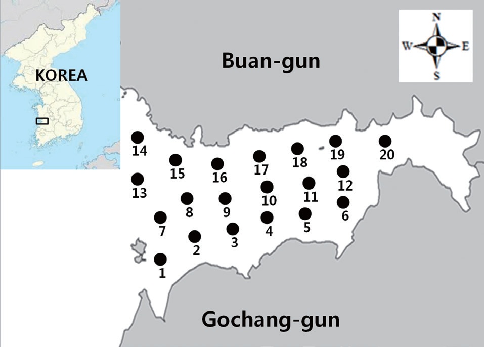
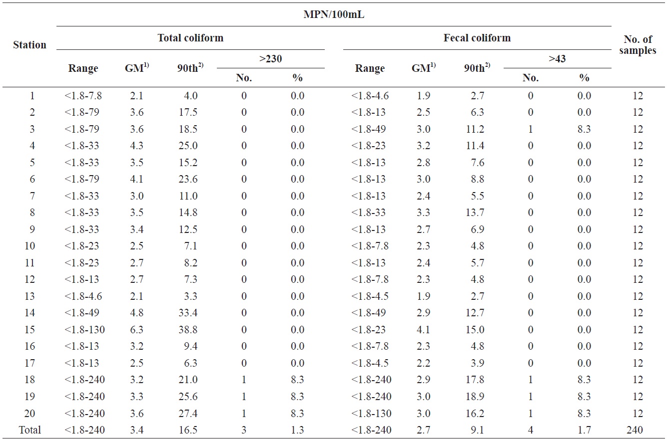
    A sanitary survey in an important shellfish-growing area of Gomso Bay, west coast of Korea, was conducted to evaluate bay conditions and compliance with bacteriological criteria for shellfish production. Seawater samples were collected 12 times from May to December 2012 from 20 sampling stations established in the survey area (Fig. 1). The values determined by the MPN-method for both the total coliform bacteria and fecal coliform bacteria in the 240 seawater samples ranged from less than 1.8 to 240 MPN/100 mL, respectively (Table 2). The geometric means and estimated 90th percentile values for total coliform bacteria ranged from 2.1 to 6.3 MPN/100 mL, and from 3.3 to 38.8 MPN/100 mL, respectively, whereas those for fecal coliform bacteria ranged from 1.9 to 4.1 MPN/100 mL,

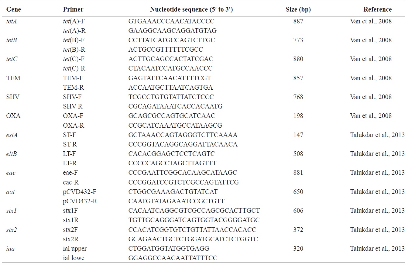
    [Table 1.] Oligonucleotide primers used in this study

    label

    Oligonucleotide primers used in this study

    and from 2.7 to 18.9 MPN/100 mL, respectively. Notably, the levels of total coliform and fecal coliform bacteria from stations near the wastewater discharge sites (stations 3, 14, 18, 19, and 20) were slightly higher than those from other sites. The bacteria levels after rainfall were also higher, than those in the dry season. Based on these data, the bacteriological quality of seawater in Gomso Bay met the criteria recommended by the National Shellfish Sanitation Program (Food and Drug Administration, 2005) and the Korea Shellfish Sanitation Program for shellfish production (Ministry of Marine Affairs and Fisheries, 2006). One strain from each seawater sample was isolated to further analyze antimicrobial resistance and virulence genes in isolated E. coli strains.

      >  Antimicrobial susceptibility of E. coli isolates

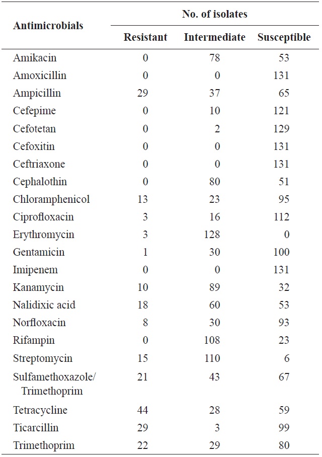
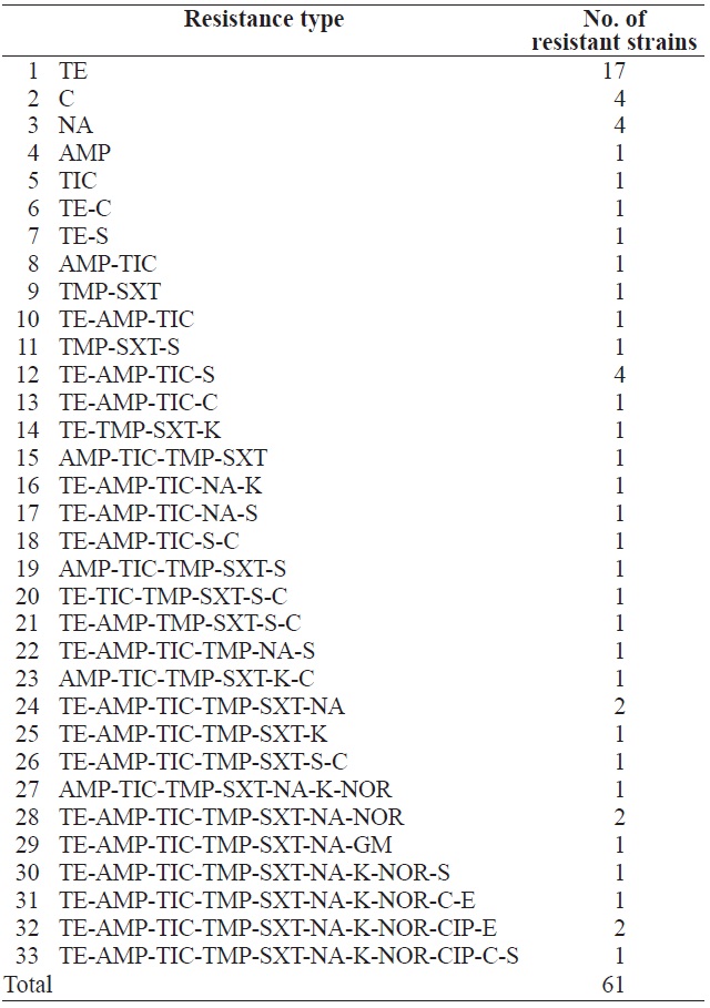
    In total, 131 isolates were tested for resistance against 22 antimicrobial agents (Table 3). Resistance to tetracycline (33.6%), ampicillin (22.1%), ticarcillin (22.1%), trimethoprim (16.8%), sulfamethoxazole/trimethoprim (16.0%), nalidixic acid (13.7%), streptomycin (11.5%), chloramphenicol (9.9%), kanamycin (7.6%), norfloxacin (6.1%), ciprofloxacin (2.3%), erythromycin (2.3%), and gentamicin (0.8%) was observed. These results are in agreement with several previous studies (You et al., 2006; Van et al., 2008; Koo and Woo, 2012; Ryu et al., 2012b). However, no isolate showed resistance to amikacin, amoxicillin, cefepime, cefotetan, cefoxitin, ceftriaxone,

    [Table 2.] Summary of bacteriological examination results of each sampling stations in Gomso Bay, from May to December 2012

    label

    Summary of bacteriological examination results of each sampling stations in Gomso Bay, from May to December 2012

    cephalothin, imipenem, or rifampin. Table 4 shows the resistance patterns of these isolates. Among the 131 E. coli isolates, 70 (53.4%) were not resistant to any of the 22 antimicrobial agents tested, whereas 61 (46.6%) were resistant to at least one antimicrobial agent. In total, 22.9% of the isolates were resistant to three or more classes of antimicrobials and defined as multidrug resistant. One strain, in particular, exhibited resistance to 11 antimicrobial agents (AMP, C, CIP, K, NA, NOR, S, SXT, TE, TIC, and TMP).

      >  Characterization of antimicrobial resistance genes in E. coli isolates

    Ampicillin-resistant isolates (n = 29) were investigated for the presence of β-lactamase-encoding genes using PCR with the primer sets specified in Table 1. We found that 27 isolates (93.1%) contained genes encoding TEM-type β-lactamases, whereas the rest had genes associated with SHV-type β-lactamases (data not shown), consistent with previous findings indicating that the former genes were among the most prevalent in ampicillin-resistant E. coli isolates derived from food and livestock (Van et al., 2008; Aslam et al., 2009; Ryu et al., 2012b).

    Among the 44 tetracycline-resistant E. coli isolates, the tetA and tetC genes were found in 35 (79.5%) and 19 (43.2%) isolates, respectively, and tetB was found in only 3 (6.8%). Fourteen isolates (31.8%) tested positive for both tetA and tetC. Our results also showed that three tetracycline-resistant isolates possessed none of the tetracycline resistance genes analyzed (tetA, tetB, or tetC) (data not shown). These findings were consistent with previous studies demonstrating that genes encoding active efflux pumps of tetracycline, such as tetA-E and tetG, occurred frequently among tetracycline-resistant E. coli isolates derived from livestock and aquaculture environments (Van et al., 2008; Aslam et al., 2009; Tang et al., 2011; Koo and Woo, 2012; Ryu et al., 2012b).

      >  Detection of virulence genes by PCR

    All 131 isolates were also examined for the presence of seven virulence genes (lt, st, eae, pCVD432, ial, stx1, and stx2) using PCR. Approximately 3.1% (n = 4) of the isolates were positive for at least one of the seven virulence genes specific to certain E. coli pathotypes. Only 0.8% (n = 1) and

    [Table 3.] Antimicrobial resistance of Escherichia coli isolated from surface seawater in Gomso Bay

    label

    Antimicrobial resistance of Escherichia coli isolated from surface seawater in Gomso Bay

    2.3% (n = 3) of the isolates were positive for the EAEC-associated plasmid (pCVD432) and the EPEC-specific attaching and effacing (eae) genes, respectively (data not shown). However, the STEC-associated stx1 and stx2 virulence genes, the ETEC-specific heat-stable toxin (ST) and heat-labile toxin (LT) genes, and the EIEC-specific invasion-associated locus (ial) gene were not detected. These results imply that none of the isolates were of the STEC, ETEC, or EIEC pathotype. The virulence gene frequency in the 131 isolates of this study was lower than that reported previously (Ram et al., 2008; Van et al., 2008; Talukdar et al., 2013).

    Discussion

    Our current study focused primarily on the prevalence of E. coli in Gomso Bay as well as the characterization of antimicrobial resistance and virulence genes in these isolates. Gomso Bay, connected to the Yellow Sea, is located between Gochang-gun and Buan-gun in Jeollabuk-do, Korea, covering approximately 76 km2 with an average width, length, and depth of 5-6 km, 17 km, and 2-3 m, respectively. The bay is

    [Table 4.] Antimicrobial resistance patterns of Escherichia coli isolated from surface seawater in Gomso Bay

    label

    Antimicrobial resistance patterns of Escherichia coli isolated from surface seawater in Gomso Bay

    a source of shellfish and other aquaculture organisms. Waste from agriculture, household and stock breeding of numerous small rivers that flow into Gomso Bay are thought to be the current source of contaminants in the area.

    Virulent or antimicrobial-resistant bacteria in drainage water can contaminate shellfish and fisheries in the bay and potentially be hazardous for human health. Many antibiotics are used for therapeutic treatment and prophylactic purposes in animals and humans; consequently, increase in antimicrobial resistance due to excess use and misuse of antimicrobials is a public health concern (World Health Organization, 2001). Therefore, antimicrobial usage in animal feed should be tightly regulated to decrease the prevalence of antimicrobial-resistant bacteria.

    In this study, 131 environmental E. coli strains isolated from surface seawater of Gomso Bay were tested for resistance against 22 antimicrobial agents. Tetracycline resistance was most prevalent, followed by that to ampicillin, ticarcillin, trimethoprim, sulfamethoxazole/trimethoprim, nalidixic acid, and streptomycin. Of the 61 isolates displaying resistance to at least 1 antimicrobial, resistance patterns varied and were classified among 33 types (Table 4).

    Ampicillin resistance in gram-negative bacteria is primarily mediated by β-lactamases, which hydrolyze the β-lactam ring and thereby inactivate antibiotics (Massova and Mobashery, 1998). In many genera of gram-negative bacteria, the most predominant β-lactamases are TEM-, SHV-, CTX-, and OXA-type enzymes (Bradford, 2001). TEM-1, a plasmid-mediated β-lactamase, is the most common β-lactamase in gram-negative bacteria, and accounts for up to 90% of the ampicillin resistance in E. coli (Livermore, 1995). In the present study, we found the TEM-type β-lactamases to be the most prevalent among 29 ampicillin-resistant isolates, consistent with previous studies (Livermore, 1995; Van et al., 2008; Aslam et al., 2009; Ryu et al., 2012b). In addition, both the tetA and tetC genes prevailed among the 44 tetracycline-resistant E. coli isolates. In the analyses of virulence genes, only 3.1% of the isolates were positive for the EAEC-associated plasmid (pCVD432) and the EPEC-specific attaching and effacing (eae) genes.

    E. coli is a facultative anaerobic bacterium found in the normal intestinal flora in humans and animals, however, some toxic strains can induce pathogenicity ranging from mild diarrhea to fatal complications (Tannock, 1995; Clements et al., 2012). While we did not investigate the mechanisms underlying the development of antimicrobial resistance in these E. coli isolates, several previous studies have established a causal relationship between antimicrobial use in aquaculture and the increase in specific antimicrobial-resistant bacterial strains (Schmidt et al., 2001; Petersen et al., 2002; Ribeiro et al., 2010; Ye et al., 2013).

    In conclusion, continuous surveillance of antimicrobial-resistant bacteria in Gomso Bay is necessary to ensure the safety of fisheries and shellfish production.

참고문헌
  • 1. Aarestrup FM 2005 Veterinary drug usage and antimicrobial resistance in bacteria of animal origin. [Basic Clin Phamacol Toxicol] Vol.96 P.271-281 google cross ref
  • 2. 1970 Recommended Procedures for the Examination of Sea Water and Shellfish google
  • 3. Aslam M, Diarra MS, Service C, Rempel H 2009 Antimicrobial resistance genes in Escherichia coli isolates recovered from a commercial beef processing plant. [J Food Prot] Vol.72 P.1089-1093 google
  • 4. Bradford PA 2001 Extended-spectrum β-lactamases in the 21st century: characterization, epidemiology, and detection of this important resistance threat. [Clin Microbiol Rev] Vol.14 P.933-951 google
  • 5. Clements A, Young JC, Constantinou N, Frankel G 2012 Infection strategies of enteric pathogenic Escherichia coli. [Gut Microbes] Vol.3 P.71-87 google
  • 6. 2010 Performance Standards for Antimicrobial Susceptibility Testing. google
  • 7. 2005 National Shellfish Sanitation Program (NSSP) Guide for the Control of Molluscan Shellfish. II. Growing Areas. P.199-204 google
  • 8. Hammerum AM, Heuer OE 2009 Human health hazards from antimicrobial-resistant Escherichia coli of animal origin. [Clin Infect Dis] Vol.48 P.916-921 google
  • 9. Koo HJ, Woo GJ 2011 Distribution and transferability of tetracycline resistance determinants in Escherichia coli isolated from meat and meat products. [Int J Food Microbiol] Vol.145 P.407-413 google cross ref
  • 10. Koo HJ, Woo GJ 2012 Characterization of antimicrobial resistance of Escherichia coli recovered from foods of animal and fish origin in Korea. [J Food Prot] Vol.75 P.966-972 google
  • 11. Lang MM, Ingham SC, Ingham BH 1999 Verifying apple cider plant sanitation and hazard analysis critical control point programs: choice of indicator bacteria and testing methods. [J Food Prot] Vol.62 P.887-893 google
  • 12. Livermore DM 1995 β-lactamases in laboratory and clinical resistance. [Clin Microbiol Rev] Vol.8 P.557-584 google
  • 13. Martinez JL 2009 The role of natural environments in the evolution of resistance traits in pathogenic bacteria. [Proc Biol Sci] Vol.276 P.2521-2530 google
  • 14. Massova I, Mobashery S. 1998 Kinship and diversification of bacterial penicillin-binding proteins and β-lactamases. [Antimicrob Agents Chemother] Vol.42 P.1-17 google
  • 15. 2006 Sanitary Criteria of Producing ? Processing Facilities and Sea Water Area for Fisheries Products. The notification of Ministry of Maritime Affairs and Fisheries google
  • 16. Petersen A, Andersen JS, Kaewmak T, Somsiri T, Dalsgaard A 2002 Impact of integrated fish farming on antimicrobial resistance in a pond environment. [Appl Environ Microbiol] Vol.68 P.6036-6042 google cross ref
  • 17. Ram S, Vajpayee P, Shanker R 2008 Contamination of potable water distribution systems by multiantimicrobial-resistant enterohemorrhagic Escherichia coli. [Environ Health Perspect] Vol.116 P.448-452 google
  • 18. Ribeiro RV, Reis EMF, Reis CMF, Freitas-Almeida AC, Rodrigues DP 2010 Incidence and antimicrobial resistance of enteropathogens isolated from an integrated aquaculture system. [Lett Appl Microbiol] Vol.51 P.611-618 google
  • 19. Rosas I, Salinas E, Martinez L, Calva E, Cravioto A, Eslava C, Amabile-Cuevas CF 2006 Urban dust faecal pollution in Mexico City: antibiotic resistance and virulence factors of Escherichia coli. [Int J Hyg Environ Health] Vol.209 P.461-470 google cross ref
  • 20. Rowe-Magnus DA, Guerout AM, Mazel D 2002 Bacterial resistance evolution by recruitment of super-integron gene cassettes. [Mol Microbiol] Vol.43 P.1657-1669 google
  • 21. Ryu SH, Lee JH, Park SH, Song MO, Park SH, Jung HW, Park GY, Choi SM, Kim MS, Chae YZ, Park SG, Lee YK 2012a Antimicrobial resistance profiles among Escherichia coli strains isolated from commercial and cooked foods. [Int J Food Microbiol] Vol.159 P.263-266 google
  • 22. Ryu SH, Park SG, Choi SM, Hwang YO, Ham HJ, Kim SU, Lee YK, Kim MS, Park GY, Kim KS, Chae YZ 2012b Antimicrobial resistance and resistance genes in Escherichia coli strains isolated from commercial fish and seafood. [Int J Food Microbiol] Vol.152 P.14-18 google
  • 23. Sapkota A, Sapkota AR, Kucharski M, Burke J, McKenzie S, Walker P, Lawrence R 2008 Aquaculture practices and potential human health risks: current knowledge and future priorities. [Environ Int] Vol.34 P.1215-1226 google
  • 24. Schmidt AS, Bruun MS, Dalsgaard I, Larsen JL 2001 Incidence, distribution, and spread of tetracycline resistance determinants and integron-associated antibiotic resistance genes among motile aeromonads from a fish farming environment. [Appl Environ Microbiol] Vol.67 P.5675-5682 google
  • 25. Stokes HW, Hall RM 1989 A novel family of potentially mobile DNA elements encoding site-specific gene-integration functions: integrons. [Mol Microbiol] Vol.3 P.1669-1683 google
  • 26. Talukdar PK, Rahman M, Rahman M, Nabi A, Islam Z, Hoque MM, Endtz HP, Islam MA 2013 Antimicrobial resistance, virulence factors and genetic diversity of Escherichia coli isolates from household water supply in Dhaka, Bangladesh. [PLoS One] Vol.8 P.e61090 google
  • 27. Tang X, Tan C, Zhang X, Zhao Z, Xia X, Wu B, Guo A, Zhou R, Chen H 2011 Antimicrobial resistances of extraintestinal pathogenic Escherichia coli isolates from swine in China. [Microb Pathog] Vol.50 P.207-212 google
  • 28. Tannock GW. 1995 Normal Microflora: An Introduction to Microbes Inhabiting the Human Body. google
  • 29. Turner SM, Scott-Tucker A, Cooper LM, Henderson IR 2006 Weapons of mass destruction: virulence factors of the global killer enterotoxigenic Escherichia coli. [FEMS Microbiol Lett] Vol.263 P.10-20 google cross ref
  • 30. Van TTH, Chin J, Chapman T, Tran LT, Coloe PJ 2008 Safety of raw meat and shellfish in Vietnam: an analysis of Escherichia coli isolations for antibiotic resistance and virulence genes. [Int J Food Microbiol] Vol.124 P.217-223 google cross ref
  • 31. 2001 Global Strategy for Containment of Antimicrobial Resistance. google
  • 32. Ye L, Lu Z, Li X, Shi L, Huang Y, Wang HH 2013 Antibiotic-resistant bacteria associated with retail aquaculture products from Guangzhou, China. [J Food Prot] Vol.76 P.295-301 google cross ref
  • 33. You JY, Moon BM, Oh IG, Baek BK, Li LG, Kim BS, Stein BD, Lee JH 2006 Antimicrobial resistance of Escherichia coli O157 from cattle in Korea. [Int J Food Microbiol] Vol.15 P.74-78 google cross ref
OAK XML 통계
이미지 / 테이블
  • [ Table 1. ]  Oligonucleotide primers used in this study
    Oligonucleotide primers used in this study
  • [ Fig. 1. ]  Location of sample collection stations in Gomso Bay, Republic of Korea, from May to December 2012.
    Location of sample collection stations in Gomso Bay, Republic of
Korea, from May to December 2012.
  • [ Table 2. ]  Summary of bacteriological examination results of each sampling stations in Gomso Bay, from May to December 2012
    Summary of bacteriological examination results of each sampling stations in Gomso Bay, from May to December 2012
  • [ Table 3. ]  Antimicrobial resistance of Escherichia coli isolated from surface seawater in Gomso Bay
    Antimicrobial resistance of Escherichia coli isolated from surface seawater in Gomso Bay
  • [ Table 4. ]  Antimicrobial resistance patterns of Escherichia coli isolated from surface seawater in Gomso Bay
    Antimicrobial resistance patterns of Escherichia coli isolated from surface seawater in Gomso Bay
(우)06579 서울시 서초구 반포대로 201(반포동)
Tel. 02-537-6389 | Fax. 02-590-0571 | 문의 : oak2014@korea.kr
Copyright(c) National Library of Korea. All rights reserved.