Freshwater green algae are characterized by having high species diversity (John 1994). The freshwater green algae include a greater diversity of cellular organization, morphological structures and reproductive processes than any other algae (Bold et al. 1978). The order Chlorococcales occur in the plankton and benthos of mesotrophic or eutrophic and waters and the highest frequency and abundance. Especially from spring to fall, their reproduction is so extensive that they can bring about a strong green coloration of the water (Hindak 1977). Thus, they play an important role in primary and secondary successional processes (John and Tsarenko 2002).
Studies in freshwater algae in Korea were initiated by Kawamura (1918), who first reported the genus
Families Botryococcaceae and Dictyosphaeriaceae are divided or merged according to a few scholars. Prescott (1962) included the family Dictyosphaeriaceae in the family Oocystaceae, which is composed of unicellular species or colonies. However, Smith (1950) separately classified the family Dictyosphaeriaceae, as cells in the genus
Genera belonging to the family Characiaceae are solitary or are joined in radiate colonies, and may be attached on other filamentous algae or free floating. This family is very small, and includes the genus
The reported taxa belong to the family Botryococcaceae and Characiaceae in Korea comprise a total of 6 genera and 13 species, which are
The purpose of this study is to reviewed about newly recorded species in algal flora of Korea (Kim and Kim 2012) of the family Botryococcaceae and Characiaceae, Chlorococcales and Chlorophyceae, representing 8 genera and 20 species collected from swamps, ponds, reservoirs, lakes and rivers in Korea from May 2011 to January 2012 (Table 1). We present taxonomic information, illustrations, classifications, references, synonyms, basionyms and distributions in Korea.
The samples of Chlorococcal algae were collected at 21 stations including ponds, swamp, reservoirs, lakes and rivers from May 2011 to January 2012 (Table 1). Sampling stations were located throughout the country. All samples were collected using 10 µm or 20 µm mesh-sized plankton nets with vertical and/or horizontal towing, or submerged benthic or soil algae with spoid or brush. Chlorococcal algae samples were immediately fixed with Lugol’s iodine solution (0.5%) for immobilizing the cells to facilitate microscopic examination. To examine the fine structures and cellular shapes, and to identify and classify the Chlorococcal species, a temporary slide was made using the follow steps: 1) the phytoplankton samples (Chloroccocal algae) were mixed with glycerin in micro tubes. 2) The mixed samples were placed, drop-wise on slide glass, and were fixed in position with cover slides. Permanent slides were made using the follow steps: 1) the phytoplankton samples (Chloroccocal algae) were mixed with liquid glycerol gelatin for mounting histochemical slides (Sigma-Aldrich, St. Louis, MO, USA). 2) The mixed sample was placed drop-wise on slide glass and was fixed in position with a cover slide. 3) It was cemented the margin of a cover glass with manicure (Thecashop, Seoul, Korea). The temporary and permanent slides were observed at × 200 to ×1,000 magnification using light microscopy (LM) ( Axioskop 20 and Axio Imager A2; Carl Zeiss) with an attached digital camera ( Axiocam HRc; Carl Zeiss ) being used to capture images. The scale bar in illustrations represents 10µm.
At each station, physical and chemical factors of water were recorded during the sampling periods. Water temp. (Water temperature,℃) and EC (electric conductivity) was measured in situ using a portable thermometer and EC meter (Orion 5-star; Thermo Scientific, Waltham, MA, USA) and a pH was measured in situ using a pH meter (Ultrabasic-5; Denver Instrument, Bohemia, NY, USA) respectively. This study used the data of total nitrogen (TN) and total phosphate (TP) concentrations at each sampling station from the water information system of the Ministry of Environment (NIER 2013).
Chlorococcal algae identification were mainly based on Komarek and Fott (1983), John and Tsarenko (2002), Hindak (1977, 1980, 1984, 1988), Prescott (1962), Hirose and Yamagishi (1977) and Yamagishi and Akiyama (1984–1996).
Family Botryococcaceae and Characiaceae, of the order Chlorococcales are composed of 8 genera, 20 species in this study. 10 taxa of these families are newly recorded in algal flora of Korea and reviewed in this study :
Genus Dictyosphaerium G.S. West
Dictyosphaerium pulchellum Wood 1873
Cells spherical, 3-10 µm in diameter.
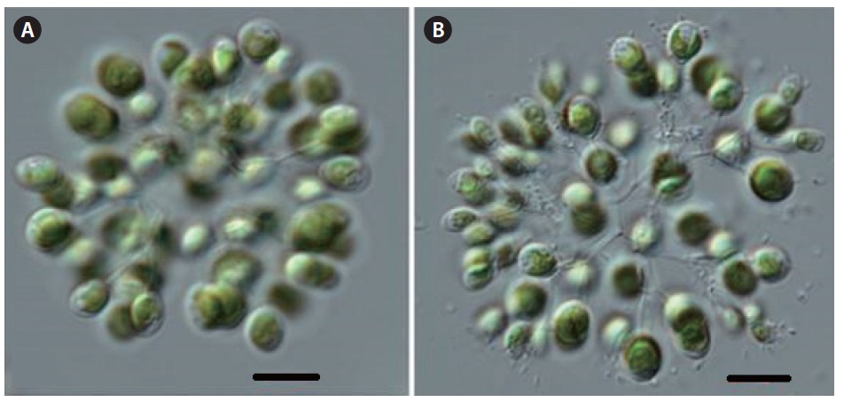
Dictyosphaerium elegans Nageli 1849 (
Synonym:
Illustration: Colonies free-floating, and composed of 16, 32 cells, enclosed with a hyaline, gelatinous envelope. Cells ovoid to ellipsoid or reverse egg-form having one end slightly narrow than the other, attached with the narrow end to radially, di- or tetrachotomously branched gelatinous threads, lying sparsely near periphery of the gelatinous envelope, grouping generally in four cells which
[Table 1.] Sampling sites of Botryococcaceae and Characiaceae, Chlorococcales, 2011 and 2012.
Sampling sites of Botryococcaceae and Characiaceae, Chlorococcales, 2011 and 2012.
are distant from one another. A single chloroplast is cup-shaped, without a pyrenoid. Cells 2-7 µm in diameter, and 3-8 µm long.
Information of sampling sites: This species inhabits lakes and reservoirs as plankton, and was collected from Lake Seoho (03 Sep 2011: water temp. 24.1℃, pH 7.3; EC 569 µs cm-1, TN 6.515 mg L-1, TP 0.278 mg L-1 ), Ugeum reservoir (29 Aug 2011: water temp. 25.9℃, pH 7.5, EC 128 µs cm-1, TN 1.675 mg L-1, TP 0.043 mg L-1 ).
Key reference: Bachmann (1913).
Remark: This taxon is already reported in Algal flora of Korea by Kim and Kim (2012).
Dictyosphaerium ehrenbergianum Nageli
Cells are oval, ellipsoidal. Cells 3-7 µm in diameter, and 4-10µm long.
Dictyosphaerium reniforme Bulnheim 1859 (
Illustration: Colonies free-floating, spherical to broad ellipsoid, and composed of 16-64 or more cells, enclosed with a hyaline, gelatinous envelope. Cells reniform in side view and broad-ellipsoid in face view attached by their convex side to the radially, di- or tetrachotomously branched gelatinous threads, lying near the periphery of the gelatinous envelope, grouping generally in four cells. Single chloroplast, cup-shaped, lying on the convex side of the cells, with a pyrenoid. Cells 3-10 µm in diameter, and 6-20 µm long.
Information of collected sites: This species inhabits lakes and reservoirs as plankton, and was collected from Lake Soeho (03 Sep 2011: water temp. 24.1℃, pH 7.3, EC 569 µs cm-1, TN 6.515 mg L-1, TP 0.278 mg L-1 ).
Key reference: Bulnheim (1859).
Remark: This taxon is already reported in Algal flora of Korea by Kim and Kim (2012).
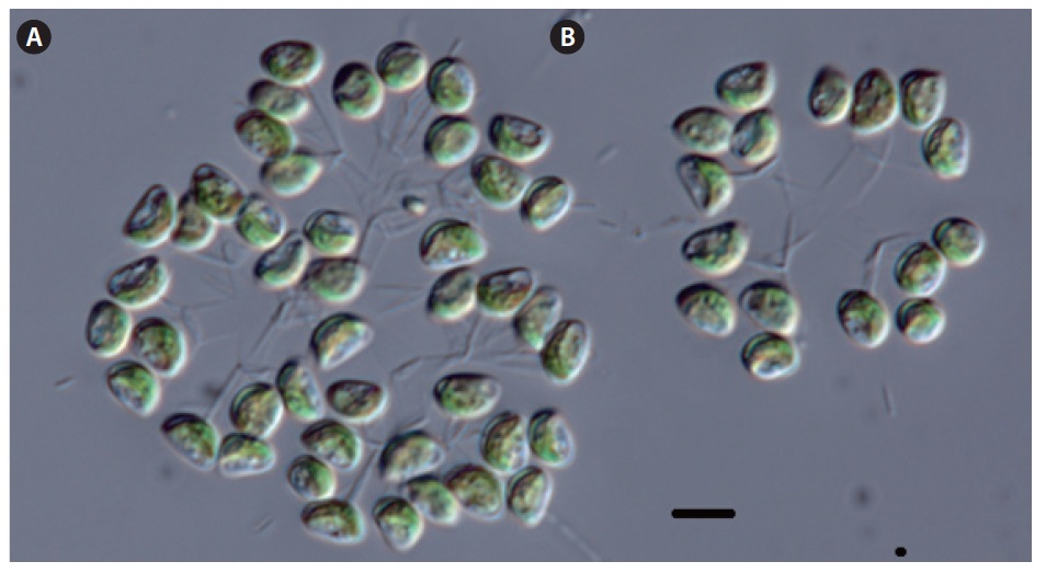
Dictyosphaerium simplex Korshikov 1953 (
Synonym:
Illustration: Colonies free floating, composed of 4-64 celled, only rarely containing more cells, with distinct and regular stalk branching, 4-celled colonies 25-40 µm in diameter, 64 celled to 50 µm in diameter. Mucilaginous envelopes hyaline, not stratified, 5-8 µm round cells. Cell shape varies depending on age. Mature cells spherically oval or oval, attached to stalks laterally, by the medium part of the longer wall on di- or tetrachotomously branched gelatinous threads, lying near the periphery of the gelatinous envelope, grouping generally in four cells. Sporadically a continuous layer of dense mucilage to 2 µm thick is formed around cells, or a fine layer of regularly distributed short rod-like colorless spines. Chloroplast cup-shaped, with one big oval pyrenoid. Cells 8-15 µm in diameter, and 13-20 µm long.
Information of sampling sites: This species inhabits lakes and reservoirs as plankton, and is collected from Lake Seoho (03 Sep 2011: water temp. 24.1℃, pH 7.3, EC 569 µs cm-1, TN 6.515 mg L-1, TP 0.278 mg L-1), Ugeum reservoir (29 Aug 2011: water temp. 25.9℃, pH 7.5, EC 128 µs cm-1, TN 1.675 mg L-1, TP 0.043 mg L-1).
Key reference: Korsikov (1953).
Remark: This taxon is already reported in Algal flora of Korea by Kim and Kim (2012).
Dictyosphaerium elongatum Hindak 1977 (
Synonym:
Illustration: Colonies free-floating, spherical to ellipsoid, composed of 4, 16, 32, or 64 celled or more celled with distinct and regular gelatinous threads branching, gelatinous envelopes hyaline, not stratified. Cells long, asymmetrically oval-fusiform, attached to the branched gelatinous threads by the median portion of cells. Single chloroplast, cup-shaped included a pyrenoid. Cells 6-10 µm in diameter, and 15-20 µm long.
Information of sampling sites: This species inhabit in lakes and reservoirs as plankton, and was collected from Lake Soeho (03 Sep 2011: water temp. 24.1℃, pH 7.3, EC 569 µs cm-1, TN 6.515 mg L-1, TP 0.278 mg L-1), Ugeum reservoir (29 Aug 2011: water temp. 25.9℃, pH 7.5, EC 128 µs cm-1, TN 1.675 mg L-1, TP 0.043 mg L-1).
Key reference: Hindak (1977).
Remark: This taxon is already reported in Algal flora of Korea by Kim and Kim (2012).
Genus Dimorphococcus A. Braun
Dimorphococcus lunatus A. Braun
Cells reniform to cordate with rounded ends. Cells 4-15 µm in diameter, and 9-25 µm long
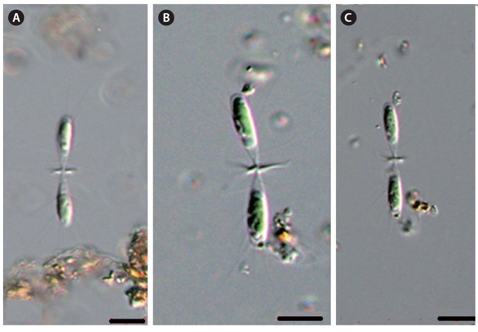
Genus Dichotomococcus Korshikov
Dichotomococcus curvatus Korshikov 1939
Cells elongate-ovoid to ellipsoid, broader at the base than at the poles, in radially arranged in groups of 2-4 apposed at their bases, cell groups at the periphery of wide, homogeneous mucilage envelope. Cells 2-4 µm in diameter, and 5.5-11 µm long.
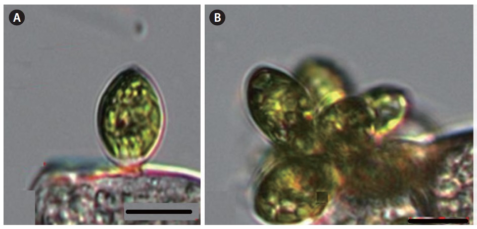
Genus Botryococcus Kutzing 1849
Botryococcus braunii Kutzing 1849
Colonies globes or ovoid, botryoidal, cells elongated, ovoid or ellipsoid. Cells 3-6 µm in diameter, and 6-15 µm long.
>
Family Characiaceae (Nageli) Wille in Warming 1884
Genus Characium A. Braun in Kutzing 1849
Characium rostratum Rabenhorst ex Printz 1914
Cells elongated fusiform, slightly curved. Cells with stipe 5-8 µm in diameter , and 28-36 µm long.
Characium pringsheimii (A. Braun) Komarek 1855
Cells narrowly elongated ovoid to fusiform. Cells with stipe 7-10 µm in diameter, and 18-33 µm long.
Characium ambiguum Hermann in Rabenhorst 1863
Cells lanceolate, fusiform or ensiform shaped. Cells with stipe 4-8 µm in diameter, and 25-32 µm long.
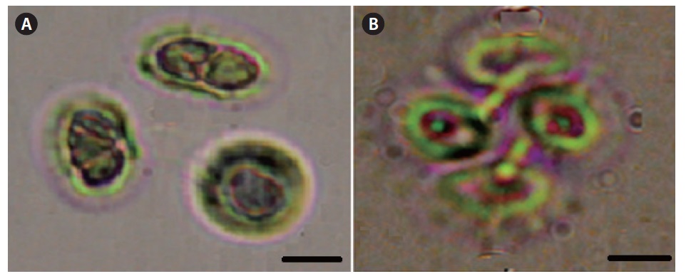
Characium conicum Korshikov 1953 in Komarek et Fott 1983 (
Synonym:
Illustration: Cells narrowly wide-ovoid to fusiform, erect but with a short oblique tip, stipe short, chloroplast a parietal, with a pyrenoid. Cells 10-15 µm in diameter, and 15-30 µm long.
Information of sampling sites: This species attached on filamentous green algae or plants, and were collected from Samrak park (03 Oct 2011: Water temp. 20.0℃, TN 2.030 mg L-1, TP 0.112 mg L-1).
Key reference: Komarek and Fott (1983).
Remark: This taxon is already reported in Algal flora of Korea by Kim and Kim (2012) and was collected by Han Soon Kim.
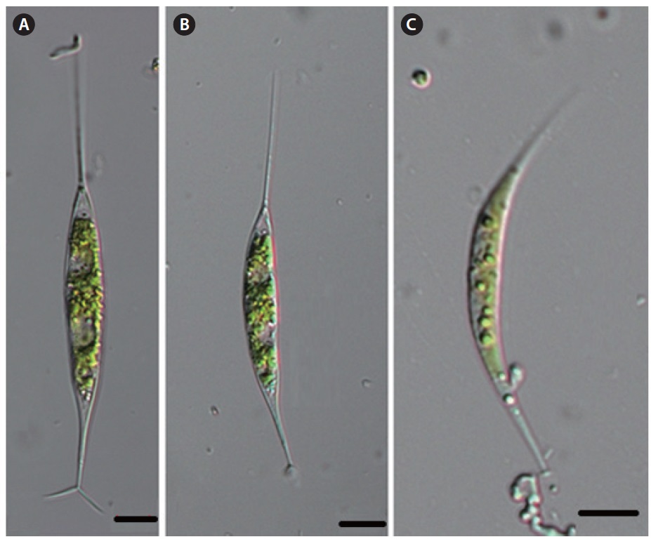
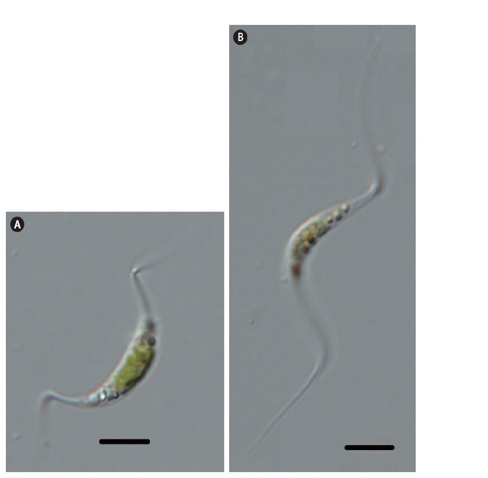
Genus Ankyra (G.M. Smith) Fott
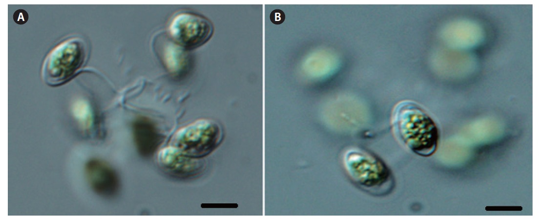
Ankyra ancora (G.M. Smith) Fott 1975 (Fig. 6A, 6B)
Basionym:
Synonym:
Illustration: Cells solitary, spindle-like, fusiform, commonly straight, but sometimes slightly covered. The anterior pole tapers to a long bristle bearing either a pair of leaf-like appendages or a cluster of horse tail-like bristles. The posterior pole tapers gradually and bifurcates the processes as an anchor-like shape. Chloroplasts single, with a pyrenoid. Cells (included stipe) 5-16 µm in diameter, and 130-150 µm long.
Information of sampling sites: This species inhabits lakes and reservoirs as plankton, and is collected from the Nakdong river (25 Oct 2011: water temp. 20.0℃, pH 8.0, TN 2.044 mg L-1, TP 0.073 mg L-1), Samrak Park (03 Oct 2011: water temp. 20.0℃, pH 8.0, TN 2.030 mg L-1, TP 0.112 mg L-1)
Key reference: Fott (1957).
Remark: This taxon is already reported in Algal flora of Korea by Kim and Kim (2012) and was collected by Han Soon Kim.
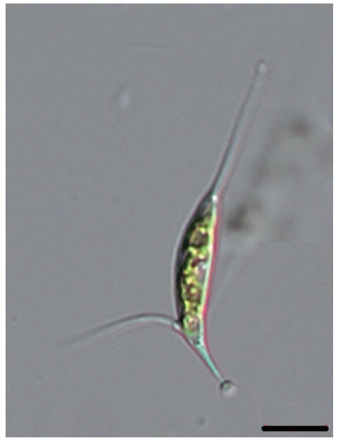
Ankyra judayi (G.M. Smith) Fott (
Basionym:
Synonym:
Illustration: Cells solitary, spindle-like, fusiform, commonly straight, but sometimes slightly covered. The anterior pole tapers to a long bristle which bears either a
pair of leaf-like appendages or a cluster of horse tail-like bristles. The posterior pole tapers gradually and bifurcates the processes as an anchor-like shape. Chloroplasts a parietal plates, with a pyrenoid. Cells (included stip) 6 µm in diameter, and 18-80 µm long.
Information of sampling sites: This species inhabits lakes and reservoirs as plankton and, collected from the Nakdong river (25 Oct 2011: water temp. 20.0℃, pH 8.0, TN 2.044 mg L-1, TP 0.073 mg L-1), Samrak park (03 Oct 2011: water temp. 20.0℃, pH 8.0, TN 2.030 mg L-1, TP 0.112 mg L-1).
Key reference: Fott (1957).
Remark: This taxon is already reported in Algal flora of Korea by Kim and Kim (2012) and was collected by Han Soon Kim.
Ankyra calcarifera (Kisselev) Fott (
Basionym:
Illustration: Cells solitary, spindle-like, fusiform, commonly straight, but sometimes slightly covered. The anterior pole tapers, to a long bristle bearing either a pair of leaf-like appendages or a cluster of horse tail-like bristles. The posterior pole tapers gradually and bifurcates the processes as anchor-like shape, a crooked bristle is on the convex side of the cell. Chloroplast a parietal, with a pyrenoid. Cells 5-7 µm in diameter, 40-50 µm long, and bristle on the convex side wall 30 µm long.
Information of sampling sites This species inhabit lakes and reservoirs as plankton, and was collected from the Nakdong river (25 Oct 2011: water temp. 20.0℃, pH 8.0, TN 2.044 mg L-1, TP 0.073 mg L-1), Samrak park (03 Oct 2011: Water temp. 20.0℃, pH 8.0, TN 2.030 mg L-1, TP 0.112 mg L-1).
Key reference: Fott (1957).
Remark: This taxon is already reported in Algal flora of Korea by Kim and Kim (2012) Han Soon Kim.
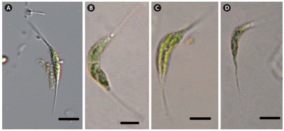
Genus Paradoxa Svirenko 1928
Paradoxa multiseta Svirenko 1928 (
Illustration: Cells fusiform, the anterior pole tapering gradually to a long bristle, bearing either a pair of leaf-like appendages or a number of bristles, 18-45 µm long and 3-8 µm wide, The leaf-like appendages are 1-6 µm long and up to 2.3 µm in diameter at their base, and give the cell an anchor-like shape.
Information of sampling sites: This species inhabits lakes and reservoirs as plankton, and was collected from the Nakdong river (25 Oct 2011: water temp. 20.0℃, pH 8.0, TN 2.044 mg L-1, TP 0.073 mg L-1).
Key reference: Svirenko (1928).
Remark : This was collected by Han Soon Kim (2011) from the Nakdong river. This genus
Genus Schroederia Lemmermann
Schroederia setigera (Schroder) Lemmermann
Cells spindle form, fusiform and crescent form. Cells with setae 4-6 µm in diameter, and 25-60 µm long.
Schroederia spiralis (Printz) Korshikov 1953 (
Basionym:
Illustration: Cells solitary, spindle-form, fusiform, crescent form. The poles extend into long, fine setae, spirally twisted with setae, without a small disk. Chloroplast a single, parietal, laminate, with a pyrenoid. Cells (with setae) 25-60 µm long, and 4-6 µm in diameter.
Information of collected sites: This species inhabits lakes and reservoirs as plankton, and was collected from Lake Seoho (20 Nov 2011: water temp. 8.1℃, pH 7.9, TN 2.730 mg L-1, TP 0.055 mg L-1), Donghwa stream (23 Sep 2011: water temp. 22.9℃, pH 7.6, TN 7.277 mg L-1, TP 0.050 mg L-1), Nammae reservoir (10 Sep 2011: water temp. 28.0℃, pH 8.4, TN 2.916 mg L-1, TP 0.083 mg L-1), Ilweul reservoir (03 Sep 2011: water temp. 30.0℃, pH 8.2, EC 636 µs cm-1), Pond at Samrak park (03 Oct 2011: water temp. 20.0℃, pH 8.0, TN 2.030 mg L-1, TP 0.112 mg L-1).
Key reference: Korshikov (1939).
Schroederia indica Philipose 1967 (
Illustration: Cells solitary, spindle-form, fusiform, crescent form. cells curved, convex on the outer side and slightly convex or swollen on the inner side. Both poles extended into long, fine setae, setae straight, point. Chloroplast a parietal, with a pyrenoid. Cells (include setae)
4-12 µm in diameter, and 28-44 µm long.
Information of Collected sites: Lake Seoho (20 Nov 2011: water temp. 8.1℃, pH 7.9, TN 2.730 mg L-1, TP 0.055 mg L-1), the Donghwa stream (23 Sep 2011: water temp. 22.9℃, pH 7.6, TN 7.277 mg L-1, TP 0.050 mg L-1), Nammae reservoir (10 Sep 2011: water temp. 28.0℃, pH 8.4, TN 2.916 mg L-1, TP 0.083 mg L-1), Ilweul reservior (03 Sep 2011; water temp. 30.0℃, pH 8.2, EC 636 µs cm-1), Pond at Samrak park (03 Oct 2011: water temp. 20.0℃, pH 8.0, TN 2.030 mg L-1, TP 0.112 mg L-1).
Key reference: Philipose (1967).
Remark: The cells of this taxon have regularly curved setae and lie in a plane. However,
Taxa belonging to these 2 families are cosmopolitan in their distribution (John and Tsarenko 2002, Komarek and Fott 1983, Prescott 1962, Yamagishi and Akiyama 1984, Dillard 1988), and inhabit swamps, ponds, reservoirs, lakes and running waters in Korea (Kim 1994, 1996, Kim and Chung 1993a, 1993b, Kim and Lee 1996). The genus
The genus
The genus
A total of 20 taxa belonging to 2 families (Botryococcaceae, Characiaceae) are identified in this study. Among them, 10 taxa are newly reported in Algal flora of Korea by Kim and Kim (2012); 1) Family Botryococcaceae :