The genus Cyclotella (Kützing) Brébisson is represented primarily in freshwater habitats, but many species, such as C. atomus Hustedt, C. baltica (Grunow) Håkansson, C. caspia Grunow, C. choctawhatcheeana Prasad, C. cryptica Reimann, Lewin et Guillard, C. desikacharyi Prasad, C. litoralis Lange et Syvertsen, C. meneghiniana Kützing, C. scaldensis Muylaert et Sabbe, C. striata (Kützing) Grunow and C. stylorum Brightwell occur in marine or brackish waters (Håkansson 2002, Prasad and Nienow 2006, Tanaka 2007).
Cyclotella are characterized by having circular valves with different ornamentation between the central area and the marginal area of the valve face. Due to the morphological similarity among species and high intra-specific variation, taxonomic discriminations of the Cyclotella species are hard to unravel (Håkansson and Kling 1994, Meyer and Håkansson 1996). In addition, further taxonomic confusion is caused by the small size of the cells and numerous similarities among the species belonging to Cyclotella sensu lato. Recently Håkansson (2002) published the genus Puncticulata Håkansson from nine Cyclotella species based on the central area of valve faces, as well as the particular locations of fultoportulae and rimoportulae. Houk and Klee (2004) established the genus Discostella Houk et Klee from all stelligeroid Cyclotella taxa according to the position of the mantle fultoportulae and rimoportula on marginal striae.
In Korea, Lee and Lee (1988) firstly examined six Cyclotella species from southern and western coastal waters of Korea using scanning electron microscopy. Lee et al. (1994) studied the fine structure of Cyclotella pseudostelligera Hustedt. Lee et al. (1995a) reported 15 Cyclotella species of diatoms in Korea. Lee et al. (1995b) originally described Cyclotella orientalis from the Nakdong River. Lee (1995) made a check-list and added two Cyclotella species in Korean waters. Cho (1996) founded nine Cyclotella species at Nakdong River, C. woltereckii Hustedt was firstly reported in Korea. Recently, Chung et al. (2010) first reported Cyclotella atomus var. marina from a diatom assemblage attached to an eelgrass sample at the Yeosu
coast in Korea.
Although 21 species of Cyclotella diatom have been recorded in Korea (Lee et al. 1995a, Lee 1995, Cho 1996, Chung et al. 2010), several Cyclotella species have been transferred into the other cyclotelloid genera, such as Discostella and Puncticulata, following the recent complication of cyclotelloid diatoms (Håkansson 2002, Houk and Klee 2004): four Cyclotella species (C. bodanica Eulenstein, C. comta (Ehrenberg) Kützing, C. comta var. glabriuscula Grunow, C. radiosa (Grunow) Lemmermann) were transferred into the genus Puncticulata; five Cyclotella species (C. comta var. glabriuscula Grunow, C. orientalis Lee, Chung et Gotoh, C. pseudostelligera Hustedt, C. stelligera (Cleve et Grunow) Van Heurck, C. woltereckii Hustedt) were transferred into the genus Discostella.
We present the fine structure, description, distribution and taxonomic remarks of five Cyclotella species in Korean waters. Of the five species discussed in this paper, we include three species which are newly recorded in Korea.
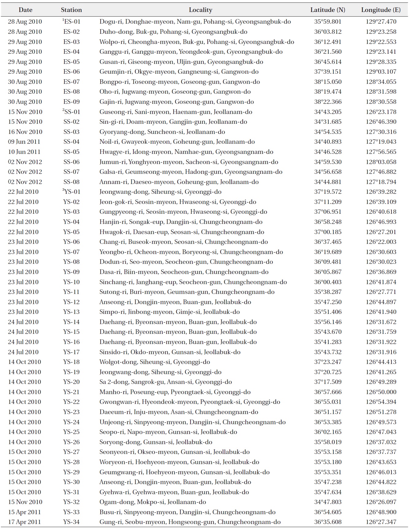
Sample collections were carried out at 51 sites from July 2010 to June 2013 in Korean coastal waters (Table 1). Phytoplankton were collected using a 20 μm mesh net by horizontal and/or vertical towing. Samples were immediately fixed with neutralized formalin (5% final concentration). Using a method presented by Hasle and Fryxell (1970), organic matter in the diatom cell was removed. Prepared diatom material was observed under a light microscope (Axioskop 40; Carl Zeiss, Oberkochen, Germany) and a scanning electron microscope (JSM-5600LV; JEOL, Tokyo, Japan), and was photographed with an MRc5 camera (Carl Zeiss). Diatom size analysis was completed with image calculation software (AxioVision AC version 4.5; Carl Zeiss). The general terminology was adopted from the proposals on Diatom Terminology (Anonymous 1975, Ross et al. 1979), and some special terms for Cyclotella followed Håkansson (2002) and Tanaka (2007).
1a. Two satellite pores on mantle fultoportula ..................2
1b. Three satellite pores on mantle fultoportula ...............4
2a. No central fultoportula ..............C. atomus var. marina
2b. Several central fultoportula ...........................................3
3a. Mantle fultoportula on recessed interstria ...C. litoralis
3b. Mantle fultoportula on thickened interstria...C. baltica
4a. Space of mantle fultoportula on every 2nd interstria ...........................................................................C. meduanae
4b. Space of mantle fultoportula on every interstria ..............................................................................C. meneghiniana
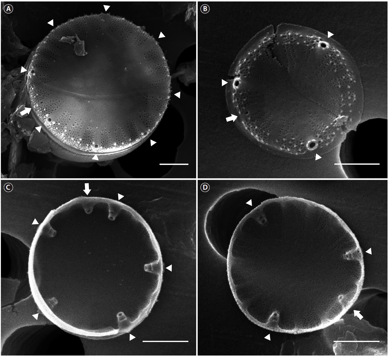
Cyclotella atomus var. marina Tanimura, Nagumo et Kato 2004 (Fig. 1)
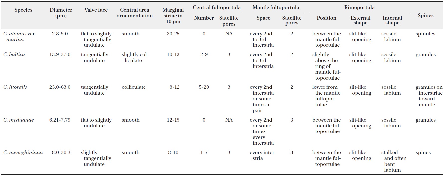
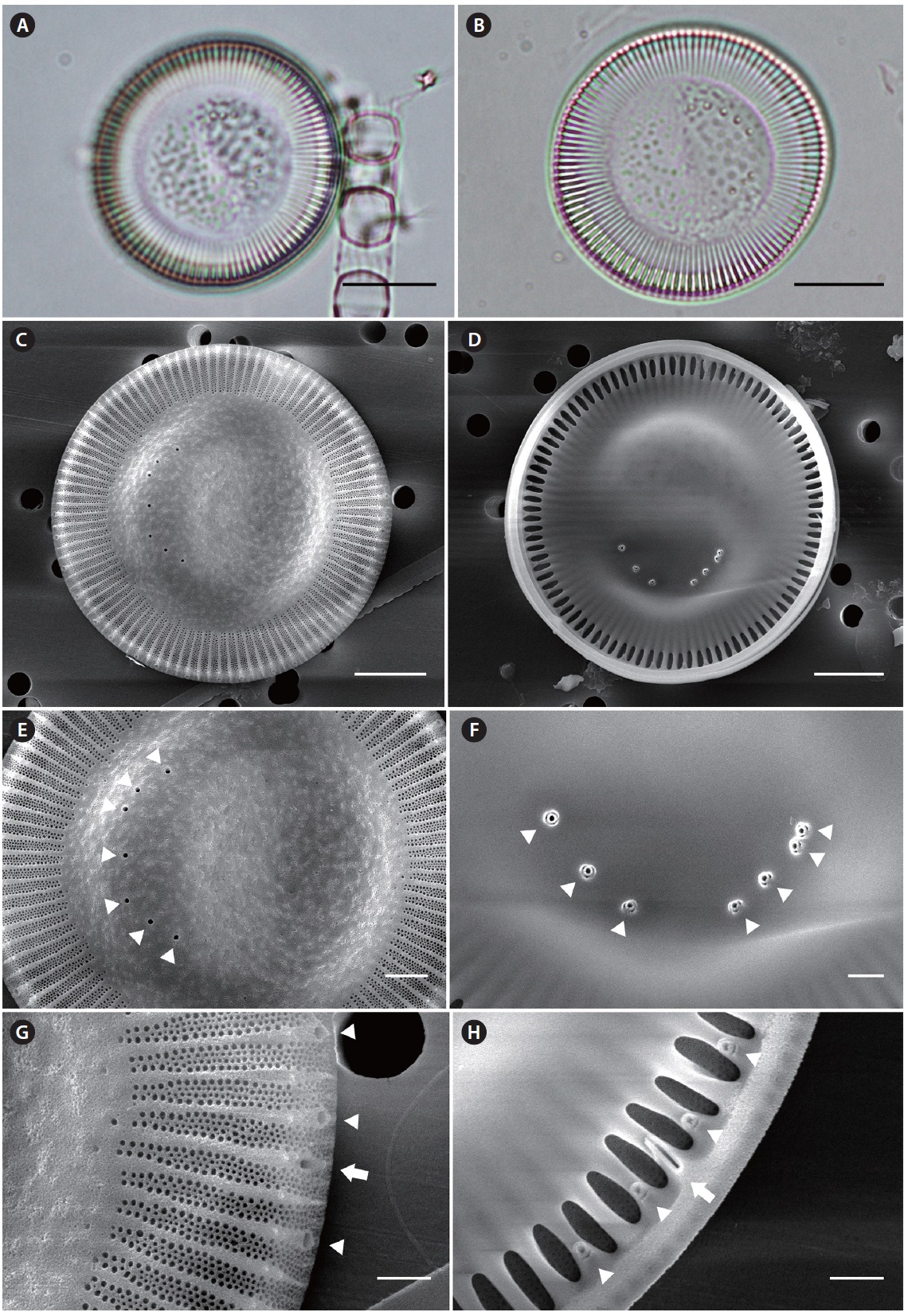
Description: Valves are 2.8-5.0 µm in diameter. The central area is flat to slightly tangentially undulate without colliculate ornamentation (Fig. 1A and 1B). The marginal striae density is 20-25 in 10 µm. Valve face fultoportula is absent. Mantle fultoportulae located on every 2nd to 3rd interstria (Fig. 1A-1D), internally the tubulus is surrounded by two satellite pores (Fig. 1C and 1D). A single rimoportula located on the ring of mantle fultoportulae, internally the rimoportula has a narrow sessile labium (Fig. 1C and 1D).
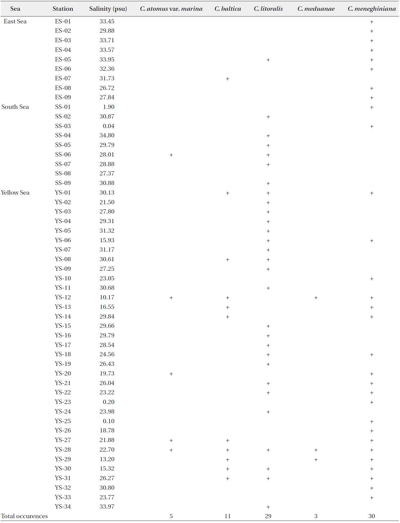
Distribution: Cyclotella atomus var. marina was found from a diatom assemblage attached to an eelgrass sample taken from waters with a salinity of approximately 26, in the region of Yulimri, Yeosu City, Korea in July 1998 (Chung et al. 2010). However, the species occurred total 5 times as a planktonic in this study and reported once and four times from the South Sea and the Yellow Sea, respectively (Table 3).
Remarks: Cyclotella atomus var. marina is distinguished from other variety of C. atomus by the absence of a valve face fultoportula (Chung et al. 2010).
Cyclotella baltica (Grunow) Hakansson 2002 (Fig. 2)
Basionym: Cyclotella striata var. baltica Grunow in Van Heurck 1882, pl. 92, figs 13-15, non Cyclotella litoralis Lange et Syvertsen sensu Håkansson 1996.
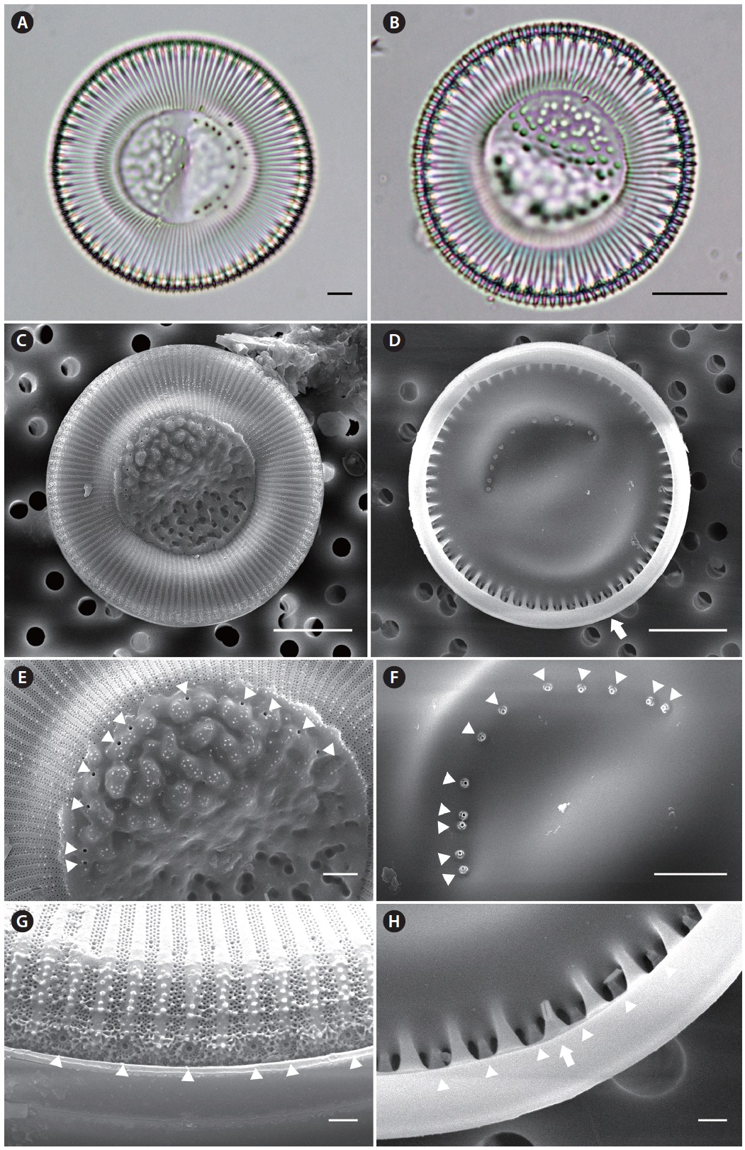
Description: Valves are 13.9-37.0 µm in diameter. The central area tangentially undulate with a slightly colliculate ornamentation (Fig. 2A-2C and 2E). The marginal striae density is 10-15 in 10 µm. Valve face fultoportula number two to nine (Fig. 2C-2F), internally surrounded by three satellite pores (Fig. 2D and 2F). Mantle fultoportulae located on every 2nd to 3rd interstria (Fig. 2G and 2H),
internally the tubulus is surrounded by two satellite pores (Fig. 2H). A single rimoportula located slightly above the ring of the mantle fultoportulae, internally the rimoportula has a sessile labium (Fig. 2H).
Distribution: Grunow (1882) collected C. baltica from the Baltic. Tanaka (2007) collected this species from Hokkaido to Kyushu in Lake Abshiri, Mikawa Bay, and regarded as a brackish to marine water species. Cyclotella baltica occurred total 11 times in this study, and was observed as a planktonic from the East Sea (one time) and the Yellow Sea (10 times), there was no occurrence from the South Sea (Table 3). This species is firstly recorded in the present study.
Remarks: Håkansson (2002) placed C. striata var. baltica in C. baltica according to the absence of the valve face fultoportula, and stated that the species were regarded as C. striata previously (Hustedt 1928, Cleve-Euler 1951, Helmcke and Krieger 1954, Helmcke et al. 1974, Takano 1976, Prasad et al. 1990) was in fact C. baltica. Tanaka (2007) described C. baltica from Japanese specimens. However, the Japanese material showed some differences from the lectotype of Håkansson (2002): a valve diameter
[Fig. 2.] Cyclotella baltica (Grunow) Hakansson. Arrow and arrowheads indicate the rimoportula and the fultoportula, respectively. (A) External valve view, (B)
Internal valve view, (C) Central area with 7 fultoportulae in the external valve view, (D) Seven fultoportulae with 3 satellite pores on the central area of valve
face in the internal valve view, (E) Externally striated marginal area with mantle fultoportulae and rimoportula, (F) Internally alveolated marginal area with
mantle fultoportulae and rimoportula. Scale bars represent: A, B, 10 μm; C, D, 5 μm; E, 2 μm; G, H, 1 μm.
and the position of external opening of rimoportula. The morphological characteristics in Korean specimens agree with the description of C. baltica in Håkansson (2002): a valve diameter, the external position of rimoportula, thestriae dense and the spacing of mantle fultoportulae. Although Korean specimens show well-developed alveoli in the internal valve view (Fig. 2B and 2F), the variation of alveoli development within Cyclotella species have been reported (Beszteri et al. 2005). Therefore, we regarded the Korean specimens as C. baltica.
Cyclotella litoralis Lange et Syvertsen 1989 (Fig. 3)
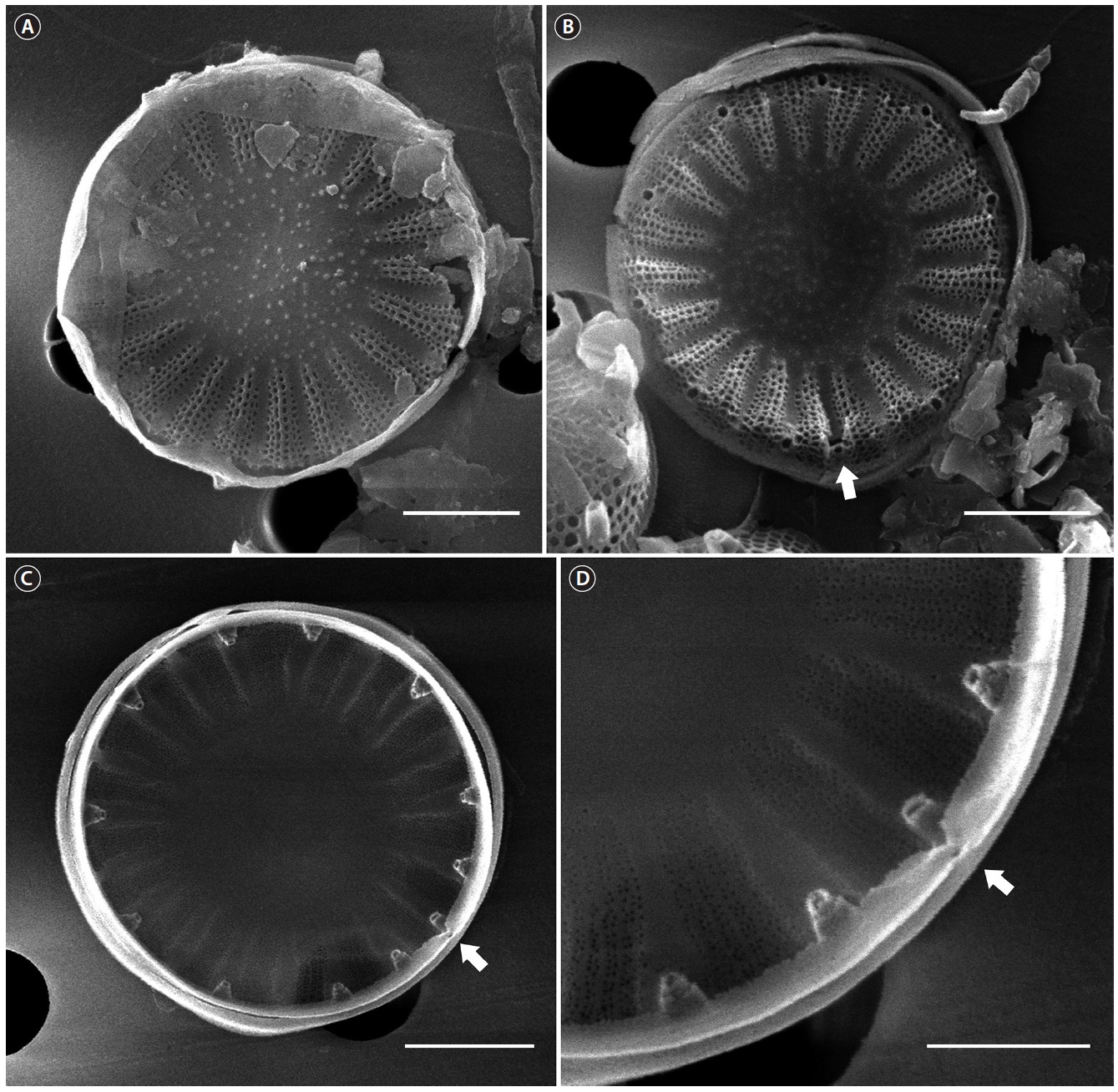
Description: Valves are 23.0-63.0 µm in diameter. The central area tangentially undulates with colliculate ornamentation (Fig. 3A-3C and 3E). The marginal striae density is 8-12 in 10 µm. Valve face fultoportula is eight to twelve Fig. 3C and 3D), internally surrounded by three satellite pores (Fig. 3F). Mantle fultoportulae located on every 2nd Fig. 3G and 3H), or sometimes, on a pair (Fig. 3G), internally the tubulus is surrounded by two satellite pores Fig. 3H). A single rimoportula is located on the fultoportulae below the mantle, internally the rimoportula has a sessile labium Fig. 3H).
Distribution: Lange and Syvertsen (1989) originally described as C. litoralis from the south western Atlantic, and Tanaka (2007) described this species in Isahaya Bay, Nagasaki Prefecture, Japan. This species was observed 29 times from all of the Sea as a planktonic in this study (Table 3). Although C. litoralis is firstly recorded in this study, Lee and Lee (1988) already observed Cyclotella sp. A from Gwangyang Bay and Jinhae Bay. However, since then there was no additional study for identification of the species.
Remarks: Cyclotella litoralis have been confused with C. striata complex (e.g., C. baltica, C. striata, C. stylorum): This species have been distinguished from C. baltica based on the spacing of the mantle fultoportulae (Håkansson 2002), as well as the number of central fultoportula (Prasad and Nienow 2006). In this study, the presence of the recessed costa is an additional characteristic for distinguishing between the two species (Fig 2B and 3B). Cyclotella striata can be distinguished from C. litoralis based on the absence of a valve face fultoportula (Håkansson 2002). The spacing of mantle fultoportula can be used to distinguish between C. litoralis and C. stylorum (Lange and Syvertsen 1989).
Cyclotella meduanae Germain 1981 (Fig. 4)
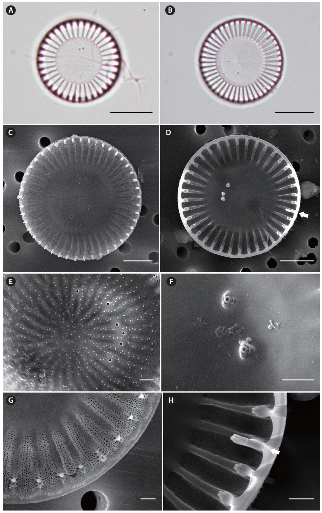
Description: Valves are 6.2-7.8 µm in diameter. The central area is flat to slightly tangentially undulate, without colliculate ornamentation (Fig. 4A and 4B). The marginal striae density is 12-15 in 10 µm. Valve face fultoportula is absent (Fig. 4A-4C). Marginal fultoportulae are located on every 2nd to 3rd interstria (Fig. 4A-4D), internally the tubulus is surrounded by three satellite pores (Fig. 4D). A single rimoportula is located on the ring of marginal fultoportulae, internally the rimoportula has a narrow sessile labium (Fig. 4D).
Distribution: Cyclotella meduanae originally described from Mayenne, France (Germain 1981). Tanaka (2007) examined the Japanese specimen from Inba Pond, Chiba Prefecture and mentioned that C. meduanae has been found mainly in eutrophic water and freshwater. In the present study, C. meduanae is newly recorded and was found 3 times as a planktonic (Table 3).
Remarks: Cyclotella meduanae has been noted to have morphological similarity with C. meneghiniana (Håkansson 2002). Håkansson (2002) mentioned that the only difference between both species is the absence or presence of the valve face fultoportula. However, we found an additional difference in the spacing of the mantle fultoportulae between C. meduanae and C. meneghiniana: in the first one, the mantle fultoportulae was located on every second interstria, while the latter on every interstria (compare Fig. 4C with Fig. 5B).
Cyclotella meneghiniana Kützing 1844 (Fig. 5)
Synonyms: Surirella melosiroides G.G.A. Meneghini ms. 1844; Cyclotella kuetzingiana var. meneghiniana (Kützing) Brun 1880; Stephanocyclus meneghiniana (Kützing) Skabichevskii 1975.
Description: Valves are 8.0-30.3 µm in diameter. The central area is slightly tangentially undulate and without colliculate ornamentation (Fig. 5A-5E). The marginal striae density is 8-10 in 10 µm. Valve face fultoportula is one to seven (Fig. 5A-5F), internally surrounded by three satellite pores (Fig. 5F). Marginal fultoportulae are located on every interstria (Fig. 5G and 5H), internally the tubulus is surrounded by three satellite pores (Fig. 5H). A single rimoportula located on the ring of marginal fultoportulae, internally the rimoportula has a stalked, and often bent, sessile labium (Fig. 5H).
Distribution: Cyclotella meneghiniana have been found in varied habitats including brackish water, and both eutrophic and oligotrophic freshwater (Håkansson 2002, Tanaka 2007). In Korea, Lee and Lee (1988) firstly recorded C. meneghiniana in the Han River and Ulsan Bay near Taehwa River estuary. Cho (1996) described C. meneghiniana from the Nakdong River. Cyclotella meneghiniana was most frequently observed in this study. This species
[Fig. 3.] Cyclotella litoralis Lange et Syvertsen. Arrow indicates the rimoportula. (A) External valve view, (B) Internal valve view, (C) Central area with 10
fultoportulae (arrowheads) in the external valve view, (D) Twelve fultoportulae (arrowheads) with 3 satellite pores on the central area of valve face in the
internal valve view, (E) Externally striated marginal area with mantle fultoportulae (arrowheads) and rimoportula, (F) Internally marginal area with mantle
fultoportulae (arrowheads) on the recessed interstria and a sessile rimoportula between two fultoportulae. Scale bars represent: A-D, 10 μm; E, 1 μm; F, 5
μm; G, H, 1 μm.
was reported total 30 times in this study, 8 times from the East Sea, 2 times from the South Sea, and 20 times from the Yellow Sea as a planktonic (Table 3).
Remarks: Cyclotella meneghiniana has been known to have a wide ecological distribution (Håkansson 2002, Finlay et al. 2002), and highly variable frustule (Håkansson and Chepurnov 1999).
Therefore, this species has been confused with several Cyclotella species such as C. ceylonica Holsinger, C. pratii Toman, C. kuetzingiana Thwaites (Håkansson 2002). Recently, Beszteri et al. (2007) investigated the cryptic diversity of C. meneghiniana based on nuclear and plastid genes. Although this study did not analyze the molecular sequences, it is needed to understand the cryptic diversity of C. meneghiniana in Korea.
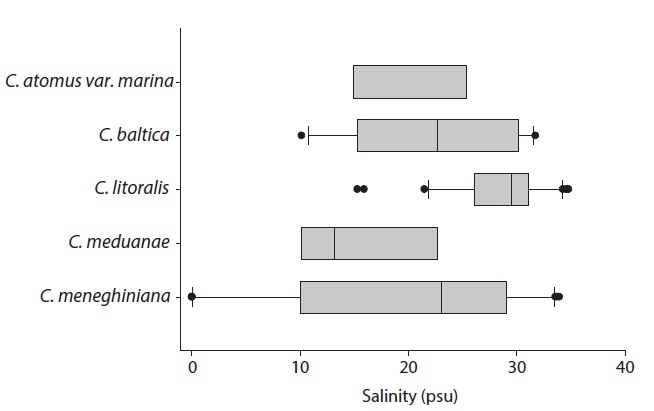
Occurrence of Cyclotella species according to the salinity
This study present the salinity range of five Cyclotella
[Fig. 5.] Cyclotella meneghiniana Kutzing. Arrow indicates the rimoportula. (A) External valve view, (B) Internal valve view, (C) Central area with 6
fultoportulae in the external valve view, (D) Two fultoportulae with 3 satellite pores on the central area of valve face in the internal valve view, (E) Externally
striated marginal area with mantle fultoportulae, (F) Internally marginal area with mantle fultoportulae on the interstria and a sessile rimoportula between
two fultoportulae. Scale bars represent: A, B, 10 μm; C, D, 5 μm; E-H, 1 μm.
Cyclotella meneghiniana Kutzing. Arrow indicates the rimoportula. (A) External valve view, (B) Internal valve view, (C) Central area with 6
fultoportulae in the external valve view, (D) Two fultoportulae with 3 satellite pores on the central area of valve face in the internal valve view, (E) Externally
striated marginal area with mantle fultoportulae, (F) Internally marginal area with mantle fultoportulae on the interstria and a sessile rimoportula between
two fultoportulae. Scale bars represent: A, B, 10 μm; C, D, 5 μm; E-H, 1 μm.
species in the Korean coastal waters (Fig. 6). Cyclotella atomus var. marina occurred in a range of 10.17-28.01 psu from five sites; C. baltica was collected at 11 sites in a range of 10.17-31.73 psu; C. litoralis was reported in a range of 15.32-34.80 psu at 29 sites; C. meduanae have a salinity range of 10.17-22.70 psu from 3 sites; C. meneghiniana occurred in a range of 0.04-34.95 psu from 30 sites. Most of species occur a wide range of 10-30 psu, which indicate the leakage of Cyclotella species into the coastal waters may be more diverse than previous consideration. (Round and Sims 1980).