검색 전체 메뉴
PDF
맨 위로
OA 학술지
Preparation of Cellulose Nanofibril/Regenerated Silk Fibroin Composite Fibers
  • 비영리 CC BY-NC
  • 비영리 CC BY-NC
ABSTRACT
Preparation of Cellulose Nanofibril/Regenerated Silk Fibroin Composite Fibers
KEYWORD
Cellulose nanofibrils , Wet spun silk fiber , Composite , Rheology , Crystallinity index , Post drawing
  • Introduction

    It has been previously reported that regenerated silk fibroin (SF) is highly compatible with blood (Sakabe et al., 1989; Um et al., 2002), has excellent cyto-compatibility (Minoura et al., 1995), and displays low inflammatory reactions in the body (Meinel et al., 2005). Additionally, regenerated SF fibers have several advantages: 1) the fiber morphology (thickness and shape) can be controlled through a variety of methods, 2) new materials, including enzymes and drugs, can be added to the regenerated silk fiber, and 3) the structure and properties can be varied depending on the application. Therefore, regenerated silk fiber has attracted the attention of researchers because of its extensive utilization in biomedical applications, including surgical sutures, tissue engineering scaffolds, drug delivery systems, and wound dressings.

    Several studies have been conducted on the wet spinning of silk, and many researchers have tried to find a new and effective solvent/coagulant system to fabricate regenerated silk fibers with desirable mechanical properties (Trabbic and Yager, 1998; Liivak et al., 1998; Um et al., 2004; Phillips et al., 2005; Ki et al., 2007; Ko and Um, 2009; Yan et al., 2010; Yoo et al., 2010; Cho et al., 2012). However, the mechanical properties of wet-spun silk filaments were not suitable for commercialization in a variety of contexts, including medical and textile applications.

    Cellulose nanofibrils (CNFs) are typically obtained from cellulose by various means, including chemical and physical treatments (Klemm et al., 2011), and subsequently used in the preparation of composite nanocomposite films to improve the mechanical properties of the matrix material. For example, CNFs are used as reinforcing agents to prepare nanocomposites, including cellulose/polyurethane (Aksoy et al., 2007), cellulose/polyvinyl alcohol (Bhatnagar and Sain, 2005), cellulose/starch (Dufresne and Vignon, 1998), cellulose/poly(ethyleneoxide) (Brown and Laborie, 2007), and cellulose/chitosan (Nordqvist et al., 2007).

    Recently, in relation to regenerated silk, the preparation of the nanocomposite material using nanosized cellulose has been studied by a plethora of researchers. Huang et al. (2011) prepared electrospun SF nanofiber mats reinforced by cellulose nanowhiskers (CNWs). They reported that the tensile strength and Young’s modulus of the reinforced SF nanofiber mats were almost twice those of unreinforced SF mats when the CNW content was 2% (w/w). Additionally, Park et al. (2012) fabricated nanofibrous bacterial cellulose nanocrystal (BCN)-embedded SF by using an electrospinning technique and found that Young’s modulus of the SF nanofibers could be increased significantly by the addition of the BCNs. Li et al. (2012) prepared SF/ poly(ethylene glycol)/CNW composite films and reported that the mechanical properties of the SF film could be improved significantly by incorporating CNWs.

    Although several researchers have reported nanocellulose-based silk composite films and electrospun fibers, an experiment concerning wet-spun nanocellulose/SF composites has not been performed until now. In this study, CNFs were added to SF to prepare CNF/SF fibers using a wet-spinning technique, and the effect of the CNF content on the rheological properties, wet-spinnability, molecular conformation of the fiber, and fiber morphology was examined.

    Materials and Methods

      >  Isolation of CNFs

    CNFs were isolated from cellulose pulp according to the modified TEMPO oxidation method (Saito et al. 2004). In short, the kraft pulp sample (2 g of cellulose) was suspended in water (150 mL) containing TEMPO (0.025 g) and sodium bromide (0.25 g). The TEMPO-mediated oxidation of the cellulose slurry was initiated by adding 5 mmol of 13% NaClO for every gram of cellulose and carried out at room temperature under gentle agitation. The pH was maintained at 10.5 by adding 0.5 M NaOH. When a decrease in pH was no longer observed, the reaction was considered complete, and the pH was then adjusted to 7 by adding 0.5 M HCl. The TEMPO-oxidized product was filtered, thoroughly washed with water, and physically fibrillated by ultrasonication in a sonicator (Sonosmasher, Jeio Tech, Korea) for 20 minute. The suspension was then centrifuged at 5000 rpm two or three times for 30 minute. The supernatant, which was in fact a suspension containing the CNFs, was decanted and collected. The yield of the CNFs was calculated as a percentage of the initial weight of the CNF suspension after drying in an oven at 105℃. The final concentration of the CNFs was adjusted to 0.1% by weight/volume.

      >  Preparation of regenerated SF

    The method for preparing the regenerated SF was introduced in a previous study (Yoo et al., 2010). Bombyx mori silk cocoons were degummed with an aqueous solution containing 0.3% sodium oleate (o.w.f.) and 0.2% sodium carbonate (o.w.f.), which was boiled for 1 hour, and rinsed thoroughly with distilled water to obtain the SF. The SF was then dissolved in a solution of CaCl2/H2O/EtOH (molar ratio=1/8/2) for 3 minute, with a liquor ratio of 1:20. The aqueous SF solution was obtained through dialysis of the SF solutions in a cellulose tube (molecular cutoff=12,000-14,000 Da) against circulating purified water for 4 days at room temperature. The purified water was obtained by a Water Purification System (RO50, Hana Science, South Korea) with a reverse osmosis membrane. The SF solutions were then dried to obtain the regenerated SF polymers.

      >  Preparation of CNF/SF composite fibers

    A 5% (w/w) regenerated SF formic acid solution was prepared by dissolving the regenerated SF in 98% formic acid. Approximately 1?5 wt% (against the dry mass of regenerated SF) of CNFs was added to the 5% SF formic acid solution to prepare CNF/SF dope solutions. Prior to wet spinning, the CNF/SF dope solutions were filtered twice through nonwoven fabric to remove any insoluble particles. The CNF/SF composite fibers were spun using a syringe and syringe pump by extruding the dope solution through a 26-gauge needle (inner diameter=0.241, with a needle length of 0.5 inch) into methanol, which acts as a coagulation bath. The flow rate of the fiber extrusion was controlled at 20 mL/h. The as-spun SF filaments were left to stand in the coagulant overnight to allow the solvent (formic acid) to diffuse out completely from the filament prior to post-drawing and drying.

      >  Rheological measurement

    The CNF/SF dope solutions for wet spinning were used for the rheological measurements. The complex viscosity of the solutions was measured by rheometry (MARS III, Hakke, Germany), using a cone and plate geometry at 25℃. An oscillation test was performed with a strain of 0.01%. The radius and angle of the cone were 60 mm and 1°, respectively.

      >  Maximum draw ratio measurement

    The maximum draw ratio was calculated from the ratio of the maximum drawn length of the fiber and the length of the as-spun fiber in the wet state. The fiber length measurements were performed on 20 different parts of the filament, and the maximum draw ratio was determined by averaging the 20 results (Cho et al. 2012).

      >  FTIR measurement and determination of crystallinity index

    Fourier transform infrared (FTIR) (Nicolet 380, Thermo Fisher Scientific, USA) spectra were obtained using the ATR method. The crystallinity index was calculated as the intensity ratio of the peaks at 1260 cm-1 and 1235 cm-1 from the FTIR spectrum, using the following equation (Bhat and Nadiger, 1980):

    image

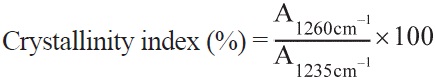
    A1235cm-1 : Absorbance at 1235 cm-1

    A1260cm-1 : Absorbance at 1260 cm-1

    To obtain the average and variation of the crystallinity index, FTIR measurements were performed on five different portions of the samples.

      >  SEM observation

    The morphology of the CNF/SF composite fibers was observed by field-emission SEM observation (FE-SEM, S-4300, Hitachi, Japan) after coating them with gold.

    Results and Discussion

      >  Rheological properties and wet spinnability of dope solution

    The rheological properties of the dope solution were examined since they strongly affect the fiber formation performance when using wet-spinning or electrospinning techniques (Cho et al., 2012; Ko et al., 2013). In the wet spinning of the regenerated silk, at least a viscosity of 70 mPa?s was needed for fiber formation in the methanol coagulant (Kim and Um, 2011; Chung and Um, 2011). This result indicates that a certain level of viscosity is necessary to fabricate the wet-spun regenerated silk fibers.

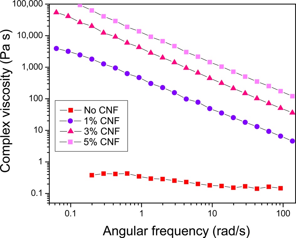
    To understand the change in the rheological properties of the regenerated silk/formic acid solution after adding the CNFs, the complex viscosity of the dope solution was measured; the results are shown in (Fig. 1). The 5% regenerated SF solution without CNFs showed almost Newtonian fluid behavior, although slight shear thinning was observed. This is consistent with the shear viscosity

    results of SF formic acid solutions (Ko et al., 2013; Kim and Um, 2011; Chung and Um, 2011). When 1% (w/w) CNFs was added to the 5% regenerated silk solution, the complex viscosity of the solution increased significantly and shear thinning was quite apparent. As the added amount of CNFs increased, the shear viscosity also increased, while the shear thinning behavior remained unchanged. The notable viscosity increase of the 1% CNF/regenerated SF solution could be due to the fact that the CNFs exist as a suspension and aggregate in solution.

      >  Post-drawing performance of wet-spun filament

    Fiber orientation is a very important characteristic since it determines the mechanical properties of the fiber. In particular, the breaking strength of the fiber tends to be higher as the fiber orientation of wet-spun filaments increases. Um et al. (2004) reported that the breaking strength of the wet-spun regenerated silk fiber could be increased significantly by drawing the fiber with a draw ratio of 4. In synthetic fiber research, many experiments have been conducted to increase the fiber drawing or takeup speed. Based on these background studies, the degree to which a fiber can be drawn is concluded to be important, and the maximum draw ratio (the ratio of the length of drawn fiber to the length of as spun fiber) has been used as a barometer to evaluate the wet spinnability of a given wetspun

    fiber (Yoo et al., 2010; Cho et al., 2010 and 2012).

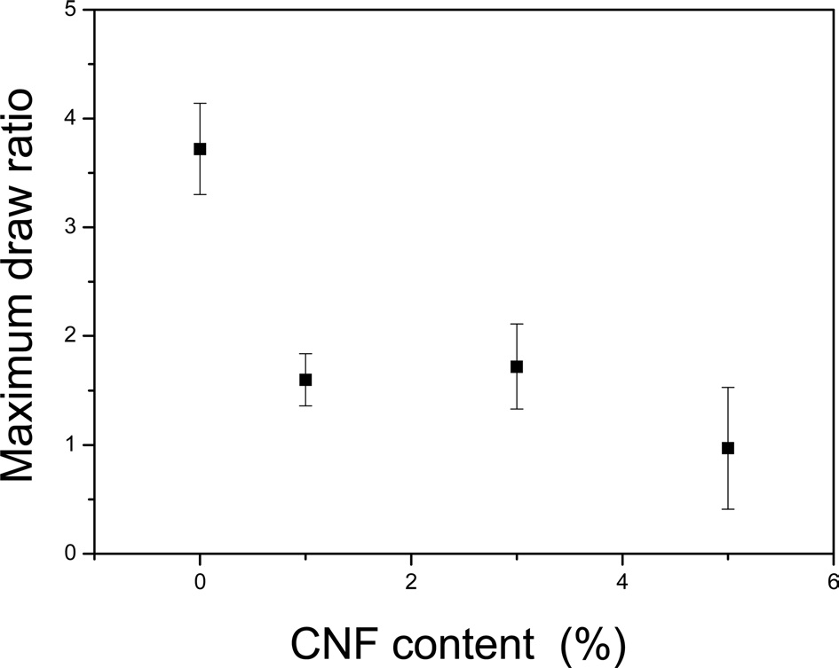
    (Fig. 2) shows the maximum draw ratio of CNF/regenerated silk filaments with different CNF contents. Without CNFs, the regenerated silk filament showed a maximum draw ratio of 3.7. However, as more CNFs were added to the silk, the maximum draw ratio decreased. This result indicated that the post drawing ability deteriorates with increasing CNF content, probably because of the inhomogeneous dope solution. Thus, the regenerated silk solution without CNFs is homogeneous and transparent. However, as the CNFs are added to the silk solution, the dope solution becomes turbid, with a higher degree of CNF and silk aggregation. In other words, the dope solution becomes inhomogeneous upon the addition of CNFs, and this solution with cellulose aggregates can prevent homogenous fiber spinning, resulting in beads and beaded fibers. The beads and beaded fibers can subsequently restrict post-drawing of the as-spun CNF/silk fibers, resulting in a decrease in the maximum draw ratio.

      >  Molecular conformation of wet-spun filaments

    The molecular conformation of the silk was examined since it strongly affects the chemical and physical properties of the silk polymers. β-sheet conformations help in the formation of crystalline regions of the silk, whereas random-coil conformations contribute to the formation of the amorphous region. Therefore, when the amount of

    β-sheets increases, the silk fiber becomes stronger, less water-soluble, and less degradable. FTIR analysis was used to examine the molecular conformation of the silk since different conformations yield absorption peaks at different positions in Amides I, II, and III.

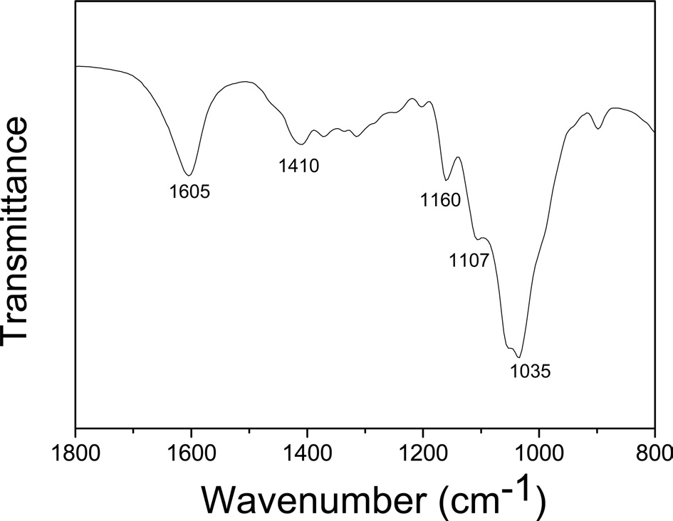
    (Fig. 3) displays the FTIR spectrum of the CNFs used in this study. Strong absorption peaks appeared at 1035 cm-1, which corresponded to stretching of the C-O and C-C bonds of cellulose, and at 1159 cm-1, which could be attributed to the antisymmetric C-O-C bridge stretching (Um et al., 2012).

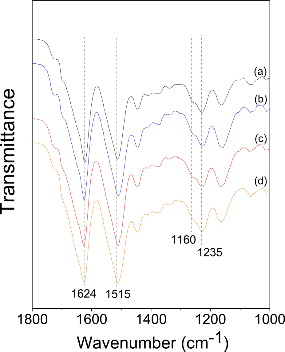
    (Fig. 4) shows the FTIR spectra of wet-spun CNF/ regenerated SF composite fibers with varying CNF contents. All the composite fibers showed strong absorption peaks at 1624 cm-1 and 1515 cm-1 and a shoulder peak at 1260 cm-1. These peaks are attributed to the β-sheet conformation, indicating that β-sheet crystallites exist in all the CNF/SF composite fibers. It has been reported that β-sheet crystallization in the SF occurs when formic acid is removed (Um et al., 2003 and 2004). Interestingly, the shoulder peak at 1260 cm-1 increased slightly as the CNF content increased.

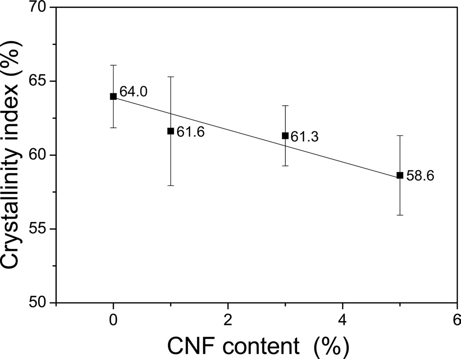
    The crystallinity index of the silk proposed by Baht and Nadiger (1980) has been used to evaluate the crystallinity of the SF since it allows for a quantitative evaluation of this characteristic (Um et al., 2001; Kim et al., 2013).

    Therefore, detailed calculations performed to determine the crystallinity index; the results are shown in (Fig. 5). The regenerated SF filament without CNFs exhibited a

    crystallinity index of 64.0%. However, the crystallinity index decreased as the amount of CNFs increased, with the 5% CNF/SF fibers showing a crystallinity index of 58.6%. This value is calculated from the IR absorption peaks in the Amide III band of SF, and it indicates the ratio of the crystalline regions to the amorphous regions of the silk. Thus, the addition of CNFs does not alter the value of the crystallinity index of the composite fibers if it has no effect on the SF. In other words, the decrease in the crystallinity index of the SF indicates that the CNFs restrict the crystallization of the SF. The SF molecules adopt the random coil conformation in formic acid solution, and β-sheet crystallization occurs in the SF fibers when formic acid is eliminated from the silk solution (Um et al., 2004) and SF is coagulated in methanol. It seems that the presence of CNFs restricts the crystallization of the SF when formic acid is removed, and methanol penetrates the coagulating dope solution during the coagulation process. β-sheet crystallization of the SF is a result of the formation of hydrogen bonds between the SF molecules originating from the many polar groups of the SF, including O-H, C=O, and N-H functional groups. CNF also has many OH groups in the cellulose chain, and hydrogen bonds may be formed between CNFs and the SF. This bonding could prevent hydrogen bonding between the SF molecules, resulting in a decrease in the crystallization of the SF/cellulose blend film (Freddi et al. 1995) and suggesting the occurrence of intermolecular interactions between fibroin and cellulose through hydrogen bonding, as evidenced from the IR spectra.

      >  Morphology of wet-spun filament

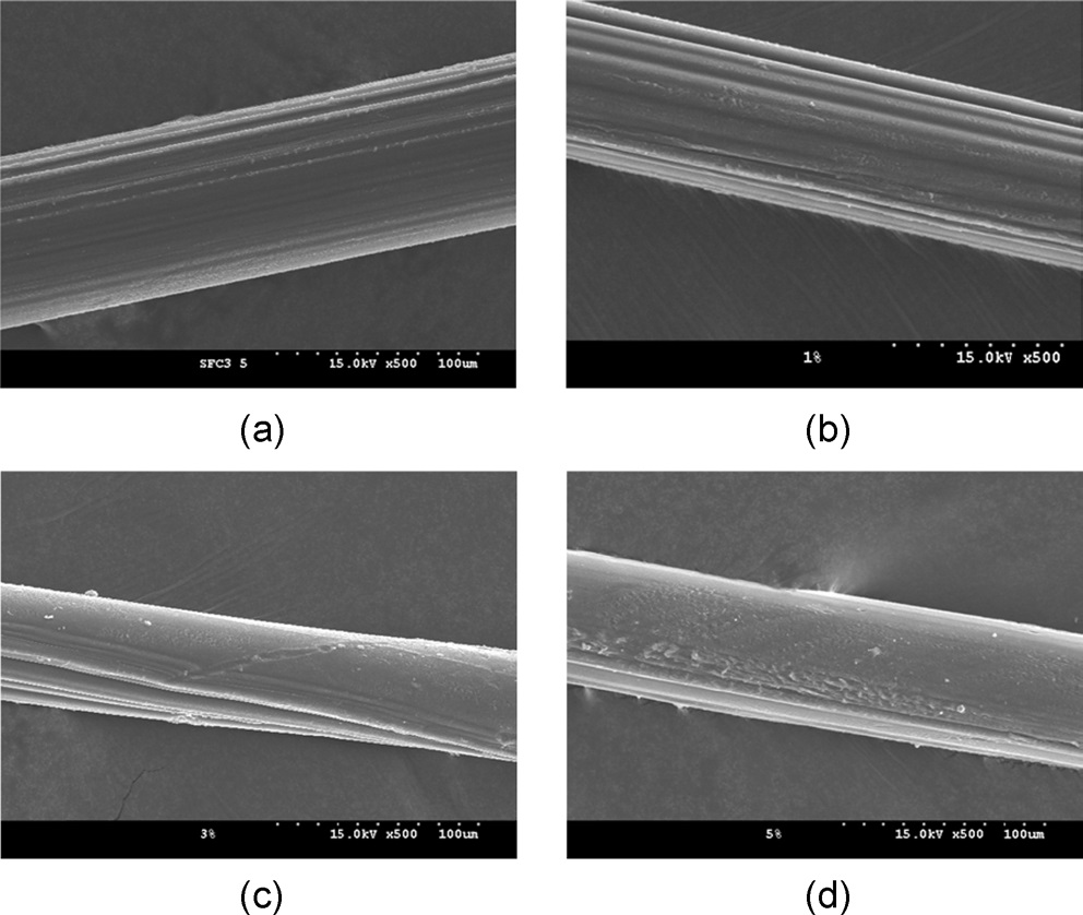
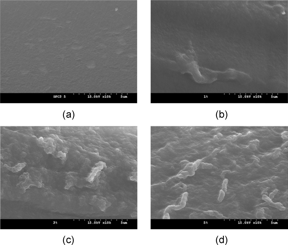
    (Figs. 6 and 7) display the surface morphology of the CNF/SF composite fibers with varying CNF contents. At a low magnification (x500, Fig. 6), the composite fiber showed stripes in the fiber axis direction, with a relatively smooth surface. Additionally, at high magnification (x10,000, Fig. 7), the regenerated SF fibers (Fig. 7a) showed smooth surfaces. However, rugged surfaces of the CNF/SF composite fibers (1?5% CNF) became visible. In particular, the rugged surfaces were more prevalent when a higher amount of CNFs was present. It seems that the rugged surface might be related to CNF aggregates, considering the fiber thickness is on the order of several hundred nanometers. This rugged surface may also be evidence for the presence of CNF aggregates in the CNF/ SF composite.

참고문헌
  • 1. Aksoy EA, Akata B, Bac N, Hasirci N (2007) Preparation and characterization of zeolite eta-polyurethane composite membranes. [J Appl Polym Sci] Vol.104 P.3378-3387 google cross ref
  • 2. Bhat NW, Nadiger GS (1980) Crystallinity in silk fibers: Partial acid hydrolysis and related studies. [J Appl Polym Sci] Vol.25 P.921-932 google cross ref
  • 3. Bhatnagar A, Sain M (2005) Processing of cellulose nanofiber reinforced composites. [J Reinf Plast Compos] Vol.24 P.1259-1268 google cross ref
  • 4. Brown EE, Laborie MPG (2007) Bioengineering bacterial cellulose/ poly(ethylene oxide) nanocomposites. [Biomacromolecules] Vol.8 P.3074-3081 google cross ref
  • 5. Cho HJ, Um IC (2010) The effect of dissolution condition on the yield, molecular weight, and wet- and electro-spinnability of regenerated silk fibroins prepared by LiBr aqueous solution. [Int J Indust Entomol] Vol.20 P.99-105 google
  • 6. Cho HJ, Yoo HJ, Kim JW, Park YH, Bae DG, Um IC (2012) Effect of molecular weight and storage time on the wet- and electrospinning of regenerated silk fibroin. [Polym Degrad Stab] Vol.97 P.1060-1066 google cross ref
  • 7. Chung DE, Um IC (2011) The relationship between the rheological properites and wet spinnability of regenerated silk fibroin solution with various molecular weights and concentrations [6th International Workshop for East Asian Young Rheologists] P.63 google
  • 8. Dufresne A, Vignon MR (1998) Improvement of starch film performances using cellulose microfibrils. [Macromolecules] Vol.31 P.2693-2696 google cross ref
  • 9. Freddi G, Romano M, Massafra MR, Tsukada M (1995) Silk fibroin/ cellulose blend films-preparation, structure, and physical-properties. [J Appl Polym Sci] Vol.56 P.1537-1545 google cross ref
  • 10. Huang J, Liu L, Yao JM (2011) Electrospinning of bombyx mori silk fibroin nanofiber mats reinforced by cellulose nanowhiskers Vol.12 P.1002-1006 google
  • 11. Ki CS, Kim JW, Oh HJ, Lee KH, Park YH (2007) The effect of residual silk sericin on the structure and mechanical property of regenerated silk filament. [Int J Biol Macromol] Vol.41 P.346-353 google cross ref
  • 12. Kim HJ, Um IC (2011) The effect of sericin content on the rheological properties and wet spinnability of regenerated silk fibroin solution [6th International Workshop for East Asian Young Rheologists] P.66 google
  • 13. Kim HJ, Chung DE, Um IC (2013) Effect of processing conditions on the homogeneity of partially degummed silk evaluated by FTIR spectroscopy. [Int J Indust Entomol] Vol.25 P.54-60 google cross ref
  • 14. Klemm D, Kramer F, Moritz S, Lindstrom T, Ankerfors M, Gray D (2011) Nanocelluloses: a new family of nature-based materials. [Angew Chem Int Edit] Vol.50 P.5438-5466 google cross ref
  • 15. Ko JS, Um IC (2009) The effect of HPMC concentration on the morphology and post drawing of wet spun regenerated SF/HPMC blend filaments. [Int J Indust Entomol] Vol.19 P.181-185 google
  • 16. Ko JS, Yoon K, Ki CS, Kim HJ, Bae DG, Lee KH (2013) Effect of degumming condition on the solution properties and electrospinnablity of regenerated silk solution. [Int J Biol Macromol] Vol.55 P.161-168 google cross ref
  • 17. Li RJ, Zhang YH, Zhu LJ, Yao JM (2012) Fabrication and characterization of silk fibroin/poly(ethylene glycol)/cellulose nanowhisker composite films. [J Appl Polym Sci] Vol.124 P.2080-2086 google cross ref
  • 18. Liivak O, Blye A, Shah N, Jelinski LW (1998) A microfabricated wet-spinning apparatus to spin fibers of silk proteins. [Structureproperty correlations. Macromolecules] Vol.31 P.2947-2951 google cross ref
  • 19. Meinel L, Hofmann S, Karageorgiou V, Kirker-Head C, McCool J, Gronowicz G (2005) The inflammatory responses to silk films in vitro and in vivo. [Biomaterials] Vol.26 P.147-155 google cross ref
  • 20. Minoura N, Aiba S, Gotoh Y, Tsukada M, Imai Y (1995) Attachment and growth of cultured fibroblast cells on silk protein matrixes. [J Biomed Mater Res] Vol.29 P.1215-1221 google cross ref
  • 21. Nordqvist D, Idermark J, Hedenqvist MS (2007) Enhancement of the wet properties of transparent chitosan-acetic-acid salt films using microfibrillated cellulose. [Biomacromolecules] Vol.8 P.2398-2403 google cross ref
  • 22. Park DJ, Choi Y, Heo S, Cho SY, Jin HJ (2012) Bacterial cellulose nanocrystals-embedded silk nanofibers. [J Nanosci Nanotech] Vol.12 P.6139-6144 google cross ref
  • 23. Phillips DM, Drummy LF, Naik RR, De Long HC, Fox DM, Trulove PC (2005) Regenerated silk fiber wet spinning from anionic liquid solution. [J Mater Chem] Vol.39 P.4206-4208 google cross ref
  • 24. Saito T, Nishiyama Y, Putaux JL, Vignon M, Isogai A (2006) Homogeneous suspensions of individualized microfibrils from TEMPOcatalyzed oxidation of native cellulose. [Biomacromolecules] Vol.7 P.1687-1691 google cross ref
  • 25. Sakabe H, Ito H, Miyamoto T, Ha WS (1989) In vivo blood compatibility of regenerated silk fibroin. [Sen-i Gakkaishi] Vol.45 P.487-490 google cross ref
  • 26. Trabbic KA, Yager P (1998) Comparative structural characterization of naturally- and synthetically-spun fibers of Bombyx mori fibroin. [Macromolecules] Vol.31 P.462-471 google cross ref
  • 27. Um IC, Kweon HY, Park YH, Hudson S (2001) Structural characteristics and properties of the regenerated silk fibroin prepared from formic acid [Int J Biol Macromol] Vol.29 P.91-97 google cross ref
  • 28. Um IC, Ki CS, Kweon H, Lee GK, Ihm DW, Park YH (2004) Wet spinning of silk polymer II. Effect of drawing on the structural characteristics and properties of filament. [Int J Biol Macromol] Vol.34 P.107-119 google cross ref
  • 29. Um IC, Kweon HY, Lee KG, Park YH (2003) The role of formic acid in solution stability and crystallization of silk protein polymer. [Int J Biol Macromol] Vol.33 P.203-213 google cross ref
  • 30. Um IC, Kweon HY, Park YH (2012) Hemicellulose removal and crystalline structure transition of flax fiber with alkali treatment. [Text Sci Eng] Vol.49 P.271-278 google
  • 31. Um IC, Kweon HY, Hwang CM, Min BG, Park YH (2002) Structural characteristics and properties of silk fibroin/polyurethane blend films. [Int J Indust Entomol] Vol.5 P.163-170 google
  • 32. Yan JP, Zhou GQ, Knight DP, Shao ZZ, Chen X (2010) Wet-spinning of regenerated silk fiber from aqueous silk fibroin solution: discussion of spinning parameters. [Biomacromolecules] Vol.11 P.1-5 google cross ref
  • 33. Yoo YJ, Kim U-J, Um IC (2010) The effect of coagulant and molecular weight on the wet spinnability of regenerated silk fibroin solution. [Int J Indust Entomol] Vol.21 P.145-150 google
OAK XML 통계
이미지 / 테이블
  • [ Fig. 1. ]  Compolex viscosity of CNF/regenerated SF dope solution with various CNF contents.
    Compolex viscosity of CNF/regenerated SF dope solution with various CNF contents.
  • [ Fig. 2. ]  Maximum draw ratio of CNF/regenerated SF filament with various CNF contents.
    Maximum draw ratio of CNF/regenerated SF filament with various CNF contents.
  • [ Fig. 3. ]  FTIR spectrum of the CNFs used in this study.
    FTIR spectrum of the CNFs used in this study.
  • [ Fig. 4. ]  FTIR spectra of CNF/regenerated SF filaments with various CNF contents: (a) 0%, (b) 1%, (c) 3%, and (d) 5%.
    FTIR spectra of CNF/regenerated SF filaments with various CNF contents: (a) 0%, (b) 1%, (c) 3%, and (d) 5%.
  • [ Fig. 5. ]  Crystallinity index of CNF/regenerated SF filaments with various CNF contents.
    Crystallinity index of CNF/regenerated SF filaments with various CNF contents.
  • [ Fig. 6. ]  SEM images of CNF/regenerated SF filament with various CNF contents at low magnification (x 500): (a) 0%, (b) 1%, (c) 3%, and (d) 5%.
    SEM images of CNF/regenerated SF filament with various CNF contents at low magnification (x 500): (a) 0%, (b) 1%, (c) 3%, and (d) 5%.
  • [ Fig. 7. ]  SEM images of CNF/regenerated SF filament with various CNF contents at high magnification (x 10,000): (a) 0%, (b) 1%, (c) 3%, and (d) 5%.
    SEM images of CNF/regenerated SF filament with various CNF contents at high magnification (x 10,000): (a) 0%, (b) 1%, (c) 3%, and (d) 5%.
(우)06579 서울시 서초구 반포대로 201(반포동)
Tel. 02-537-6389 | Fax. 02-590-0571 | 문의 : oak2014@korea.kr
Copyright(c) National Library of Korea. All rights reserved.