Gall structure and specificity in Bostrychia culture isolates (Rhodomelaceae, Rhodophyta)
- Author: West John A., Pueschel Curt M., Klochkova Tatyana A., Kim Gwang Hoon, Goë, r Susan de, Zuccarello Giuseppe C.
- Organization: West John A.; Pueschel Curt M.; Klochkova Tatyana A.; Kim Gwang Hoon; Goë r Susan de; Zuccarello Giuseppe C.
- Publish: ALGAE Volume 28, Issue1, p83~92, 15 March 2013
-
ABSTRACT
The descriptions of galls, or tumors, in red algae have been sparse. K?tzing (1865) observed possible galls of
Bostrychia but only presented a drawing. Intensive culture observations of hundreds of specimens of the genusBostrychia over many years have revealed that galls appeared in only a small subset of our unialgal cultures ofB. kelanensis, Bostrychia moritziana / radicans, B. radicosa, B. simpliciuscula , andB. tenella and continued to be produced intermittently or continuously over many years in some cultures but were never seen in field specimens. Galls appeared as unorganized tissue found primarily on males and bisexuals, but occasionally on females and tetrasporophytes. The gall cells usually were less pigmented than neighboring tissue, but contained cells with fluorescent plastids and nuclei. The galls were not transferable to other potential hosts. Galls could be produced from gall-free tissue of cultures that originally had galls even after transfer to new culture dishes. Electon microscopy of galls on one isolate (3895) showed that virus-like particles are observed in some gall cells. It is possible that a virus is the causative agent of these galls.
-
KEYWORD
Bostrychia , galls , Rhodomelaceae , Rhodophyta , unialgal culture , virus-like particles
-
Galls, or tumors, are unorganized tissue on otherwise normal plants. Galls are usually associated with abnormal cell division patterns and / or cell enlargement (Apt 1988, Scheffer 1997). Galls have not been reported extensively in red algae possibly because they are rare and not considered important, but the deformation of tissue could have fitness consequences for the host. While the cause of all red algal galls is not known several causative agents have been shown or suggested. Cyanobacteria were reported to cause galls in
Mazzaella laminarioides (Bory de Saint-Vincent) Fredericq (asIridaea laminarioides Bory de Saint-Vincent) (Correa et al. 1993). Bacteria were seen in the galls of the red algaeChondracanthus teedei (Mertens ex Roth) Kutzing (asGigartina teedii [Roth] Lamouroux) (Tsekos 1982),Grateloupia americana Kawaguchi et Wang (asPrionitis lanceolata [Harvey] Harvey), andPolyneuropsis stolonifera M. J. Wynne, D. L. McBride & J. A. West (McBride et al. 1974). Subsequently bacteria were proven to be the cause of galls onG. americana (Apt and Gibor 1989, Ashen and Goff 1996, 1998, 2000, Ashen et al. 1999). A fungus possibly causes galls ofCatenella nipae Zanardini (Zuccarello 2008). The galls ofPrionitis andCatenella were originally described as red algal parasites (Lobocolax deformans Howe andCatenellocolax leeuwenii Weber-van Bosse, respectively). Experimental infection and formation of galls was not achieved with fungi inCatenella nipae (Zuccarello 2008).Galls have also been associated with viruses. Viruslike particles (VLPs) were found in the galls of
Gracilaria epihippisora Hoyle (Apt and Gibor 1991). In this species gall tissue was capable of autonomous growth but only in an undifferentiated state. Pueschel (1995) also observed VLPs in the filamentous red algaAcrochaetium savianum (Meneghini) Nageli (asAudouinella saviana ).We have studied
Bostrychia over many years and it has become useful in research on speciation, ecophysiology, evolution and cell biology / video microscopy of reproduction (reviewed in Zuccarello and West 2011). A very large collection of over 1,000 isolates of all the recognized species ofBostrychia has been established (http://www.botany.unimelb.edu.au/West). A small subset of these isolates has produced undifferentiated tissue that persisted in culture. Galls ofBostrychia were observed in 1865, when Kutzing illustrated many species ofBostrychia and depicted a gall onB. cornifera Montagne (Kutzing 1865, Pl. 24) (Fig. 1A in this paper), later synonymized withB. moritziana (Sonders ex Kutzing) J. Agardh (King and Puttock 1989). Our observations onBostrychia galls are presented below.> Algal material and laboratory culture
Unialgal culture methods were described in West and Zuccarello (1999) and West (2005). Culture isolates were all maintained at 18-23℃, 12 : 12 LD daily photoperiods, 3-5 μmol photons m-2 s-1 cool white fluorescent or LED lighting, MPM/2 culture medium (30‰ salinity). For faster growth and reproduction cultures were placed in 10-15 μmol photons m-2 s-1 on rotary shaker (70 rpm).
Most isolates used for this research program are now available at the Korean Marine Plants Collection, Chungnam National University, 220 Gung-dong, Yuseong-gu, Daejeon, Korea.
> Bright field and fluorescence microscopy
Algal nuclei were stained with the DNA-specific fluorochrome DAPI (4′,6-diamidino-2-phenylindole, Sigma- Aldrich, St. Louis, MO, USA) using a heat fixation method. Algal thalli were dipped in 5 μg mL-1 DAPI solution in seawater for 5 min, and then the cover slips were slightly heated over a boiler for a few seconds. After staining, algal thalli were mounted on slides in the DAPI solution and were examined under a UV filter.
Micrographs were taken with Olympus DP50 digital camera affixed to an Olympus BX50 microscope (Olympus, Tokyo, Japan) using Viewfinder Lite and Studio Lite computer programs (Better Light Inc., Placerville, CA, USA) or with a Zeiss GFL bright field microscope (Carl Zeiss AG, Oberkochen, Germany) using a Canon G3 camera (Canon Inc., Tokyo, Japan) and Photoshop CS4 computer program (http://www.adobe.com/au/).
> Transmission electron microscopy (TEM)
Galls were fixed in phosphate buffered saline (PBS) buffer containing 2% glutaraldehyde at 4℃ for 2 h. The glutaraldehyde was then rinsed out with PBS buffer and the cells were postfixed with 2% osmium tetroxide at 4℃ for 1.5 h. Thereafter, the cells were rinsed out with PBS buffer and were dehydrated in a graded acetone series, embedded in Spurr’s epoxy resin (Spurr 1969) and polymerized overnight in a 70℃ oven (Polysciences Inc., Warrington, PA, USA). Sections stained with uranyl acetate and Reynolds’s lead citrate (Reynolds 1963) were viewed and photographed on a Phillips Bio Twin Transmission Electron Microscope (Phillips Electron Optics, Eindhoven, Netherlands). We were able to carry out TEM studies on only one
B. simpliciuscula isolate (3895).
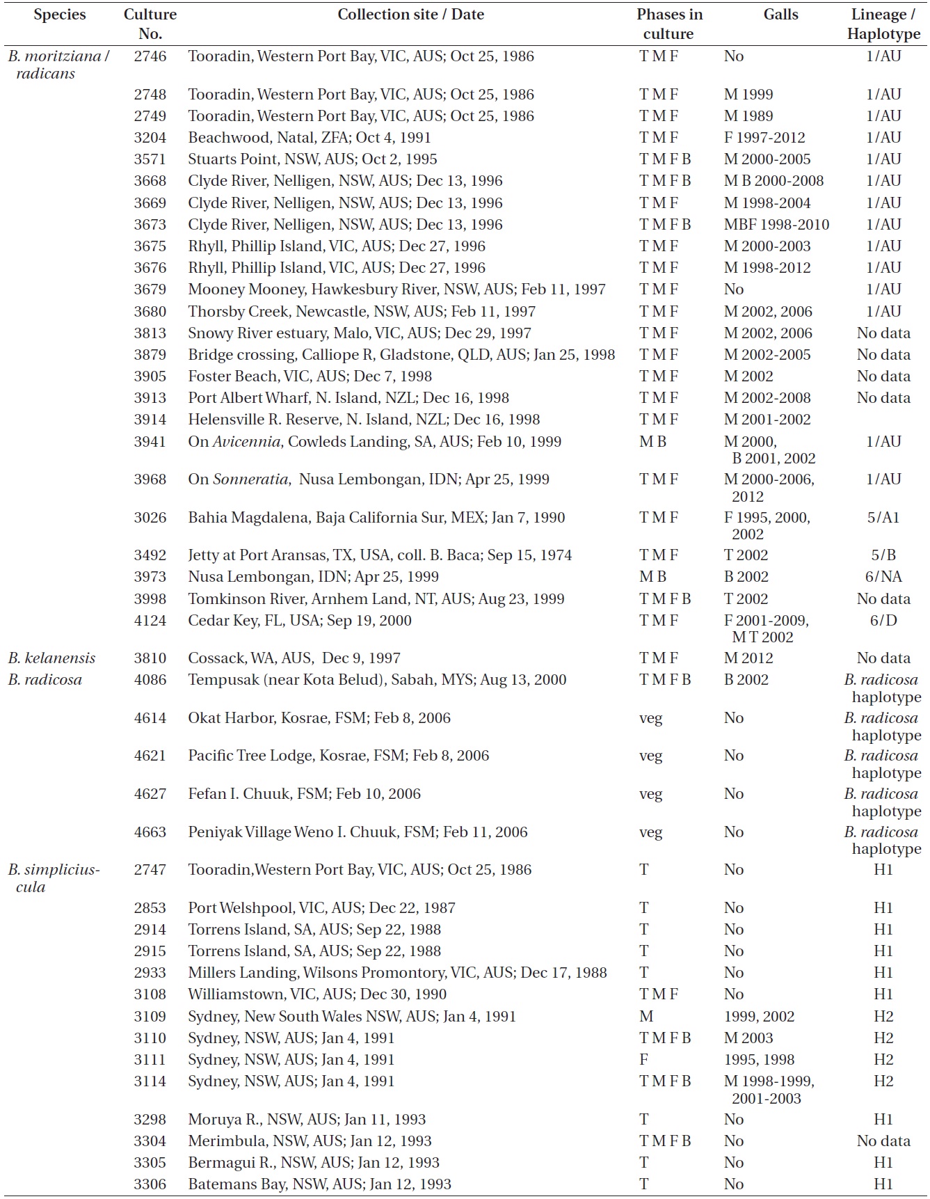
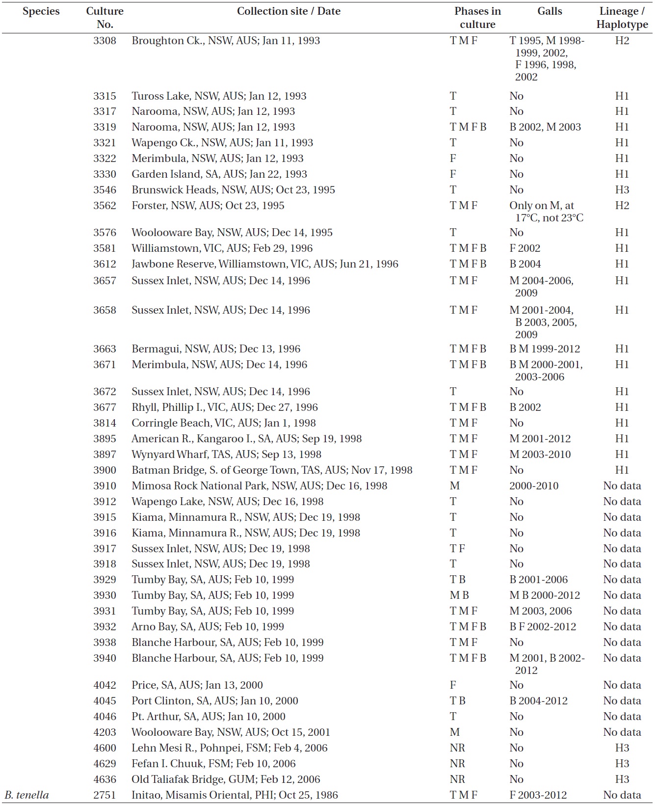
Refer to Table 1 for the time periods (years) in which galls were seen in the various isolates. These galls were predominantly found in isolates from Australia where most isolates were obtained. Only a small percentage of our isolates had galls and they were only observed in
B. simpliciuscula Harvey ex J. Agardh,B. moritziana / radicans complex,B. radicosa (Itono) J. A. West, G. C. Zuccarello & M. H. Hommersand,B. kelanensis Grunow, andB. tenella (Lamouroux) J. Agardh.Bostrychia simpliciuscula
B. simpliciuscula is a polyphyletic species consisting of three lineages (Zuccarello et al. 1999, Zuccarello andWest 2006). Galls were found on 15 of 87 (17%) isolates of lineages H1 and H2, exclusively with Australian isolates. Galls were usually associated with viable spermatangial stichidia, either on unisexual or bisexual plants, although some were evident on the non-reproductive sectors. Tetrasporophytes and female gametophytes occasionally had galls. This has been true over 26 years of culture for some isolates. Galls never developed on young gametophytes, only on reproductively mature gametophytes. On isolate 3562 no galls formed on the tetrasporophytes, females or males grown at 23℃. However, galls developed in the vicinity of spermatangial stichidia of males but not on females or tetrasporophytes when grown at 17℃. Isolate 3562, having numerous galls, was placed in a culture (on a shaker and in brighter light) for 3-6 weeks with the male of another isolate (3108) having a long history without galls. No galls developed on isolate 3108. This is only a partial test of Koch’s postulates (http://en.wikipedia.org/wiki/Koch’s postulates).
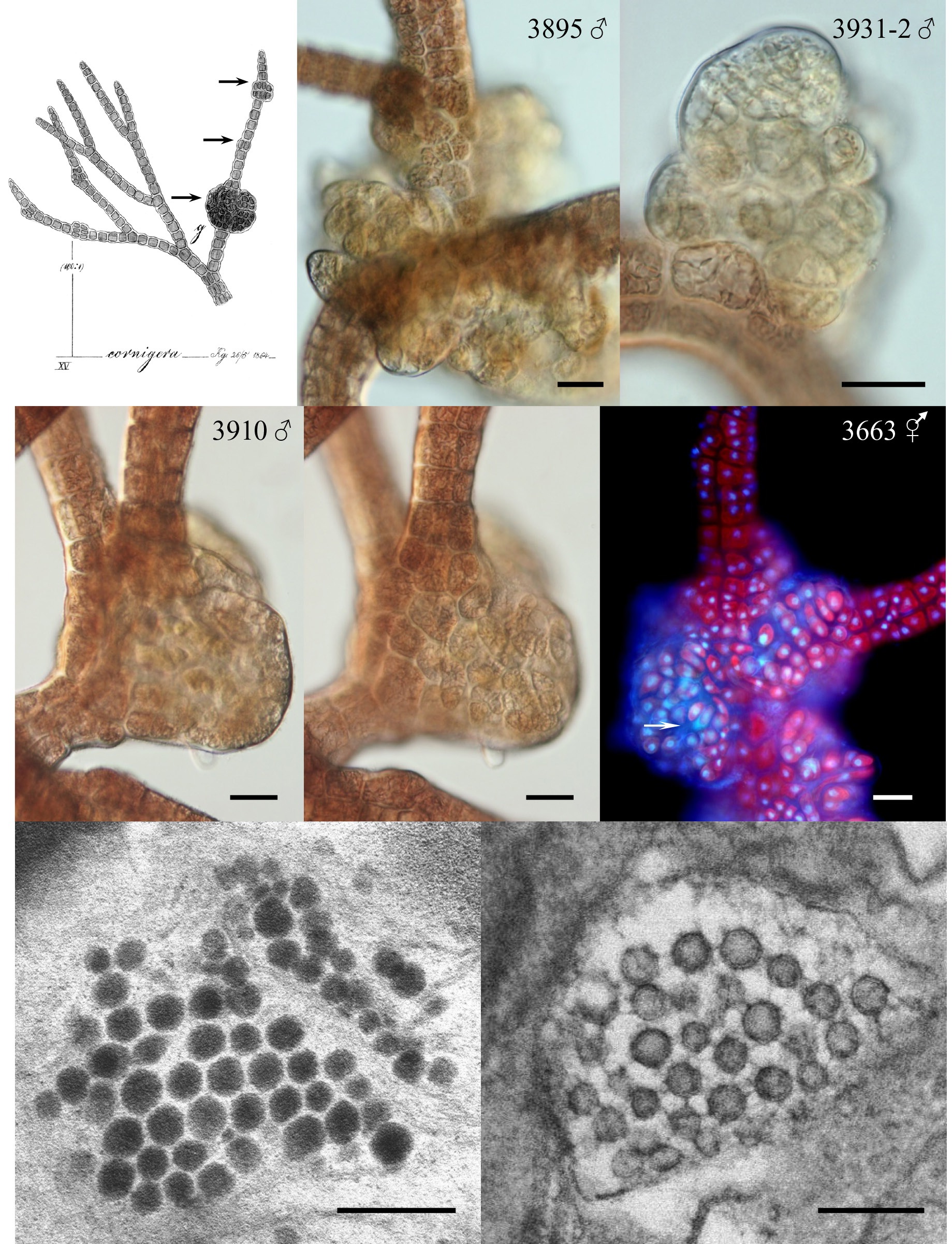
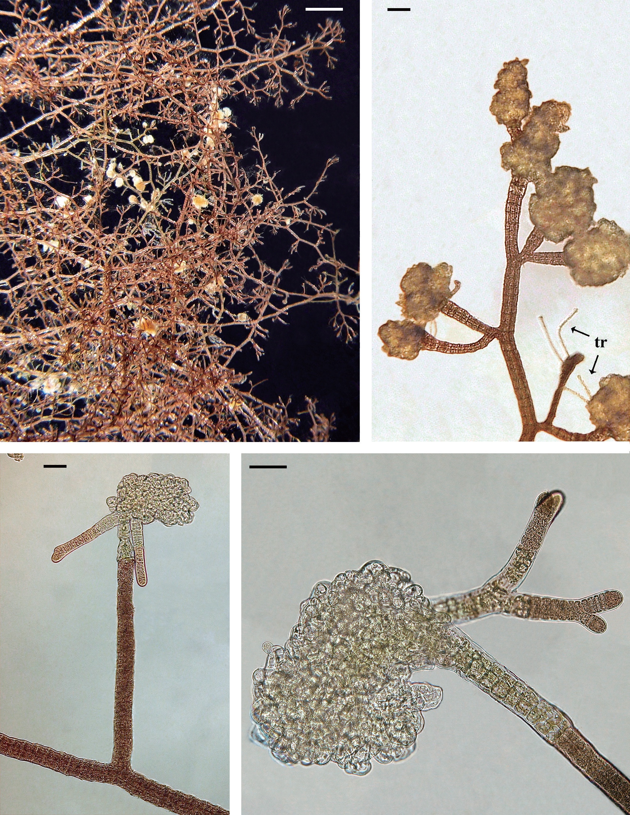
Galls varied in appearance and size. The initial stages appeared as enlarged proliferating cells. Cell divisions appeared random and very different from the polysiphonous tier cell division pattern of the normal host (Fig. 1B-E). The cells in galls were often smaller than tier cells, of various shapes and sizes and had enlarged vacuoles. Gall cells were less pigmented than normal host tissue cells (Fig. 1B). They appeared to have viable nuclei that often were larger than tier-cell nuclei (Fig. 1F). In smaller galls the cells all appeared viable but as the galls expanded and the cell number increased some dead cells were seen (arrow in Fig. 1F). In some larger galls new shoots arose within the cell mass (no photo).
In many isolates galls were present intermittently, however, galls were continuously present on the male and bisexual phases of 3663 (Fig. 1F) from 1999-2012 and on the male phase of 3895 from 2001-2012 (Fig. 1B). Isolate 3932 was unusual because galls appeared on the bisexual or female thalli but not on males from 2002-2012.
Electron microscopic (TEM) observations. While TEM fixation of cellular structure was difficult our micrographs did show VLPs in gall cells of isolate 3895 (Fig. 1G & H). These VLPs are of two distinct morphologies (staining differently) approximately 70-75 nm in size and hexagonal in shape.> Bostrychia moritziana / radicans
From 390 isolates of the
B. moritziana / radicans species-complex 24 (6%) produced galls. This species complex consists of seven different lineages (Zuccarello and West 2003, 2006, 2011) and galls were found on isolates from three lineages (1, 5, and 6). Again the preponderance of galls was found on isolates from Australia.In 26 years of culture isolate 2748 produced galls only once (May 1999) on the male spermatangial stichidia and these were very similar in overall structure to those seen in
B. simpliciuscula . On the female of isolate 2749 galls were noted only in 1989. By contrast, isolate 2747 from the same locality never had galls. Isolate 3026 female formed galls at irregular intervals in 1995, 2000, and 2002 whereas the tetrasporophyte and male showed no gall development during 22 years of culture. Isolate 3492 (Texas, USA) was isolated in 1974 and did not show galls until July, 2002 and these developed primarily on cladohaptera of tetrasporophytes.
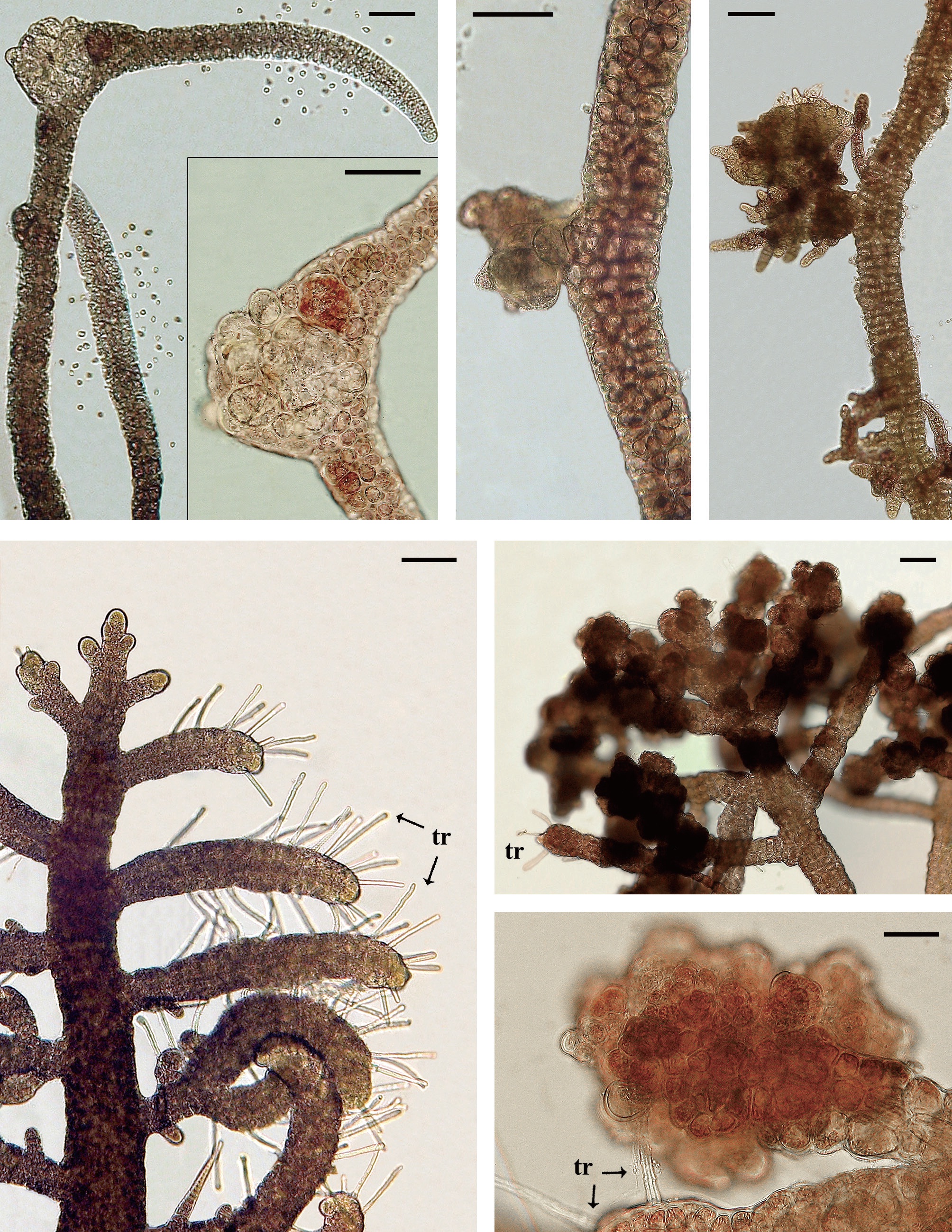
Isolate 3204 female (South Africa) was collected in 1991 and had numerous galls on procarpic lateral branches from 1997-2012 (Fig. 2A & B). Males and tetrasporophytes had no galls. Isolate 4124 female (Florida, USA) was collected in 2000 and developed galls with mostly colorless living cells on the tips of vegetative laterals. Regeneration of viable branches frequently occurred from gall tissue (Fig. 2C & D). Galls were evident for almost 8 years (2001-2009). Males and tetrasporophytes produced galls briefly in 2002.
Galls were observed on 16 males, 4 females, 4 bisexuals, and 3 tetrasporophytes of all isolates in Table 1.
Isolate 4086 (Sabah, Malaysia) was obtained in August 2000 and showed gall formation on the nodes and gametangial sectors of the bisexual gametophytes first in May, 2002. Isolate 4178 (New Caledonia) males developed galls in August, 2012 (Fig. 3A & B). Initially the gall cells enlarged and retained fully pigmented chloroplasts (Fig. 3A) but as the galls matured pale hypertrophied cells with enlarged vacuoles were evident (Fig. 3B). Other isolates of
B. radicosa from Thailand (4207) and Micronesia (4614, 4621, 4627, and 3662) did not develop galls.In 31 isolates of
B. kelanensis from Australia, Guam, India, Indonesia, Malaysia, and Micronesia only one male (3810) from Western Australia developed galls. Grouped cells divided and enlarged, projecting from tier cells (Fig. 3C) in spermatangial sectors of lateral branches. Eventually irregularly shaped masses developed numerous short branch apices (Fig. 3D).In 86 isolates of
B. tenella from many different geographic regions only isolate 2751 (Philippines) developed galls and that was only after 17 years in culture. Galls occurred continuously from 2003-2012 on the procarp bearing female, but never on the males or tetrasporophytes. In normal females the lateral branches were uniform in shape and heavily corticated, bearing numerous procarps (Fig. 3E). Many gall structures appeared as irregular club-shaped enlargements around the procarps (Fig. 3F). The variably-shaped cells contained fully pigmented chloroplasts (Fig. 3G). No branch shoot proliferation occurred from these galls.While the presence of galls is infrequent in
Bostrychia culture isolates, their similar morphology and the presence of VLP in gall cells ofB. simpliciuscula implicate viruses are the causative agent. Galls have been observed previously in other red algal cells with VLP suggesting them as a causative agent (Apt and Gibor 1991). Tumorous growth is commonly caused by viruses in higher plants (Francki et al. 1985, Scheffer 1997).Galls were not observed in laboratory culture on any other
Bostrychia species although this could be due to limited sampling. We have many more isolates of theB. moritziana / radicans (390),B. simpliciuscula (87), andB. tenella (89) than of other species but it could be that these species were more susceptible to the causative agent of gall formation.It is noteworthy that almost all species with galls lack cortication except for well-corticated
B. tenella in which only one female (2751) had galls.We have never observed galls on any
Bostrychia species in the field although we have examined thousands of specimens, however Kutzing (1865) observed and illustrated possible galls on field specimens.The ability of galls to form from healthy tissue separated from other gall tissue, suggests that the causative agent (possibly a virus) may be latent in cells of some
Bostrychia isolates. Latent bacteria are known in higher plants (Francki et al. 1985), and the stimulation of their effects (e.g., cell proliferation) could be due to stressors in the cells. This is seen in our experiments in which galls were induced in low temperature conditions. The inability of galls to be transmitted from one isolate to another inBostrychia and inGracilaria (Apt and Gibor 1991) may also indicate that much of the transmission is vertical within a single host. We did not test gall transmission to progeny of gall bearing sexual parents.Although not tested extensively three antibiotics (Penicillin G, Ciprofloxacin, and Rifampin) were routinely added to various cultures without any effect on gall presence. During antibiotic treatments various bacteria were also clearly present on the hosts as well.
The greater susceptibility of males and bisexuals to gall formation in
B. simpliciuscula may be due to the numerous spermatangia formed on male branches and the frequent release of spermatia opening more surface areas to attachment by viruses and bacteria.We know very little about causative agents of the galls seen in
Bostrychia , the effects of environmental stressors on gall formation, the transmission of the causative agent or the effects of galls on the fitness of their hosts. The study of potential pathogens of marine red algae should be pursued more critically.-
[Fig. 1.] Morphology of galls in Bostrychia. (A) Drawing of B. cornifera (currently B. moritziana) with several ‘galls’ developing on a monosiphonous branch (arrows). Reproduced from Kutzing (1865). (B-E) B. simpliciuscula microscopic images of galls developing on polysiphonous axis and laterals in different isolates (3895, 3931, and 3910). (D & E) Through-focus images of the same gall (3910). (F) B. simpliciuscula, fluorescent DAPI staining of the gall cells nuclei (blue color, DAPI-stained nuclei; red color, plastid autofluorescence; arrow points to the dead cells inside the gall). (G & H) B. simpliciuscula, transmission electron microscopy images of virus-like particles in gall tissues of isolate 3895. Scale bars represent: B-F, 50 μm; G & H, 200 nm.
-
[Table 1.] Bostrychia moritziana / radicans, B. kelanensis, B. radicosa, B. simpliciuscula, and B. tenella isolates observed. Rubisco spacer lineages of B. moritziana / radicans and B. simpliciuscula from Zuccarello and West (2003, 2006), Zuccarello et al. (1999) or during this study
-
[Table 1-1.] continued
-
[Fig. 2.] Galls on Bostrychia moritziana / radicans isolates 3204 and 4124. (A) Habit image of 3204 female with numerous white galls on branches bearing procarps. (B) Galls at tips of procarp bearing branches. Trichogynes (tr) visible projecting from gall at lower right. (C) 4124 female with gall at tip of vegetative lateral. Two regenerating shoots developed from gall cells. (D) 4124 female gall with mass of colorless cells, a few appear dead with collapsed protoplasts but most appear to be viable living cells. Healthy, branched shoot regenerated from gall tissue. Scale bars represent: A, 1 mm; B & D, 80 μm; C, 60 μm.
-
[Fig. 3.] Galls on Bostrychia radicosa, isolate 4178 (A & B); B. kelanensis, isolate 3810 (C & D); B. tenella isolate 2751 (E-G). (A) B. radicosa, elongate spermatangia bearing lateral branches with two galls visible on left. Released spermatia near branch tips also visible, small developing gall with normally pigmented cells visible on far left. (B) B. radicosa, high magnification, enlarged cells lightly pigmented with enlarged vacuoles. (C) B. kelanensis, developing gall with normally pigmented cells projecting from branch. (D) B. kelanensis, older galls with extensive branch formation. (E) B. tenella, normal procarpic branches with abundant trichogynes (tr). (F) B. tenella, numerous galls on procarpic branches, trichogynes (tr). (G) B. tenella, high magnification of gall cells, tier and cortical cells with fully pigmented chloroplasts, trichogynes (tr). Scale bars represent: A & B, 50 μm; C & D, 77 μm; E & F, 100 μm; G, 25 μm.