Biomineralization on the stalk of the eustigmatophyte Pseudocharaciopsis (Eustigmatophyceae)
- Author: Wujek Daniel E.
- Organization: Wujek Daniel E.
- Publish: ALGAE Volume 27, Issue2, p135~137, 15 June 2012
-
ABSTRACT
The stalks of the eustigmatophyte
Pseudocharaciopsis minuta were examined by light and scanning electron microscopy. Light microscopy revealed orange-red granules at the base of the stalk. Energy dispersive X-ray microanalysis of the bases indicated that they were mainly composed of manganese. Manganese has not been previously reported from eustigmatophytes. This study indicates that the Eustigmatophyceae needs further study into many aspects of the biology.
-
KEYWORD
biomineralization , Eustigmatophyceae , manganese , Pseudocharaciopsis , stalk
-
During an investigation of the chrysophyte alga
Lagynion delicatulum (O’Kelly and Wujek 2001) cells of the eustigmatophyte algaPseudocharaciopsis minuta (Braun) Hibberd contaminating our culture were isolated and placed in culture. Of particular interest was the presence of orange-red deposits at the base ofPseudocharaciopsis minuta ’s stalk.Extra-cellular biomineralization occurs in many algal groups: the Chrysophyceae (Preisig 1986) including colorless forms (Lee and Kugrens 1989), the Synurophyceae (Siver 2003), the Prymnesiophyceae (Nicholls 2003), the Euglenophyceae (Dunlap and Walne 1985, Rosowski 2003), the Chlorophyceae
sensu lato (Wefer 1980, Dunlap and Walne 1993), and the Bacillariophyceae (Sheath and Wehr 2003) including stalked forms (Novarino 1993). Such mineralization is seen among the bacteria (Heldal and Tumyr 1983, Omelon et al. 2006). A general review of biomineralization in photosynthetic organisms can be found in Raven and Giordano (2009).Characterization of mineralized structures is an important prerequisite to experimental studies on mechanisms of biomineralization in the algae. As the mineralization in
Pseudocharaciopsis occurs extracellular, the composition of granules at the cell’s none-living base provides an opportunity using energy dispersive X-ray elemental analysis system (EDS) to identify the type of metals present. Reported for the first time is the deposition of manganese on the stalk ofPseudocharaciopsis .Pseudocharaciopsis minuta appeared in enrichment cultures containingLagynion delicatulum , plant material, primarilyElodea canadensis from Lake Namunamu, North Island, New Zealand (39°53′ S, 175°28′ E), collected in February 1989. Unialgal cultures were established and maintained in Bold’s 3N Bristol’s medium with added soil water extract (BBM) (Starr and Zeikus 1987). Experimental cultures were grown in 10 × 100 mm plastic Petri dishes under long-day (16 : 8) photoperiods at 23℃; coolwhite fluorescent lamps yielded photons at 6-10 μmol m- 2 s-1. Under these conditions, significant populations of vegetative cells developed on coverslips placed in the Petri dishes. These cells were used for all critical observations and manipulations, as cells adhering to the dish could not be removed intact.
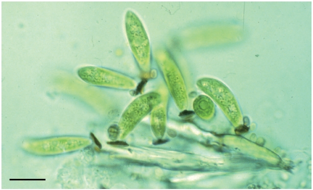
Living unstained cells were observed with a Zeiss Axiophot photomicroscope (Zeiss, Gottingen, Germany) equipped with differential interference contrast optics. Photographs were taken on Kodak Technical Pan 2415 35 mm film (Kodak, Rochester, NY, USA). Stalks bearing mineral deposits on stalks were studied by examining cells which had been affixed to coverslips, air-dried, mounted on aluminum stubs, and observed in a scanning electron microscope (Stereoscan 250 Mk3; Cambridge Instruments, Cambridge, UK) equipped with a Link AN10/55 EDS (Oxford Instruments, Oxford, UK).
Light microscope observations of untreated wet mo- unts showed the presence of orange-red granules at the base of the
Pseudocharaciopsis stalk (Fig. 1). No observable granules were present on any other parts of the cell.
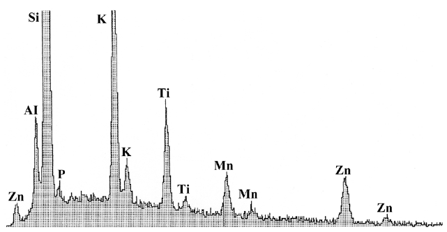
When the base of the stalks were examined with the scanning electron microscope and EDS analysis, the analyses showed manganese to be the predominant element associated with the base (Fig. 2). There was little variation in the relative proportion of Mn in the stalks from oldest to youngest. Other elements not associated with the glass or mounting stub or other parts of the cells were not detected. Iron was not detected, although low amounts could be masked by the Mn peak. Similarly other elements such as calcium were not evident.
Extracellular deposition of Mn in other algal groups has been reported in the Volvocales (Schulz-Baldes and Lewin 1975), the Charophyceae (Raven et al. 1986), and the Chrysophyceae (Dunlap et al. 1987). The lack of other minerals in the granules might be due to on the amount of iron or calcium in the culture medium, as has been demonstrated in the chrysophyte
Anthophysa (Lee and Kugrens 1989). Dunlap and Walne (1987) showed that the addition of iron to the media ofTrachelomonas cultures resulted in the presence of iron in the lorica. However it should be noted that BBM medium does contain relatively high amounts of either iron or calcium.The prevalence of Mn in the stalk of
Pseudocharaciopsis minuta is particularly interesting in light of the reports that Fe is the principal element of stalks in other algae. The Mn is probably present in the form of a carbonateor oxide.
This study supports the Eustigmatophyceae bear further investigation for the presence of metal elements in the remaining eight genera that produce mucilage but lack a stalk. As Santos (1996) points out, the chemical composition of the cell walls of the Eustigmatophyceae, and I can now add the mucilage, remain unclear. The presence of Mn on the stalks of
Pseudocharaciopsis raises questions as to the possible functions of these stalks and Mn metabolism.-
[Fig. 1.] Light microscope image of Pseudocharaciopsis minuta. Scale bar represent: 10 μm.
-
[Fig. 2.] X-ray spectrum of the stalk and base of Pseudocharaciopsis minuta. Manganese (Mn) is the main heavy element shown. Other elements present (Al, aluminum; Ti, titanium; K, potassium; Si, silicon; Zn, zinc) comprise the cover-slip on which the cells grew attached, the aluminum (Al) stub on which the cover slip was attached and the residue from an incompletely washed sample.