The genus Spirostomum is assigned to the family Spirosto-midae, order Heterotrichida, class Heterotrichea within the phylum Ciliophora. The family Spirostomidae is characteriz-ed by a highly contractile body, holotrichous somatic cilia-tion, long oral region in the anterior half and habitats in fresh-water and brackish water. Within the family Spirostomidae four genera have been recognized until now (Lynn, 2008). The genus Spirostomum Ehrenberg, 1833 is differentially diagnosed by a long collecting canal in the contractile vac-uole, a truncated posterior end and a long buccal field on the body edge (Curds et al., 1983). Since the establishment of the genus Spirostomum by Ehrenberg, 1833, nine species have been described worldwide (Claparede and Lachmann, 1858; Kahl, 1932; Shigenaka, 1959; Dragesco and Dragesco-Kerneis, 1986; Foissner et al., 1992). In this study, two Spiro-stomum species were isolated and reported here for the first time in Korea. We provide morphological descriptions of two Spirostomum species for the study of Korean ciliate diversity.
Spirostomum caudatum was collected from a freshwater pond near the Simnidaebat (Bamboo Forest) located near the bank of the Taehwa River, Ulsan (35 ? 32′55′′N, 129? 18′40′′E), Korea on 9 March 2009. Spirostomum teres was collected from a freshwater pond in Gulhwa-ri, Beomseo-eup, Ulju-gun, Ulsan (35? 33′31′′N, 129? 15′08′′E), Korea on 16 May 2011. The surface sediments (~10 cm) including water were collected and transferred to Petri dishes with debris, then maintained in the laboratory for several days at room tempe-rature. Meanwhile, a few wheat grains were added to the raw culture for the enrichment of the bacteria and ciliates (Li et al., 2010).
Phylum Ciliophora Doflein, 1901
Subphylum Postciliodesmatophora Gerassimova and Servin, 1976
Class Heterotrichea Stein, 1859
Order Heterotrichida Stein, 1859
Family Spirostomidae Stein, 1867
1*Genus Spirostomum Ehrenberg, 1833
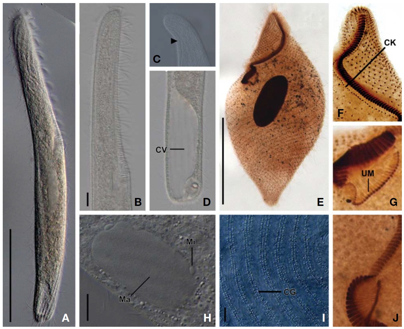
2*Spirostomum caudatum (Muller, 1786) Delphy, 1939 (Tables 1, 2, Fig. 1)
Enchelis caudata Muller, 1786: 34.
Spirostomum filum Penard, 1922: 200; Dragesco and Drage-sco-Kerneis, 1986: 375.
Spirostomum caudatum: Delphy, 1939: 144; Repak and Isq-uith, 1974: 328; Foissner et al., 1992: 324.
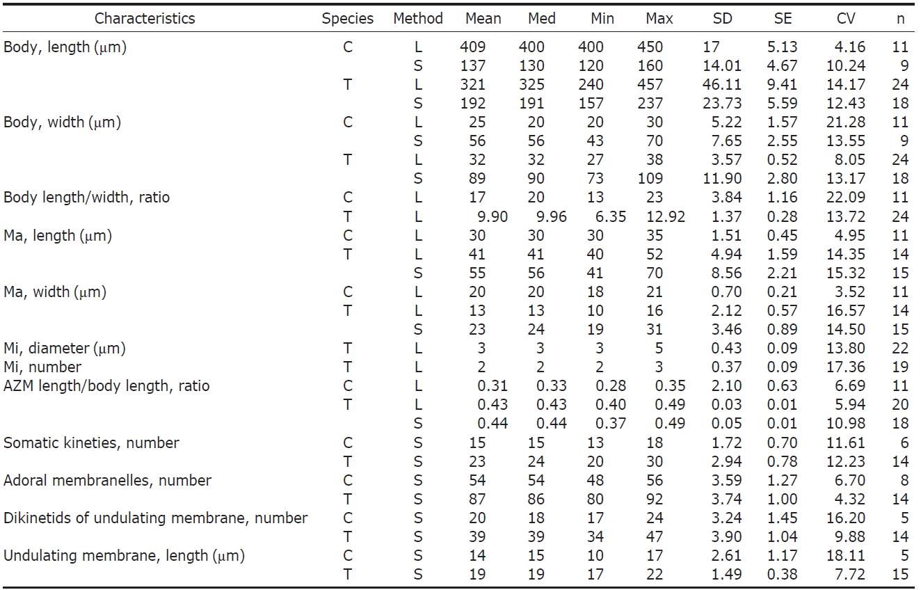
Diagnosis. Body size 400-450×2 0-30 μm in vivo; body shape cylindrical, slender with tail-like posterior end, ellip-soidal macronucleus, 13-18 somatic kineties, adoral zone about 30% of body length; and 48-56 adoral membranelles.
Description. Body size 400-450× 20-30 μm in vivo. Body shape cylindrical, slender with tail-like posterior end, ante-rior end slightly beaked, length to width ratio about 17 : 1(Fig.1 A, C), body fusiform when contracted (Fig.1 B, F). Macronucleus ellipsoidal, located near mid-body, size about 30× 20 μm in vivo (Fig.1 A, B, F). Contractile vacuole locat-ed in tail-like posterior region (Fig.1 D). Body flexible. Cyto-plasm colored slightly yellowish brown. Cortical granules arranged in 2-3 irregular rows between somatic kineties (Fig.1E). Movement relatively slow, usually gliding on the bottom.Somatic kineties arranged 13-18 longitudinally in impregna-
tion but arranged spirally when contracted (Fig.1 B, F). Ado-ral zone occupied 28-35% of body length (Fig.1 A); adoral zone of membranelles near proximal end twisted one time in impregnated specimens and consisted of 48-56 membranelles (Fig.1 B, F). Undulating membrane consisted of 17-24 dikinetids, near proximal end of adoral zone (Fig.1 G). Circum-oral kinety arranged on right side of adoral zone, dikinetids densely in one row (Fig.1 G).
Distribution. Africa, Europe and Asia (Korea [present stu-dy]).
Remarks. The Korean population of Spirostomum caudatum is in good agreement with the subsequent redescriptions in body size, tail apparatus, number of somatic ciliary rows, and presence of cortical granules (Foissner et al., 1992) (Table 2).
Spirostomum caudatum (Muller, 1786) is similar to S. teres Claparede and Lachmann, 1858, and S. yagiui Shigenaka, 1959, in respect to its single macronucleus. However, S. cau-datum and S. teres are distinguished by the shape of the pos-terior end (tail-like vs. blunted), the ratio of the oral length to body length (20-30% vs. 40-50%), and the arrangement of the cortical granular rows (irregular vs. regular) (Table 2) (Foissner et al., 1992).
Spirostomum caudatum and S. yagiui are different in the ratio of the oral length to body length (28-35% vs. about 50%), the number of adoral membranelles (48-56 vs. 130-140), the shape of the macronucleus (ellipsoid vs. rod-shape), the num-ber of micronuclei (1 vs. 3-7) and the habitat (freshwater vs.salt-water) (Table 2) (Shigenaka, 1959; Dragesco and Dra-gesco-Kerneis, 1986; Foissner et al., 1992).
1*Spirostomum teres Claparede and Lachmann, 1858(Tables 1, 2, Figs. 2,3)
Spirostomum teres Claparede and Lachmann, 1858: 233;Stein, 1867: 190; Kahl, 1932: 440; Wang and Nie, 1935:471; Dragesco and Dragesco-Kerneis, 1986: 378; Foissner et al., 1992: 332; Al-Rasheid, 1999: 130.
Diagnosis. Body size 240-460× 30-40 μm in vivo; shaped long and slender; macronucleus ellipsoidal; 2-3 micronuclei; cortical granules regularly arranged in 2-3 rows between somatic kineties; 20-30 somatic kineties arranged longitudi-nally; adoral zone of membranelles covered about 40-50% of body length with 80-92 adoral membranelles.
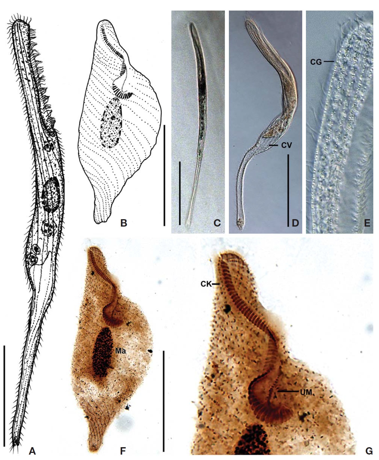
Description. Body size 240-460× 30-40 μm, usually about 330× 30 μm in vivo. Body shaped long and slender with tapered anterior and blunted posterior ends, length to width ratio about 10 : 1 (Figs.2 A, 3A), and body fusiform when contracted. Macronucleus ellipsoidal, located at mid-body, size about 56×24 μm in impregnated specimens, 2-3 ellip-soidal micronuclei, attached to a macronucleus, about 3 μm in diameter (Fig.3 E, H). Contractile vacuole located terminal-ly, occupied about 1/4-1/7 of body length with a long canal extending anteriorly (Figs.2 A, 3C, D). Body flexible. Cyto-plasm colorless. Cortical granules arranged regularly in 2-3 rows between somatic kineties, colorless, about 0.5 μm in diameter in vivo (Fig.3 I).
Movement relatively slow, usually gliding on the bottom.Somatic kineties arranged longitudinally 20-30 in number, but spirally when contracted, consisted of dikinetids, com-menced along the apical end to left side of the adoral zone of the membranelles (Figs. 2B, C, 3E), somiatic cilia about 8μm in length. Adoral zone of membranelles occupied 40-50% of body length (Figs. 2A, 3A), proximal end twisted one time in impregnated specimens (Fig. 3J), and consisted of 80-92 membranelles with each about 10 μm in length (Figs. 2B, C, 3B). The undulating membrane consisted of 34-47 dikinetids (Fig. 3G), near the proximal end of the adoral zone, length about 19 μm. A circumoral kinety arranged at right side of adoral zone, with dikinetids densely in one row (Fig. 3F).
Distribution. Africa, Asia (China, Korea [present study], Saudi Arabia, Turkey), Europe.
Remarks. The Korean population of Spirostomum teres closely resembles the Asian, African and European popula-tions with respect to the ratio of the adoral zone membranelles/body length, the shape of the macronucleus and micronuclei, the number of somatic ciliary rows, and the habitat (Dragesco and Dragesco-Kerneis, 1986; Foissner et al., 1992). However, this Korean population slightly differs from the German population in the color of the cortical granules (lemon-yellow to colorless vs. lemon-yellow), from the African population in the number of adoral membranelles (80-92 vs. about 120) and the number of somatic kineties (20-30 vs. 12-24), and from the Baltic Sea and Arabian Gulf populations (freshwater vs. salt water). The populations of S. teres have been record-ed in different salinities of habitats so far (Table 2) (Clapa-rede and Lachmann, 1858; Kahl, 1932; Dragesco and Dra-gesco-Kerneis, 1986; Foissner et al., 1992; Al-Rasheid, 1999;enler and Yildiz, 2004). Therefore, it will be needed to dis-close the variation caused by environmental gradients using independent criteria like molecular markers or others.
Spirostomum teres and S. yagiui are different with respect to the ratio of body length/width (6-13 : 1 vs. 12-18 : 1), the number of adoral membranelles (80-92 vs. 130-140), the shape of the macronucleus (ellipsoid vs. rod-shape), the num-ber of micronuclei (2-3 vs. 3-7) and the habitat (freshwater vs. salt-water) (Table 2) (Shigenaka, 1959; Foissner et al.,1992).
Korean name: 1*나선입섬모충속, 2*꼬리나선입섬모충
Korean name: 1*막대나선입섬모충