The genus
According to a recent study, this genus includes 17 describ-ed species, although the generic status of some species has been questioned (Jo, 1988; Serejo, 2004; Serejo and Lowry, 2008; Cheng et al., 2011). Moreover, this genus has been known as two cryptic species complexes such as
Recently, the mitochondrial cytochrome c oxidase subunit 1 (CO1) gene, which can distinguish species boundaries within most animal phyla, is very useful for DNA barcoding (Haji-babaei et al., 2006; Clare et al., 2007; Elsasser et al., 2009; Zemlak et al., 2009). Therefore, information derived from the CO1 gene sequence from different species is important for animal taxonomy.
In this paper,
Talitrids were collected under rotting hay on river mouth or sea shore in Korea between 2009 and 2010. They were imme-diately fixed on-site with 95% ethyl alcohol. The specimens were observed under both a stereomicroscope (Model SZX-ILLB2-200; Olympus, Tokyo, Japan) and a light microscope (Model DM 2500; Leica, Wetzlar, Germany). All dissected appendages were drawn using a microscope with a drawing tube attachment. Images were recorded using a microscope digital camera (Model Moticam 2000; Motic Incorporation Ltd., Hong Kong, China), and produced with Helicon Focus software (Model Helicon Focus; Helicon Soft Ltd., Kharkov, Ukraine). Classification system of crustacean setae follows Garm (2004) and Zimmer et al. (2009). All specimens exam-ined here were deposited in the Department of Biological Sciences, Inha University, South Korea.
Total genomic DNA was extracted from the pereopods of each specimen using a RED-Extract-N-Amp Tissue PCR Kit (Sig-ma, St. Louis, MO, USA) according to the manufacturer’s instructions. The target DNA segment of the mitochondrial CO1 gene was amplified using the primers LCO1490 and HCO2198 (Folmer et al., 1994).
Optimized PCR conditions were as follows: denaturation at 94℃ for 2 min; followed by 10 cycles of denaturation at 94℃ for 10 sec, annealing at 37℃ for 30 sec, extension at 72℃ for 60 sec; then followed by 25 cycles of denaturation at 94℃ for 10 sec, annealing at 48℃ for 30 sec, extension at 72℃ for 60 sec; and final extension step at 72℃ for 7 min.
The QIAquick PCR Purification Kit (Qiagen, Valencia, CA, USA) was used for the purification of PCR products. An ABI 3700 sequencer (Applied Biosystems, Foster City, CA, USA) was used for sequencing, and sequences obtained in this study were all deposited in GenBank.
Order Amphipoda Latreille, 1816
Family Talitridae Rafinesque, 1815
Genus Platorchestia Bousfield, 1982
1*
Material examined. Korea: Busan: 7♂♂ and 5♀♀, Saha-gu, Dadae-dong, Dadaepo Beach (35? 02′ N, 128? 57′ E), 26 Jun 2010, Kim MS; Gyeongsangbuk-do: 10♂♂ and 11♀♀, Yeongdeok-gun, Byeonggok-myeon, Songcheon Stream
(36? 33′ N, 129? 25′ E), 10 Jun 2010, Kim MS; Incheon: 15♂♂ and 13♀♀, Ganghwa-gun, Gilsang-myeon, Onsu-cheon Stream (37? 38′ N, 126? 31′ E), 10 Sep 2009, Kim MS; 6♂♂ and 7♀♀, Ongjin-gun, Bukdo-myeon, Isl. Yongyudo, Wangsan Beach (37? 27′ N, 126? 21′ E), 8 May 2009, Kim MS; 13♂♂ and 11♀♀, Jung-gu, Sinheung-dong, Yeo-nangyo Bridge (37? 26′ N, 126? 37′ E), 23 Mar 2010, Kim MS; Ulsan: 12♂♂ and 10♀♀, Buk-gu, Myeongchon-dong, Taehwa River (35? 33′ N, 129? 20′ E), 13 Oct 2009, Kim MS.
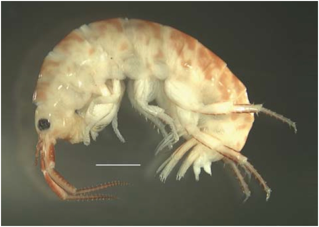
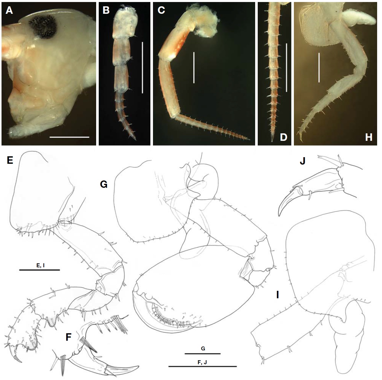
Description (male). Body (Figs. 1, 2A) 9.3 mm. Eyes sub-round and black, width 0.4 times head diameter.
Antenna 1 (Figs. 1, 2B) not reaching peduncular article 5 of antenna 2; peduncular articles 1-3 subequal in length; peduncular articles 1-2 marginally bare ventrally, ventral margin of peduncular article 3 with one cuspidate seta; flagel-lum 6-articulate, each article with two groups of 2-3 simple setae distally.
Antenna 2 (Figs. 1, 2C, D), 0.31 times as long as body length; peduncular articles 4-5 with length ratio of 1 : 1.3, weakly incrassate, bearing many short cuspidate setae, distal margin with simple setae; flagellum 1.44 times of peduncular article 5, with 16 articles, flagellum 2 proximal articles incom-pletely fused; each article with short simple setae distally.
Gnathopod 1 (Fig.2 E, F) cuspidactylate and subchelate; coxal plate 1 with two rows of many simple and cuspidate setae on ventral margin; basis straight, with eight cuspidate setae on anterior margin and four cuspidate setae on posterior margin; merus with nine cuspidate setae on posterior margin;carpus with two groups of 2-3 cuspidate setae on dorsal mar-gin, posterior margin with tumescent hump, seven lateral, and six medial setae; propodus 0.63 times as long as carpus, with four groups of 1-4 cuspidate setae on dorsal margin and tumescent hump posteriorly, lateral margin with 12 setae, medial margin with 11 setae; dactyl with one simple seta on inner margin and two simple setae at hinge of unguis.
Gnathopod 2 (Fig.2 G) powerfully subchelate; coxal plate 2 with many simple setae on ventral margin, posterior cusp obtuse; basis straight, with six cuspidate setae on anterior margin, posterior margin with seven cuspidate setae; pro-podus oval in shape, posterior margin marginally bare and palmar margin fringed with many short simple and cuspi-date setae, with two protrusions, these protrusions wide and shallow, anterior one about 1.9 times as long as posterior one; dactyl strongly curved, inner margin with many simple setae, distal part narrowed.
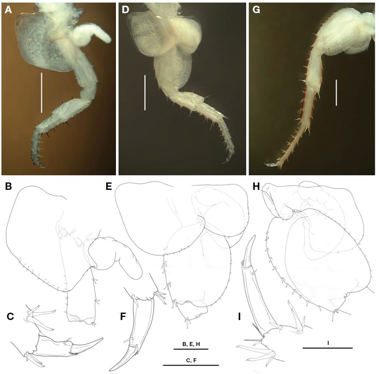
Pereopods 3-4 (Figs.2 H-J, 3A-C) cuspidactylate; coxal plate 3 sub-rectangular, with many simple setae on ventral margin, posterior margin with cusp; basis straight, with nine
cuspidate setae on anterior margin, posterior margins with three groups of 1-2 cuspidate setae; merus to propodus with several groups of 1-3 cuspidate setae on anterior and poste-rior margins; dactyl with two simple setae at hinge of unguis; pereopod 4 resembling pereopod 3, but posterior corner of coxal plate 4 weakly expanded and dactyl slightly thickened and pinched posteriorly.
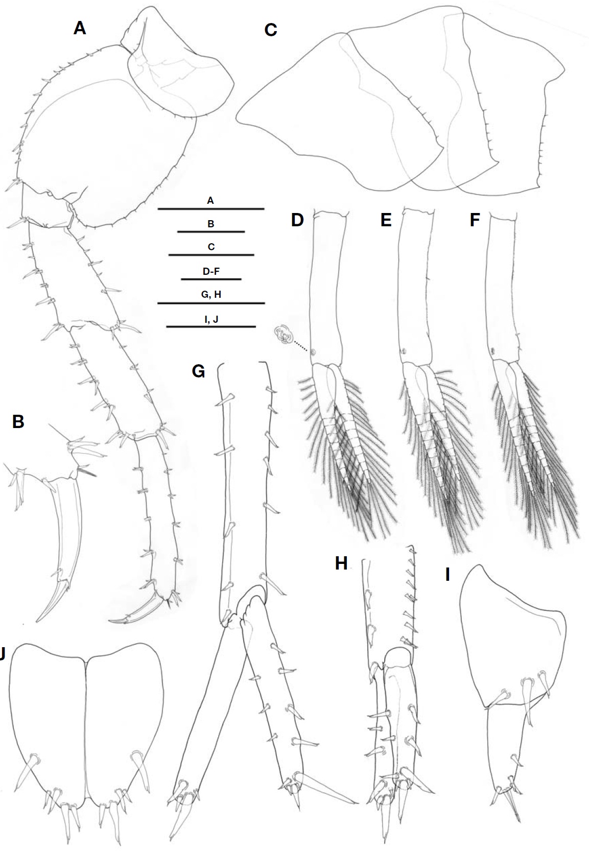
Pereopods 5-7 (Figs.3 D-I, 4A, B) cuspidactylate; coxal plates 5-6 bilobate, anterior lobe of coxal plate 5 larger than posterior one, while anterior lobe of coxal plate 6 very small and posterior lobe lacking process; coxal plate 7 non-lobate; basis of pereopods 5-7 oval in shape, with 13-15 simple setae on posterior margin, anterior margin with 5-12 groups of 1-3 cuspidate setae; merus to dactyls resembling pereopod 3.
Gills 2-6 (Figs.2 G, H, I, 3A, B, D, E, G, H) present; gill 2 elongated and curved in middle; gills 3-4 similar in resem-blance; gill 5 broader than gills 3-4; gill 6 as large as gill 2.
Epimeral plates 1-3 (Fig.4 C) with weakly pointed posterior angles, ventral margin marginally bare and posterior margin with 5-7 simple setae.
Pleopods 1-3 (Fig.4 D-F) well developed, subequal in length; peduncles of all pleopods with two retinaculae distally, both rami with about 8-10 articles, armed with plumose setae; peduncle of pleopod 1 marginally bare; peduncle of pleopod 2 with one simple seta in middle part of outer margin; pedun-cle of pleopod 3 with two simple setae in distal part of outer margin.
Uropod 1 (Fig.4 G), peduncle with two rows of 4-6 marginal cuspidate setae and distolateral cuspidate seta; outer ramus about 0.79 times of peduncle, marginally bare, distal margin
with four cuspidate setae; inner ramus with three, four, and five cuspidate setae on inner, outer, and distal margins, res-pectively.
Uropod 2 (Fig.4 H), peduncle with three cuspidate setae on outer margin, inner margin with 10 cuspidate setae; outer ramus with two marginal and four distal cuspidate setae, inner ramus with two, two, and five cuspidate setae on in-ner, outer, and distal margins respectively.
Uropod 3 (Fig.4 I), peduncle slightly broadened dorsally, with three dorsal cuspidate setae; ramus slightly shorter than peduncle, with one marginal cuspidate seta, distal margin with two long simple and three short cuspidate setae.
Telson (Fig.4 J) subtriangular in shape, distally notched,
with medial suture-line on dorsal surface; each lobe with two groups of 1-2 lateral cuspidate setae and two apical cus-pidate setae.
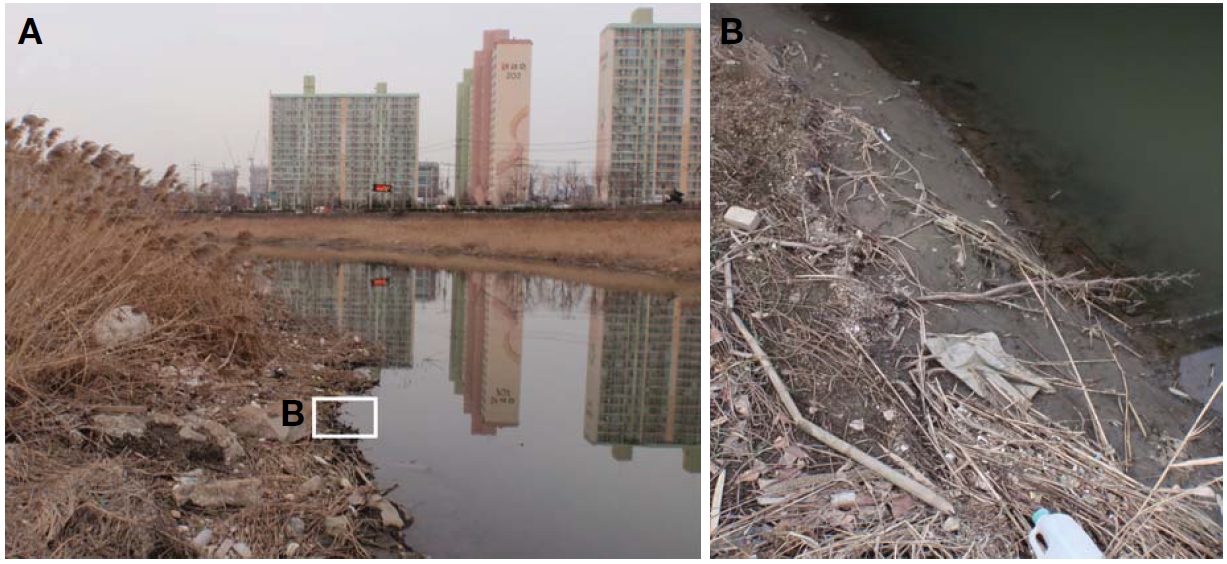
Habitat.This species lives in river mouth or sea shore. They were usually found under rotting hay or in silt beside the water (Fig.5 ).
Molecular data. CO1 gene sequences (GenBank accession no: JN712919-JN712930) were obtained from 12 individuals of
[Table 1.] Morphological differences between Korean Platorchestia monodi those from other countries
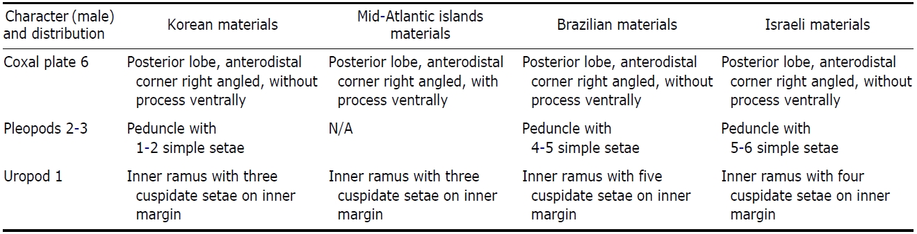
Morphological differences between Korean Platorchestia monodi those from other countries
not found.
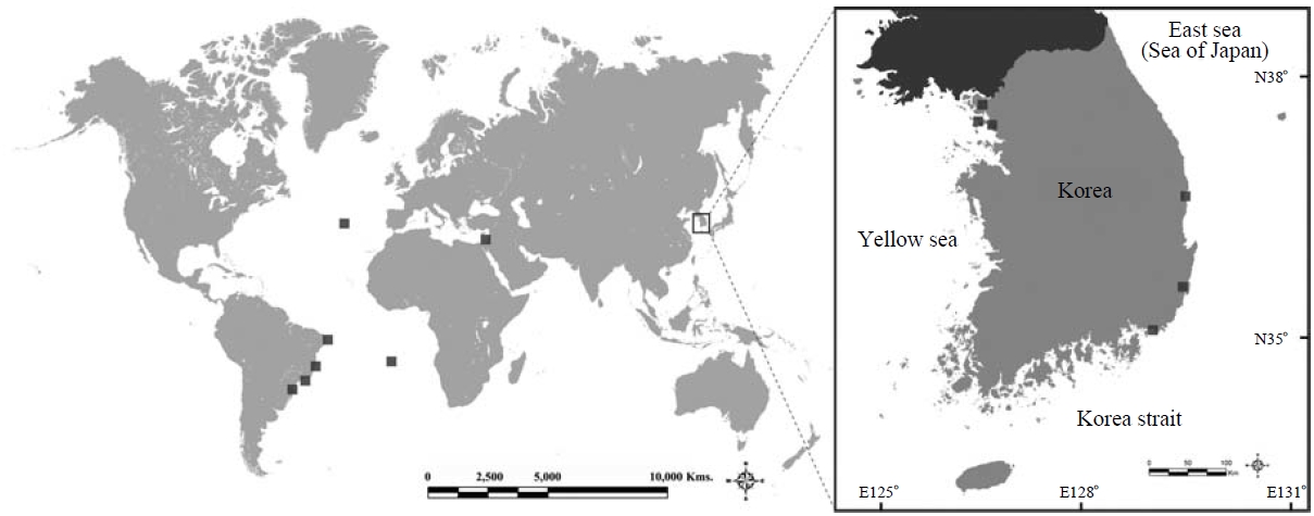
Remarks. Mateus et al. (1986) first reported this species from the sea shores of the Azores Island in Portugal. According to a recent study, present species is mainly distributed from the Atlantic region such as Mid-Atlantic islands, Brazilian coast, and Israel (Mateus et al., 1986; Stock and Biernbaum, 1994; Morino and Ortal, 1995; Stock, 1996; Serejo, 2004). In this paper, this species was reported for the first time from the Pacific region (Fig.6 ).
This species is characterized by the following features: (i) antenna 2 (♂), peduncular articles 4-5 weakly incrassate; (ii) gnathopod 1 (♂), dactyl cuspidactylate; (iii) propodus of gnathopod 2 (♂), posterior margin marginally bare and palmar margin with two protrusions, these protrusions wide and shallow; (iv) coxal plate 6, anterodistal corner of poste-rior lobe rounded; and (v) pereopod 7 (♂) not sexually dimor-phic (merus and carpus not expanded).
In general, Korean
The partial CO1 sequence of this species was determined for the first time and registered at GenBank. This genetic data would be helpful in the study of the speciation processes, radiation patterns, and cryptic species diversity.
Korean name: 1*기수도약옆새우 (신칭)