Genera Anteholosticha Berger, 2003 and Metaurostylopsis Song et al., 2001 belong to the family Urostylidae Butschli, 1889. This family is characterized by having one or more marginal rows of left and right marginal cirri and the midventral cirri which arranged as a “zig-zag” file (Borror, 1979; Borror and Wicklow, 1983; Lynn, 2008).
Diverse and polyphyletic species in the Holosticha com-plex (Gao et al., 2010) were split up into four genera, Holosti-cha, Anteholosticha, Caudiholosticha and Biholosticha by Berger (2003). Recently Li et al. (2009) added another genus Nothoholosticha to this complex, and therefore there are five genera in the Holosticha complex at present. The genus Ante-holosticha can be distinguished from other genera in Holo-sticha complex by its lack of caudal cirri and several other apomorphic characters, e.g., anterior end of left marginal row curved rightwards, proximalmost membranelles widened (Berger, 2003). Recently a Chinese population of A. pulchra was reported by Li et al. (2007).
The genus Metaurostylopsis was established by Song et al. (2001). The characteristics of this genus are as follows: clear-ly differentiated frontal, buccal, transverse cirri and frontoter-minal cirral row present; caudal cirri absent; more than one left and right marginal row (Song et al., 2001). Metaurosty-lopsis struederkypkeae was newly established by Shao et al. (2008).
Here, we redescribe two urostylid ciliates, A. pulchra and M. struederkypkeae, collected from Korea. For precise iden-tification and description, we conducted live observation, protargol impregnation and small subunit ribosomal DNA(SSU rDNA) sequence analysis.
Anteholosticha pulchra was isolated from Incheon Harbor (salinity, 30.6‰; temperature, -0.6 ?C; 37 ? 6′N, 126 ? 35′E), Incheon, Korea at January of 2011. Metaurostylopsis strue-derkypkeae was collected from the lagoon of Youngrang (salinity, 14.8‰; temperature, 18.3 ?C; 38 ?13′N, 128 ? 35′E), Sokcho, Korea at June of 2011. These two populations were maintained in petri dishes with rice grains to enrich bacteria growth as a food resource. The specimens were observed in vivo under a light microscope (Leica DM2500, Wetzlar, Ger-many) at a magnification of 50× to 1,000×. Protargol im-pregnation was performed to reveal the infraciliatures (Foiss-ner, 1991). Drawing was performed under a light microscope at a magnification of 1,000×. Terminology and classification were mainly followed according to Lynn (2008).
DNA extraction, amplification and sequencing of nearly complete SSU rDNA were performed according to Jung et al. (2011). Our sequences and retrieved sequences from Gen-Bank were aligned using BioEdit (Hall, 1999), and pairwise genetic distance based on Kimura two-parameter inference was obtained using MEGA 4.0 (Kumar et al., 2004).
Korean name: 1*홍색옛전열하모충 (신칭)
Phylum Ciliophora Doflein, 1901
Class Spirotrichea Butschli, 1889
Order Urostylida Jankowski, 1979
Family Urostylidae Butschli, 1889
Genus Anteholosticha Berger, 2003
1*Anteholosticha pulchra (Kahl, 1932) Berger, 2003 (Table 1, Figs.1 A-E, 2)
Keronopsis pulchra Kahl, 1932: 573, fig.104 ; Kahl, 1933:109, fig. 16.
Holosticha (Keronopsis) pulchra: Berger, 2001: 36.
Anteholosticha pulchra: Berger, 2003: 377; Berger, 2006: 435, fig.90 ; Li et al., 2007: 113, figs. 4-7
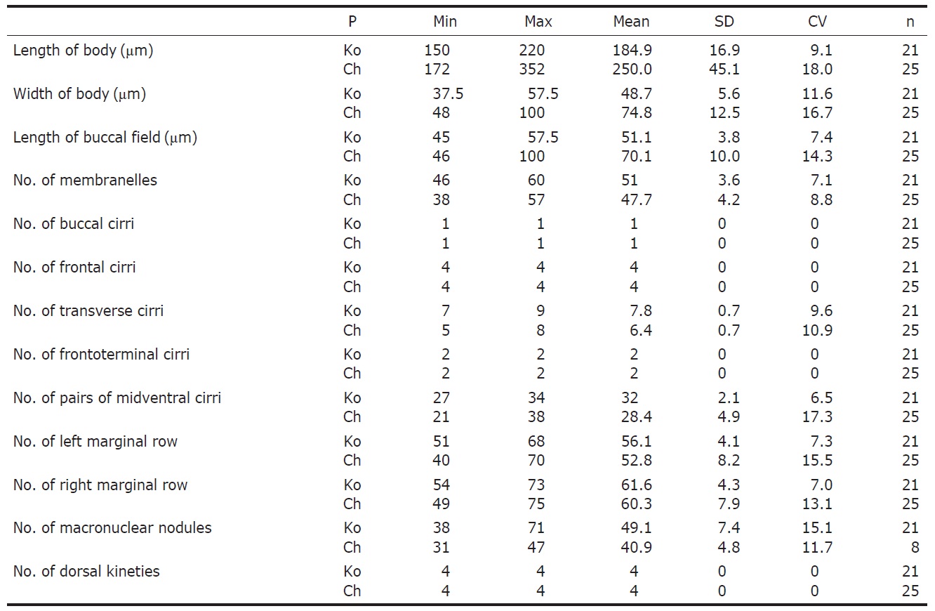
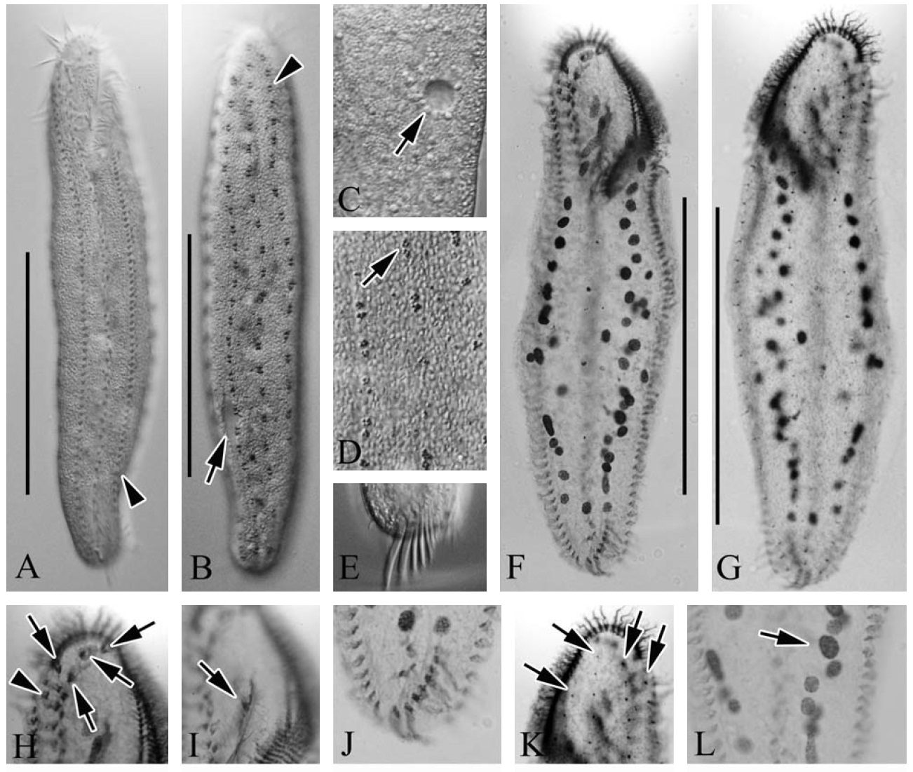
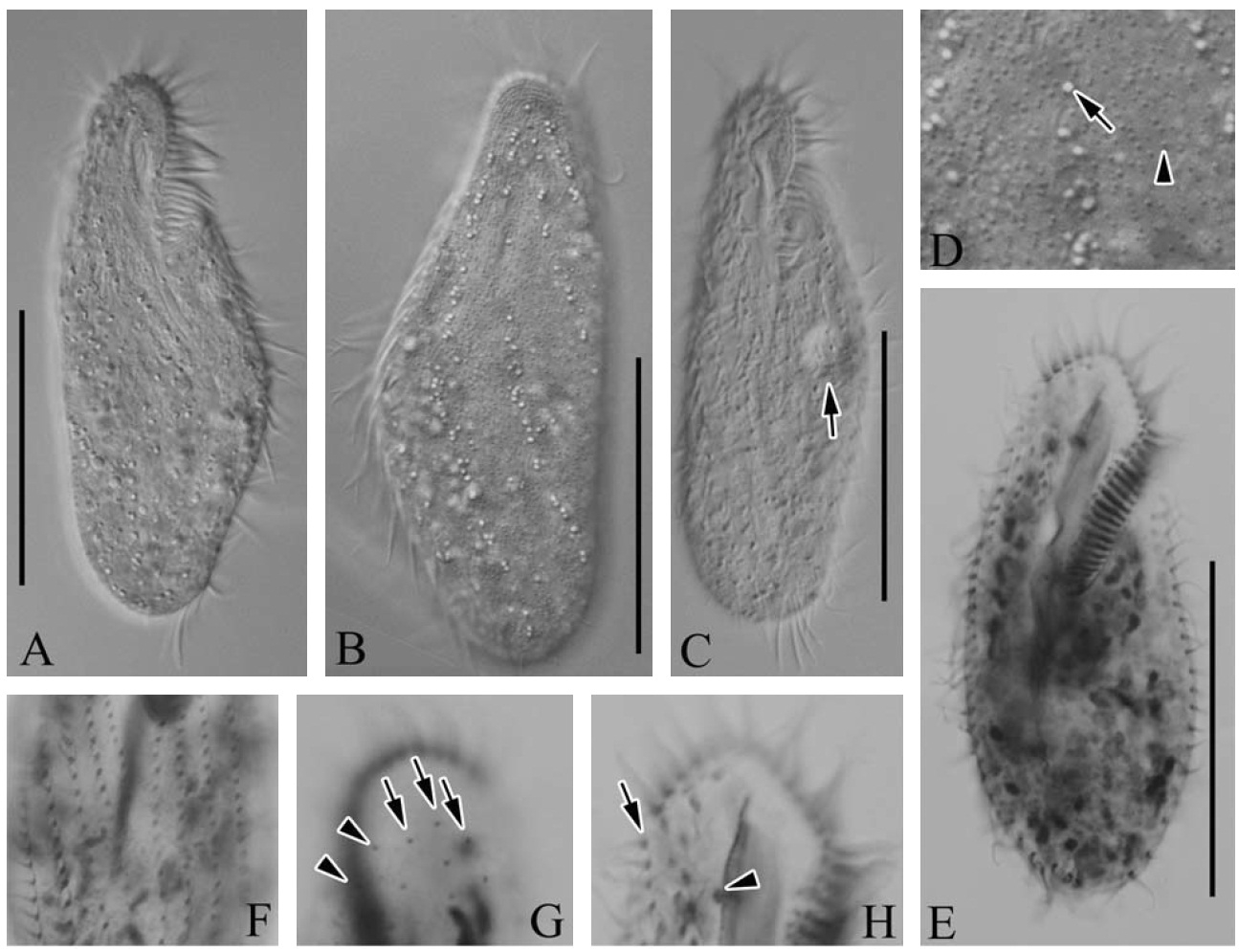
Description. Live cell size usually 190-300×30-55 μm, slen-der in shape (Figs.1 A, 2A, B), flexible but not contractile, an-terior and posterior ends rounded, contractile vacuole approxi-
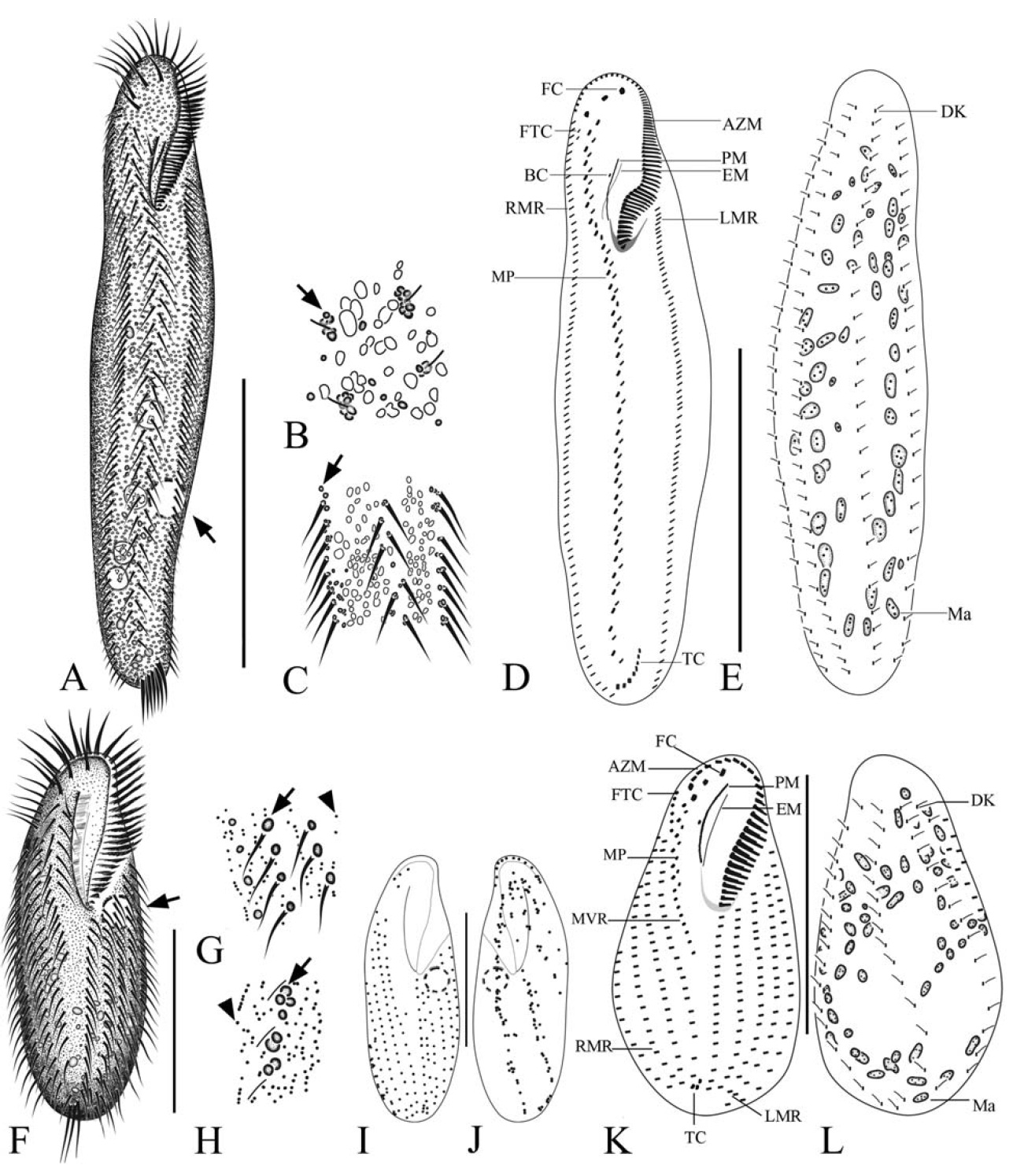
mately 10 μm in diameter, positioned on left side of posterior 1/4 of cell (Figs.1 A, 2B, C, arrow). Spherical-reddish corti-cal granules at cirral bases and around dorsal bristles, ca. 1μm across, some sparsely distributed throughout cell surface (Figs.1 B, C, 2D, arrow). Four frontal cirri each located in anterior ventral area, ca. 18 μm (Fig.2 H, arrows), and two frontoterminal located near anterior end of right marginal row (Fig.2 H, arrowhead) and seven to nine transverse cirri (Fig.2 E, J), respectively ca. 10 and 20 μm in length, with one left and right marginal row located on left and right side (left marginal cirri ca. 56, right marginal cirri ca. 61). Buccal cirrus positioned near paroral membrane (Fig.2 I, arrow), cau-dal cirri absent. Four dorsal kineties extended from anterior to posterior area (Fig.2 G, K, arrows) and approximately 49 macronuclear nodules scattered in body (Figs.1 E, 2L).
46-60 membranelles in adoral zone, ca. 25-35% of body length, and thick buccal lip. Long midventral pairs extending to transverse cirri, in a “zig-zag” pattern, 27-34 pairs of cirri. Food vacuoles colorless and sparsely located on cell.
Distribution. Germany (Kiel, the Baltic Sea), China (Laiz-hou, the Yellow Sea), and Korea (this study).
Remarks. Polyphyly of genus Anteholosticha was shown by molecular phylogenetic analysis based on the SSU rDNA. Therefore this genus should be divided into subgroups acc-
ording to the relativeness of both morphology and phylogeny (Berger, 2006; Gao et al., 2010; Yi and Song, 2011). Among ca. 40 species known from this genus, following five species showed closest relationship with Anteholosticha pulchra in morphology: A. gracilis, A. xanthichroma, A. australis, A.sigmoidea and A. monilata.
Anteholosticha gracilis is most similar to A. pulchra be-cause both species have four frontal and two frontoterminal cirri as marine ciliates. However Anteholosticha pulchra dif-fers from A. gracilis mainly in size (190-300×30-55 μm vs.100-150×30-40 μm in vivo), position of contractile vacuole (posterior 1/4 vs. anterior 1/3), color of cortical granules (re-ddish vs. yellow-greenish), number of dorsal kineties (four vs. three), adoral membranelles (46-60 vs. 24-30) and pair of midventral cirri (27-34 vs. 17-24) (Hu and Suzuki, 2004). An-teholosticha pulchra can be distinguished from other species by the position of contractile vacuoles: it locates around 1/4 region from the posterior margin in A. pulchra; around 1/3 from the anterior margin in A. gracilis; ahead of mid-body in A. xanthichroma, A. australis, A. sigmoidea and A. monilata (Foissner and Didier, 1981; Foissner, 1982; Wirnsberger and Foissner, 1987; Blatterer and Foissner, 1988; Hu and Suzuki, 2004).
Nothoholosticha fasciola transferred from the genus Ante-holosticha by Li et al. (2009) because this species has no frontoterminal cirri. However two species are similar in fol-lowing characteristics, e.g., one buccal cirrus and long belt-like body shape. However, A. pulchra differs from N. fasci-ola in having frontoterminal cirri (vs. absent), four frontal cirri (vs. six), four dorsal kineties (vs. three), reddish cortical granules (vs. bright orange) and the contractile vacuole posi-tion (located around 1/5 region from the posterior margin in N. fasciola) (Li et al., 2009).
The Korean population corresponds well with Chinese population reported by Li et al. (2007), and showed high sim-ilarity each other in morphometric characteristics (Table 1).
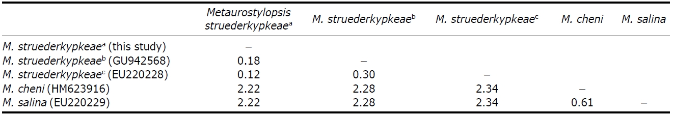
The SSU rDNA sequence of Korean A. pulchra is 1,650 bp in length (GenBank accession no: JN880476). Inter-specific genetic variation among five species (A. pulchra, A. gracilis, A. monilata, A. pseudomonilata, and N. fasciola) are ranged from 3.3% to 6.6% (Table 3).
1*Genus Metaurostylopsis Song, Petz and Warren, 2001
2*Metaurostylopsis struederkypkeae Shao et al., 2008 (Table 2, Figs.1F-L, 3)
Metaurostylopsis struederkypkeae Shao et al., 2008: 289, figs. 1-20
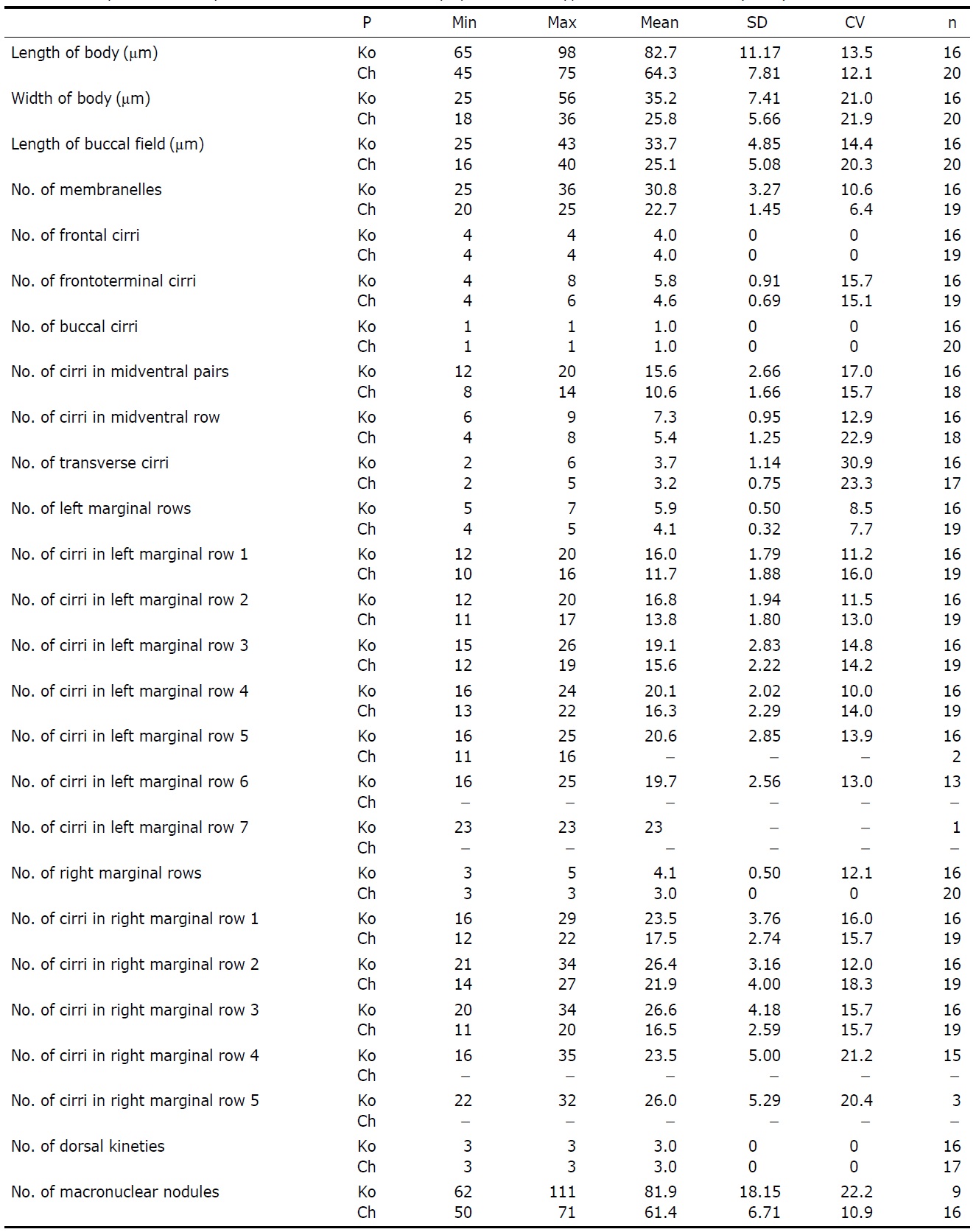
Description. Live cell size usually 80-110× 40-50 ㎛ (Figs.1F, 3A-C), slender in shape and slightly contractile, with an-terior and posterior ends rounded, and contractile vacuole approximately 10㎛ in diameter located on left side of equa-torial level of cell (Fig.3 C, arrow). Two types of cortical granules: one small and reddish, approximately 0.5㎛ across, spread irregularly on surface (Figs.1 G, H, 3D, arrowhead), and other one yellow-green, approximately 1.5 ㎛ across, situated at base of cirri, with some cortical granules clustered along cirral rows and dorsal kineties (Figs.1 G, H, 3D, arrow). Color of cells appeared reddish under low magnification due to numerous small cortical granules. Lengths of transverse and frontal cirri 15 ㎛ and 13 ㎛, respectively. Other soma-tic cirri, midventral, left and right marginal cirri, ca. 10 ㎛ in length. Four frontal cirri and four to eight frontoterminal cirri located near distal portion of adoral membranelles (Fig.3H, arrow). Two to six transverse cirri, and one buccal cirrus located in middle of next paroral membrane (Fig.3 H, arrow-head). Left and right marginal rows composed of five to seven and three to five, respectively (Fig.3F). 12-20 midventral pairs, and six to nine unpaired midventral cirri between mid-ventral pairs and transverse cirri, three dorsal kineties (Fig.3G, arrows), and separately two dikinetids (Fig.3 G, arrow-heads) on anterior dorsal side. Macronuclear nodules, ovoid and ellipsoid shape, ca. 82. Membranelles in adoral zone con-sisted of 25-36 membranelles, and portion of adoral zone is ca. 35-45% in length. Paroral and endoral membrane are equal level in length.
Distribution. China (Qingdao, the Yellow Sea) and Korea (this study).
Remarks.There are six species in this genus: Metaurostylop-sis marina Song et al., 2001, M. rubra Song and Wilbert, 2002, M. songi Lei et al., 2005, M. salina Lei et al., 2005, M. struederkypkeae Shao et al., 2008, and M. cheni Chen et al., 2011.
Metaurostylopsis struederkypkeae and M. rubra have red-dish cell color. However M. struederkypkeae is distinguished from M. rubra by the number of right marginal rows (3-5 vs. 6-7) and the type of cortical granules (two vs. one) and size (80-110 × 40-50 ㎛ vs. 150-300× 50-90 ㎛ in vivo) (Song and Wilbert, 2002). Metaurostylopsis struederkypkeae is distinguished from M. marina by the body shape (slender vs. oval), the cell color (reddish vs. colorless) and the type of cortical granules (two vs. one) (Song et al., 2001).
The Korean population of M. struederkypkeae corresponds with the original description by Shao et al. (2008) and mor-phometric comparisons highly overlap (Table 2); however, there are a few differences. The Korean population has more
number of left and right marginal rows, midventral pairs, and macronuclear nodules than those of the Chinese population (Shao et al., 2008).
The SSU rDNA sequence of M. struederkypkeae is 1,652 bp in length (GenBank accession no: JN880477). The inter-specific pairwise distances between M. struederkypkeae and its relatives (M. cheni and M. salina) are more than 2%, while intra-specific variation are less than 0.3% among two Korean and one Chinese M. struederkypkeae populations (Table 4).
Korean name: 1*사미주하모충 (신칭), 2* 작은홍색사미주하모충 (신칭)