검색 전체 메뉴
PDF
맨 위로
OA 학술지
Stimulative Effects of Hominis Placental Pharmacopuncture Solution Combined with Zinc-oxide Nanoparticles on RAW 264.7 Cells - ZnO HPPS more easily stimulates RAW 264.7 cells -
  • 비영리 CC BY-NC
  • 비영리 CC BY-NC
ABSTRACT
Stimulative Effects of Hominis Placental Pharmacopuncture Solution Combined with Zinc-oxide Nanoparticles on RAW 264.7 Cells - ZnO HPPS more easily stimulates RAW 264.7 cells -
KEYWORD
Hominis Placenta , zinc oxide , pharmacopuncture , RAW 264.7 cells
  • 1. Introduction

    Nanotechnology is referred to as a science that makes a new system through which matter is manipulated on a nanoscale [1], and great advances have been made in the field of nanotechnology over the last decade. It is one of the most advancing technologies of the 21st century and is thought to occupy a leading position in industry. Engineered nanomaterials are already being used in various industrial fields such as sporting goods, cosmetics, and electronics and will be increasingly applied to medical fields such as diagnosis, cancer treatment, and drug delivery [2,3]. Regardless of these extensive applications, many concerns about the safety and the potential risks of engineered nanomaterials associated with human health have been raised [4].

    Zinc oxid (ZnO) is one of the most exciting materials because of its versatile properties such as its semiconducting, biosafe, and biocompatible natures. ZnO is also considered to be a “GRAS” (generally recognized as safe) subst-ance by the Food and Drug Administration (FDA). Because of these properties, ZnO can be safely used in medical fields such as cancer therapy and drug delivery vehicles [5].

    Hominis Placenta extracts have been used in traditional Korean medicine to improve physiological function. Recently, there have also been several reports that Hominis Placenta can be applied to pathologic conditions such as wounds [6], cartilage degradation [7], osteoporosis [8], nerve injury [9], and anemia [10].

    In this study, we examined whether ZnO makes a difference when it is combined with Hominis Placenta pharmacopuncture solution (HPPS). We made ZnO NP HPPS, a combined form of ZnO nanoparticles (NPs) and HPPS. Then, we compared the stimulatory effects of ZnO NP HPPS with those of ZnO NPs and HPPS. In this study, we found that ZnO NP HPPS, the combined form of ZnO NPs and HPPS, had more ability to stimulate RAW 264.7 cells, than ZnO NPs and HPPS alone.

    2. Materials and methods

       2.1. Preparation of Hominis Placenta pharmacopuncture solutions (HPPS)

    HPPS was prepared by using the following protocol provided by the Korean Pharmacopuncture Institute. Briefly, Hominis Placenta was hydrolyzed with HCL at 101℃, and the extracts were heated at 80-100 ℃ for 10 hrs, autoclaved, and filtered with a 0.1 μm filter membrane. The final product was kept refrigerated until usage.

       2.2. Synthesis of ZnO nanoparticles (ZnO NP)

    ZnO NPs were synthesized by using a low-temperature solution method with zinc acetate dihydrate (ZnAc), hexamethylenetetramine (HMTA), and LiOH [11]. For synthesizing fluorescein isothiocyanate (FITC)-conjugated ZnO NPs, a 3 ml FITC solution (3 mM) was added to a solution of 0.1 M ZnAc and 0.05 M HMTA. Subsequently, a 1.0 M LiOH solution was slowly added to the above solution to pH 10 and was refluxed at 90℃ for 3 hrs. The precipitates were washed and then airdried. The structure and the crystallinity of the as-synthesized ZnO NPs were determined by using transmission electron microscopy (TEM) and Xray diffraction (XRD) in the range of 20-70° at an 8°/min scanning speed.

       2.3. Cell culture

    The RAW 264.7 cells were obtained from ATCC (American Type Culture Collection, Rockville, MD, USA), were cultured in Dulbecco’s modi ed Eagle’s medium (DMEM) containing L-glutamine (200 mg/L) supplemented with 10 % (v/v) heat-inactivated fetal bovine serum (FBS), 100 U/ml penicillin, and 100 μg/ml streptomycin, and were maintained in a humidified incubator at 37℃ with 5% CO2 prior to the experiment.

       2.4. Measurement of intracellular ROS generation

    Cells, 1 x 10⁶/ml, were cultured under the same conditions as in the flow cytometric analysis. ZnO NPs (1 μg/ml), ZnO NP HPPS and 10 uM 2’, 7’-dichlorofluorescin diacetate (DCFH-DA) were added to cell plates. For 30 mins, the cells were maintained under cell culture conditions. Then, reactive oxygen species (ROS) was measured by using a Victor3 system (Perkin Elmer, Waltham, MA, USA). H2O2 (1mM) was added to the cells to verify the ROS experiment.

       2.5. Reporter construct and luciferase assay

    To measure the NF-κB transcriptional activity, we created an NF-κB reporter construct, and stably transfected it into RAW 264.7 cells. The reporter construct harbors four tandem copies of a 36-base enhancer from the 5’HIV-long terminal repeat(containing two NF-κB binding sites, GGGACTFTTTCC) placed upstream of the HSV minimal thymidine kinase promoter, which were cloned into pEGFPLuc. Then, the reporter construct was introduced into RAW 264.7 cells by transfection using lipofectamine, and the transfected cells were selected under G418 (600 μg/ml). Candidate cell lines harboring the NF-κB reporter construct were tested for luciferase activity. The cell line used in this study was cultured in DMEM medium containing 10% FBS and 1% penicillin/streptomycin antibiotics (PSA) at 37℃ under a 5% CO2 atmosphere. G418 was added as to a final 50 μg/ml concentration to maintain the transfected cells. ZnO NPs or ZnO NP HPPS was added to the cells at a concentration of 1 μg/ml, and the treated cells were incubated for 8 hrs. Cells were harvested by centrifugation at 13000 rpm and 4℃, and cell lysate was prepared. Ten μl of the cell lysate was used for the luciferase activity per the protocol of the manufacturer (Promega, Madis-on, WI, USA). NF-κB activities were measured by using an infini-te M200 (TECAN Group Ltd.).

       2.6. Isolation of total RNA from cells and RT-PCR

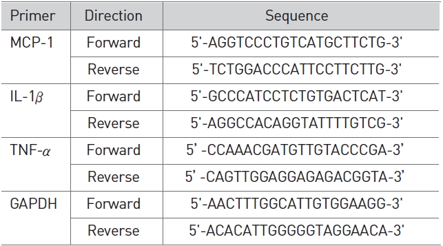
    We isolated total RNA by using Trizol reagent through the manufacturer’s instructions (Invitrogen, Carlslab, CA, USA). After the concentration of RNA had been determined by using a spectrophotometer, we made cDNA by using reverse-transcription from 1 μg of RNA obtained using M-MLV reverse transcriptase (Promega) and amplified using polymerase chain reaction (PCR) with specific primers (Table 1). The final PCR products were separated on 1.5% agarose gel by electrophoresis. Images of the bands were captured and quantified by using Image J (Bio-Rad, Hercules, CA, USA).

       2.7. Cellular uptake and FACS analysis

    RAW 264.7 cells (1 × 10⁶ cells/ml) were plated in a 96-well plate and were incubated in 5% CO2 at 37℃. After a 24 hrs incubation, the culture medium was replaced with 100 μl of fresh medium containing FITC- conjugated ZnO NPs or ZnO NP HPPS and was further incubated in 5% CO2 at 37℃ for 2 and 4 hrs. After incubation, the cells were washed with PBS 3 times. Finally, the cells were resuspended in PBS for the flow cytometric analysis (FACS Canto II Analyzer, BD Bioscience, San Jose, CA, USA).

       2.8. Transmission electron microscopy (TEM)

    Cells were cultured at a density of 1 × 10⁶ cells/ml in a culture medium, each medium containing samples at a concentration of 100 μg/ml of ZnO NPs or ZnO NP HPPS. After 4 hrs, cells were washed in PBS and were centrifuged at 1000 rpm for 5 mins. Cell pallets were fixed in 2% osmium tetroxide and then dehydrated with propion aldehyde. Epoxy resins were added; then, they were sectioned for transmission microscopy (TEM) observations (HITACHI H-7600, Japan).

       2.9. Statistical analysis

    All data are expressed as means ± standard deviations of at least three independent experiments. For comparison among groups, a one-way analysis of variance (ANOVA) test was used (with the assistance of Graph Pad Software, Inc., San Diego, CA).

    3. Results

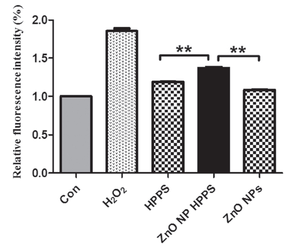
       3.1. ZnO NP HPPS significantly induced intracellular ROS production

    To measure the effects of ZnO NP HPPS on the intracellular ROS production, we treated RAW 264.7 cells with 1 μg/ml HPPS, ZnO NP, and ZnO NP HPPS for 30 mins. We observed that the amounts of intracellular ROS were higher in ZnO NP HPPS treated cells than HPPS and ZnO NP treated cells (Fig. 1). H2O2 was used as a positive control. Although HPPS or ZnO NP treatment alone induced a little of intracellular ROS, it was not statistically significant.

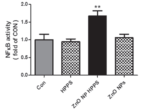
       3.2. ZnO NP HPPS significantly increased the NF-κB activation

    To evaluate the potential of ZnO NP HPPS to induce cellular activation, we measured the activation of NF-κB transcription factor. We treated the NF-κ B reporter cell line with 1 μ g/ml of HPPS, ZnO NP, and ZnO NP HPPS in for 8 hrs, and then measured the NF-κ B activity by luciferase assay. Either HPPS or ZnO NP did not increase the luciferase activity but ZnO NP HPPS significantly increased the luciferase activity (p< 0.01).

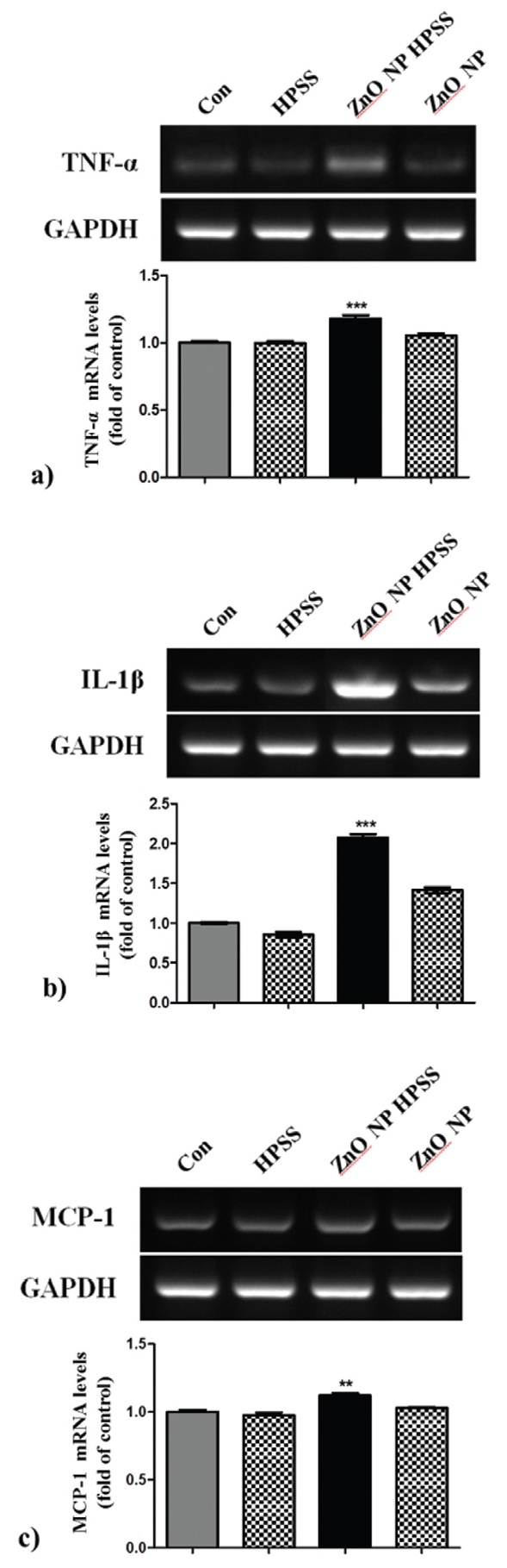
       3.3. ZnO NP HPPS increased the expression of NF-κB dependent cytokine mRNA

    To evaluate whether ZnO NP HPPS could modulate TNF-α, IL-1β, MCP-1 cytokine production, we performed semi-quantitiative RT-PCR. Total RNA was extracted from the cells treated with HPPS, ZnO NP, and ZnO NP HPPS for 4 hrs at the concentration of 1 μg/ml. In cases of TNF-α, IL-1β, MCP-1, no changes were obs-erved in the HPPS and ZnO NP treated groups when compared to the control, while significant increases of mRNA expression, especially IL-1β, were identified in the ZnO NP HPPS treated groups (Fig. 3). The results clearly show that ZnO NP HPPS affects production of MCP-1, TNF-α, IL-1β cytokines.

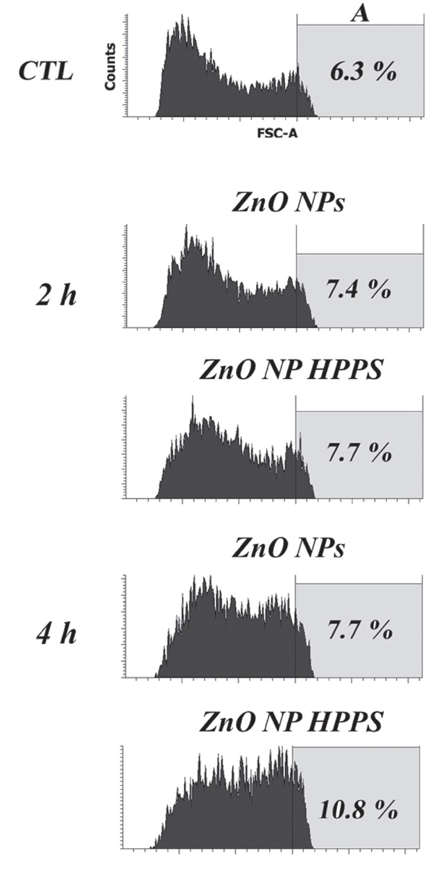
       3.4. Uptake of ZnO NP HPPS and ZnO NPs (by FACS analysis)

    To examine the cellular uptake and the permeation of the ZnO NP HPPS through the cell membrane, we treated the RAW 264.7 cells with 1 μg/ml of FITC-conjugated ZnO NP and ZnO NP HPPS for 2~4 hrs, and then performed flow cytometric analysis. The cells treated without ZnO NP were used as negative controls. As shown in Fig. 4, there are almost negligible fluorescence differences between ZnO NP and ZnO NP HPPS for 2 hrs treatment. However, a slight increase of fluorescence intensity was detected in 4 hrs treatment, suggesting that the amount of cellular uptake was increased.

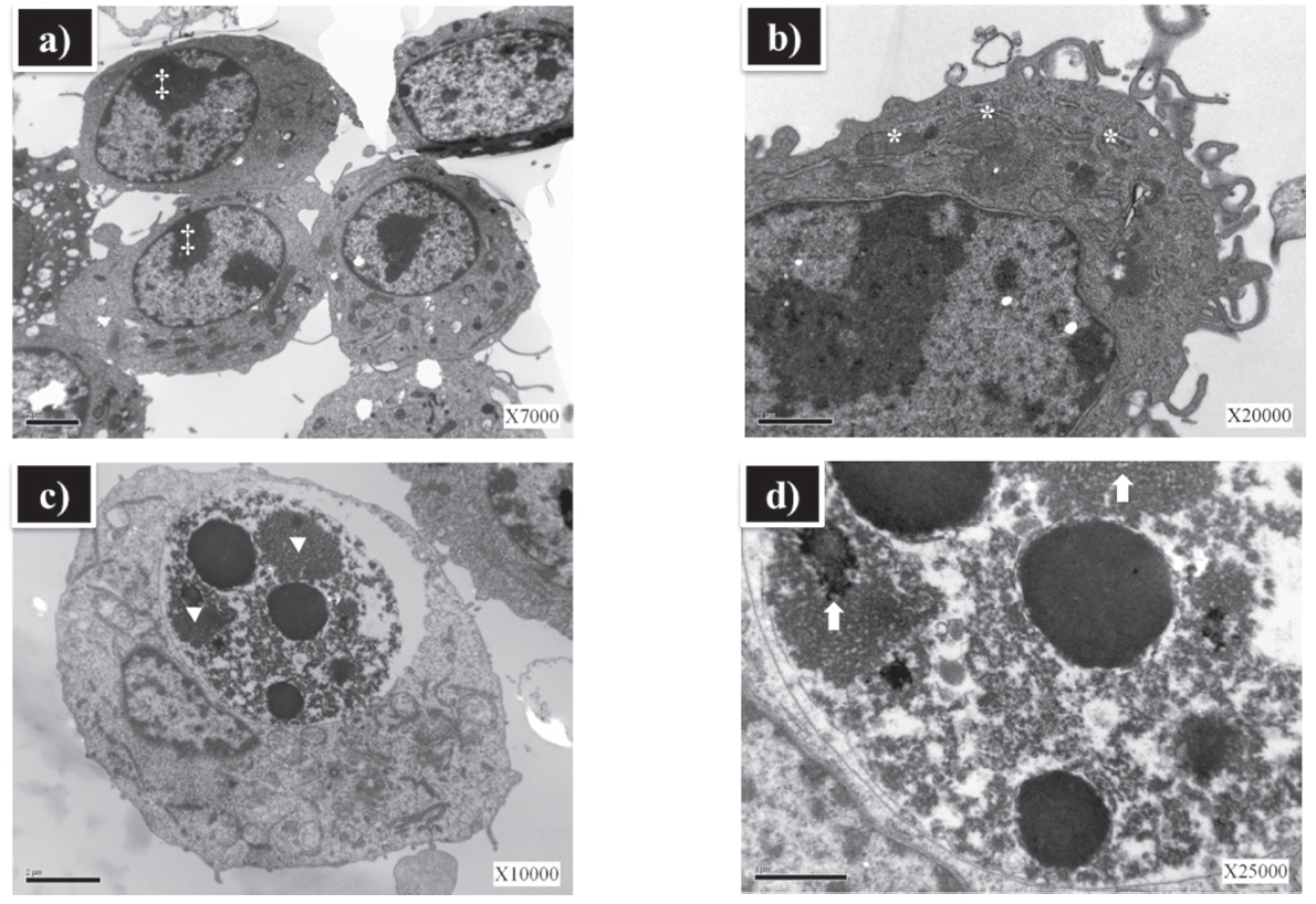
       3.5. ZnO NP HPPS caused structural alterations in RAW 264.7 cells

    For the analysis of subcellular morphologic changes, ZnO NP and ZnO NP HPPS treated cells were examined by transmission electron microscopy (TEM). RAW 264.7 cells were treated with either ZnO NP or ZnO NP HPPS for 4 hrs. There were no ultrastructural alterations in the cells treated with ZnO NP. The integrity of subcellular structures such as plasma and nuclear membrane were normal (Fig. 5a, b). However, significant alterations were observed in the cells treated with ZnO NP HPPS. Variations in size and shape of the cells were prominent, and large inclusions were observed in the lysosome (Fig. 5c, d).

    4. Discussion

    Nanotechnology is an engineering system that manipulates matter at a molecular scale. Engineered nanomaterials have already been used in industry such as sports, cosmetics, and electronics. In the last decade, great advances have been made in nanomedicinal field. Recent studies have focused on using nanoparticles for imaging, tissue engineering, and drug delivery [12-15].

    Delivering therapeutic agents into cells can be highly inefficient because of diverse physicochemical properties of therapeutic agents. Recently there were many extensive investigations in the field of drug delivery systems for the purpose of improving and facilitating drug efficacy. Nanomaterials can modify the physicochemical properties of a drug so to provide an opportunity for increasing uptake and interaction with target cells. Therefore, it is important to ensure that these agents do not cause any adverse effect. However, there are growing concerns about the toxic effects of engineered nanomaterials on human health because of their unusual physicochemical properties. There are some consensuses that deal with potential risks associated with inhalation of nanoparticles [16,17].

    Nanoscale ZnO used in many applications in daily life is believed to be nontoxic and biocompatible. Recently, attentions have focused on the Zinc Oxide nanoparticles (ZnO NPs) because of its own unique biosafe and biocompatible properties [5].

    HPPS is one of the most widely used herb-acupunctural material to replenish vital essence and blood in Korean traditional medicine [18]. Several studies about therapeutic effectiveness of HPPS were reported. HPPS has a protective effect against osteoporosis [8], growth-promoting activity on the nerve regeneration [9], protective effect on radiation enteropathy [19], and capacity to regulate bone resorption [20].

    In this study, we examined whether ZnO NO HPPS, a combining form of ZnO NP and HPPS, has any special effects over HPPS. Oxidative stress was regarded as a major cytotoxic standard caused by nanomaterials [2]. Previous studies showed that ZnO NP induces ROS production [21-23]. However, our results revealed that ZnO NP did not increase intracellular ROS production, but ZnO NP HPPS up-regulated the intracellular ROS level (Fig. 1). It is well known that NF-κB is a transcription factor which is involved in cellular response to stimuli such as stress, UV, ROS, and bacterial antigens. It is a key factor that regulates genes responsible for the innate immunity [24,25]. Tsou et al. reported that ZnO NP is related to NF-κB activation [26]. However, our results show that ZnO NP did not influence the NF-κB activation. On the other hand, ZnO NP HPPS significantly activated the NF-κB transcription factor (Fig. 2) and its dependent genes (Fig. 3). Among NF-κB dependent genes, IL-1β was especially up-regulated by the treatment of ZnO NP HPPS (Fig. 3b). Our unpublished results indicate that a longer treatment of ZnO NP also increased the NF-κB activation (data not shown). From these results, we have assumed that some components in HPPS makes ZnO to easily activate RAW 264.7 cells via NF-κB. As shown in Figure 4, increase of cellular uptake was observed in the ZnO NP HPPS treated group for 4 hrs. To observe whether ZnO NP HPPS can cause any subcellular changes, we examined ultastructural alterations by using transmission electron microscopy (TEM) in the RAW 264.7 cells treated with ZnO NP and ZnO NP HPPS for 4 hrs. The cells maintained normal appearance when were treated with ZnO NP; however, the cells exhibited significant ultrastructural alterations, such as large lysosomal inclusions, condensation of chromatin around the periphery of the nucleus when treated with ZnO NP HPPS (Fig. 5).

    In conclusion, our results show that each of the HPPS or ZnO NP did not induce any remarkable changes on RAW 264.7 cells, while ZnO NP HPPS, a combined form of ZnO NP and HPPS, significantly activated the RAW 264.7 cells. ZnO NP HPPS increased the intracellular ROS production, NF-κB transcription, and the expression of its dependent genes, such as TNF-α, IL-1β, and MCP-1. ZnO NP HPPS was more readily taken up by RAW 264.7 cells. It also induced ultrastructural alterations, such as large inclusions in the lysosome. As considering these results and another work, we assume that some components in the HPPS might play a role in the internalization of ZnO NP, thereby activate the RAW 264.7 cells.

    ROS production by phagocytes normally occurs in response to a variety of infectious and proinflammatory stimuli. This process forms essential part of the innate immune system [27]. From the result that ZnO NP HPPS has effectively stimulated RAW 264.7 cells through ROS production and NF-κB activation, we assumed that ZnO NP HPPS could be able to strengthen our defense system. But there are many obstacles to overcome for the clinical application, we need to put much effort to clarify the toxicity of ZnO NP HPPS. In addition, it is presently unknown which component in the HPPS plays a major role in the cellular activation. Thus, further studies are needed to define the roles of each component of HPPS in the ZnO NP HPPS.

참고문헌
  • 1. Nohynek GJ, Lademann J, Ribaud C, Roberts MS. 2007 Grey goo on the skin? Nanotechnology, cosmetic and sunscreen safety. [Crit Rev Toxicol.] Vol.37 P.251-77 google
  • 2. Nel A, Xia T, Madler L, Li N. 2006 Toxic potential of materials at the nanolevel. [Science.] Vol.311 P.622-7 google
  • 3. Brigger I, Dubernet C, Couvreur P. 2002 Nanoparticles in cancer therapy and diagnosis. [Adv Drug Delivery Rev.] Vol.54 P.631-51 google
  • 4. Colvin VL. 2003 The potential environmental impact of engineered nanomaterials. [Nat Biotechnol.] Vol.21 P.1166-70 google
  • 5. Yuan Q, Hein S, Misra RD. 2010 New generation of chitosan-encapsulated ZnO quantum dots loaded with drug: synthesis, characterization and in vitro drug delivery response. [Acta Biomater.] Vol.6 P.2732-9 google
  • 6. Tonello G, Daglio M, Zaccarelli N, Sottofattori E, Mazzei M, Balbi A. 1996 Characterization and quantitation of the active polynucleotide fraction (PDRN) from human placenta, a tissue repair stimulating agent. [J Pharm Biomed Anal.] Vol.14 P.1555-60 google
  • 7. Kim JK, Kim TH, Park SW, Kim HY, Kim SH, Lee SY 2010 Protective effects of human placenta extract on cartilage degradation in experimental osteoarthritis. [Biol Pharm Bull.] Vol.33 P.1004-10 google
  • 8. Chae HJ, Choi KH, Chae SW, Kim HM, Shin TK, Lee GY 2006 Placenta hominis protects osteoporosis in ovariectomized rats. [Immunopharmacol Immunotoxicol.] Vol.28 P.165-73 google
  • 9. Seo TB, Han IS, Yoon JH, Seol IC, Kim YS, Jo HK 2006 Growth-promoting activity of Hominis Placenta extract on regenerating sciatic nerve. [Acta Pharmacol Sin.] Vol.27 P.50-8 google
  • 10. Hijikata Y, Kano T, Xi L. 2009 Treatment for intractable anemia with the traditional Chinese medicines Hominis Placenta and Cervi Cornus Colla (deer antler glue). [Int J Gen Med.] Vol.2 P.83-90 google
  • 11. Umar A, Rahman MM, Vaseem M, Hahn YB. 2009 Ultra-sensitive cholesterol biosensor based on low-temperature grown ZnO nanoparticles. [Electrochem Commun.] Vol.11 P.118-21 google
  • 12. Panyam J, Labhasetwar V. 2003 Biodegradable nanoparticles for drug and gene delivery to cells and tissue. [Adv Drug Deliv Rev.] Vol.55 P.329-47 google
  • 13. Pitsillides CM, Joe EK, Wei X, Anderson RR, Lin CP. 2003 Selective cell targeting with light-absorbing microparticles and nanoparticles. [Biophys J.] Vol.84 P.4023-32 google
  • 14. Sarikaya M, Tamerler C, Jen AKY, Schulten K, Baneyx F. 2003 Molecular biomimetics: nanotechnology through biology. [Nat Mater.] Vol.2 P.577-85 google
  • 15. Yang F, Murugan R, Wang S, Ramakrishna S. 2005 Electrospinning of nano/micro scale poly(L-lactic acid) aligned fibers and their potential in neural tissue engineering. [Biomaterials.] Vol.26 P.2603-10 google
  • 16. Hoet PH, Bruske-Hohlfeld I, Salata OV. 2004 Nanoparticles - known and unknown health risks. [J Nanobiotechnology.] Vol.2 P.12 google
  • 17. Maynard AD, Aitken RJ, Butz T, Colvin V, Donaldson K, Oberdorster G 2006 Safe handling of nanotechnology. [Nature.] Vol.444 P.267-9 google
  • 18. Yeom MJ, Lee HC, Kim GH, Shim I, Lee HJ, Hahm DH. 2003 Therapeutic effects of Hominis placenta injection into an acupuncture point on the inflammatory responses in subchondral bone region of adjuvant-induced polyarthritic rat. [Biol Pharm Bull.] Vol.26 P.1472-7 google
  • 19. Jang SY, Park JW, Bu Y, Kang JO, Kim J. 2011 Protective effects of hominis placenta hydrolysates on radiation enteropathy in mice. [Nat Prod Res.] Vol.25 P.1988-92 google
  • 20. Jin UH, Kim DI, Lee TK, Lee DN, Kim JK, Lee IS 2006 Herbal formulation, Yukmi-jihang-tang-Jahage, regulates bone resorption by inhibition of phosphorylation mediated by tyrosine kinase Src and cyclooxygenase expression. [J Ethnopharmacol.] Vol.106 P.333-43 google
  • 21. Sun J, Wang S, Zhao D, Hun FH, Weng L, Liu H. 2011 Cytotoxicity, permeability, and inflammation of metal oxide nanoparticles in human cardiac microvascular endothelial cells: cytotoxicity, permeability, and inflammation of metal oxide nanoparticles. [Cell Biol Toxicol.] Vol.27 P.333-42 google
  • 22. Wang Y, Aker WG, Hwang HM, Yedjou CG, Yu H, Tchounwou PB. 2011 A study of the mechanism of in vitro cytotoxicity of metal oxide nanoparticles using catfish primary hepatocytes and human HepG2 cells. [Sci Total Environ.] Vol.409 P.4753-62 google
  • 23. Park SJ, Park YC, Lee SW, Jeong MS, Yu KN, Jung H 2011 Comparing the toxic mechanism of synthesized zinc oxide nanomaterials by physicochemical characterization and reactive oxygen species properties. [Toxicol Lett.] Vol.207 P.197-203 google
  • 24. Gilmore TD. 2006 Introduction to NF-kappaB: players, pathways, perspectives. [Oncogene.] Vol.25 P.6680-4 google
  • 25. Brasier AR. 2006 The NF-kappaB regulatory network. [Cardiovasc Toxicol.] Vol.6 P.111-30 google
  • 26. Tsou TC, Yeh SC, Tsai FY, Lin HJ, Cheng TJ, Chao HR 2010 Zinc oxide particles induce inflammatory responses in vascular endothelial cells via NF-κB signaling. [J Hazard Mater.] Vol.183 P.182-8 google
  • 27. Leto TL, Geiszt M. 2006 Role of Nox family NADPH oxidases in host defense. [Antioxid Redox Signal.] Vol.8 P.1549-61 google
OAK XML 통계
이미지 / 테이블
  • [ Figure 1 ]  Effects of HPPS, ZnO NPs, and ZnO NP HPPS on the intracellular ROS production in RAW 264.7 cells. Cells were treated with 1μ g/ml HPPS, ZnO NPs, and ZnO NP HPPS for 30 mins prior to assay. The y axis represents the relative fluorescence intensity and reflects the amount of ROS. ZnO NP HPPS induces 25% and 15% more ROS than ZnO NPs and HPPS, respectively (**p <0.01).
    Effects of HPPS, ZnO NPs, and ZnO NP HPPS on the intracellular ROS production in RAW 264.7 cells. Cells were treated with 1μ g/ml HPPS, ZnO NPs, and ZnO NP HPPS for 30 mins prior to assay. The y axis represents the relative fluorescence intensity and reflects the amount of ROS. ZnO NP HPPS induces 25% and 15% more ROS than ZnO NPs and HPPS, respectively (**p <0.01).
  • [ Figure 2 ]  Effects of HPPS, ZnO NPs, and ZnO NP HPPS on the NF-κ B activity. An NF-κ B reporter cell line derived from RAW 264.7 cells was treated with 1 μ g/ml HPPS, ZnO NPs, or ZnO HPPS for 8 hrs. HPPS and ZnO NPs did not increase the luciferase activity. ZnO NP HPPS showed significantly higher activity than CTL and ZnO NP. Luciferase activity was normalized to the amount of total protein in cell lysate. ** Statistically significant difference as compared with the control (**p < 0.01).
    Effects of HPPS, ZnO NPs, and ZnO NP HPPS on the NF-κ B activity. An NF-κ B reporter cell line derived from RAW 264.7 cells was treated with 1 μ g/ml HPPS, ZnO NPs, or ZnO HPPS for 8 hrs. HPPS and ZnO NPs did not increase the luciferase activity. ZnO NP HPPS showed significantly higher activity than CTL and ZnO NP. Luciferase activity was normalized to the amount of total protein in cell lysate. ** Statistically significant difference as compared with the control (**p < 0.01).
  • [ Figure 3 ]  Effects of HPPS, ZnO NPs, and ZnO NP HPPS on the NF-κB dependent gene expression. RAW 264.7 cells were treated with 1 μg/ml HPPS, ZnO NPs, and ZnO HPPS for 4 hrs. Then, total RNA was extracted, and the expressions of NF-κ B dependent genes, including TNF-α , IL-1β , MCP-1, were analyzed by semiquantitative RT-PCR. The relative expression of each gene was determined by using a densitometric analysis of each band (**p< 0.01, ***p< 0.001).
    Effects of HPPS, ZnO NPs, and ZnO NP HPPS on the NF-κB dependent gene expression. RAW 264.7 cells were treated with 1 μg/ml HPPS, ZnO NPs, and ZnO HPPS for 4 hrs. Then, total RNA was extracted, and the expressions of NF-κ B dependent genes, including TNF-α , IL-1β , MCP-1, were analyzed by semiquantitative RT-PCR. The relative expression of each gene was determined by using a densitometric analysis of each band (**p< 0.01, ***p< 0.001).
  • [ Figure 4 ]  Uptake of ZnO NPs and ZnO NP HPPS in RAW 264.7 cells. The x axis represents the fluorescence intensity and reflects the amount of uptake, and the y coordinate shows the number of cell counts. The region of high fluorescence intensity (over 150,000) was assigned as A. A slight increase in the fluorescence intensity was observed especially in the ZnO NP HPPS treatment groups for 4 hrs.
    Uptake of ZnO NPs and ZnO NP HPPS in RAW 264.7 cells. The x axis represents the fluorescence intensity and reflects the amount of uptake, and the y coordinate shows the number of cell counts. The region of high fluorescence intensity (over 150,000) was assigned as A. A slight increase in the fluorescence intensity was observed especially in the ZnO NP HPPS treatment groups for 4 hrs.
  • [ Figure 5 ]  Cellular structural changes of RAW 264.7 cells treated with ZnO NPs (a, b) and ZnO NP HPPS (c, d). (a) The nucleus contains a moderate quantity of nuclear membrane-associated condensed chromatin (‡). (b) The nuclear and mitochondrial membranes are clearly outlined (*). (c-d) Large inclusions are observed in the lysosome (▼). The arrow in (d) indicates complex inclusions.
    Cellular structural changes of RAW 264.7 cells treated with ZnO NPs (a, b) and ZnO NP HPPS (c, d). (a) The nucleus contains a moderate quantity of nuclear membrane-associated condensed chromatin (‡). (b) The nuclear and mitochondrial membranes are clearly outlined (*). (c-d) Large inclusions are observed in the lysosome (▼). The arrow in (d) indicates complex inclusions.
(우)06579 서울시 서초구 반포대로 201(반포동)
Tel. 02-537-6389 | Fax. 02-590-0571 | 문의 : oak2014@korea.kr
Copyright(c) National Library of Korea. All rights reserved.