The genus
In this study, we collected two marine ciliates,
>
Sample collection and identification
Specimens of
Cultures were maintained in both Petri dishes and 50 mL tissue culture flasks (Greiner Bio-one, Frickenhausen, Germany).Rice grains were used for enrichment of bacterial growth in the culture, and the enriched bacteria were grazed on by ciliates as a food. Living specimens were observed under a light microscope (Leica DM2500, Wetzlar, Germany)at 50 to 1,000 magnifications. Protargol impregnation was performed in order to reveal their infraciliature (Foissner,1991).
Terminology and classification were mainly followed according to Berger (2006) and Lynn (2008).
Extraction of genomic DNAs from single specimens of each species was performed according to the manufacturer’s protocol,using a RED-Extract-N-Amp Tissue PCR Kit (Sigma,St. Louis, MO, USA). New EukA (5′-CTG GTT GAT YCT GCC AGT-3′), modified from Medlin et al. (1988) and LSU rev3 (Sonnenberg et al., 2007) primers were used for PCR amplification of the nearly complete SSU rRNA gene. Optimized conditions for this process were as follows: denaturation at 94℃ for 3 min followed by 35 cycles of denaturation at 94℃ for 30 sec, annealing at 58℃ for 30 sec, extension at 72℃ for 4 min, and then a final extension step at 72℃ for 7 min. The QIAquick® PCR Purification Kit (QIAGEN, Chatsworth,CA, USA) was used for purification of PCR products.Two internal primers were used for sequencing: 18S+810,5′-GCC GGA ATA CAT TAG CAT GG-3′ and 18S-300,5′-CAT GGT AGT CCA ATA CAC TAC-3′. An ABI 3700 sequencer (Applied Biosystems, Foster City, CA, USA) was used for sequencing. Sequencing fragments of the SSU rRNA gene were combined via BioEdit (Hall, 1999) and were aligned according to Clustal X 1.81 (Jeanmougin et al., 1998).Mega 3.1 (Kumar et al., 2004) was used for calculation of genetic distance, employing the Kimura two-parameter distance method (Kimura, 1980). SSU rRNA gene sequences belonging to the genus
Phylum Ciliophora Doflein, 1901
Class Spirotrichea Butschli, 1889
Order Urostylida Jankowski, 1979
Family Pseudokeronopsidae Borror and Wicklow, 1983
1*Genus Apokeronopsis Shao et al., 2007
2*Apokeronopsis bergeri Li et al., 2008
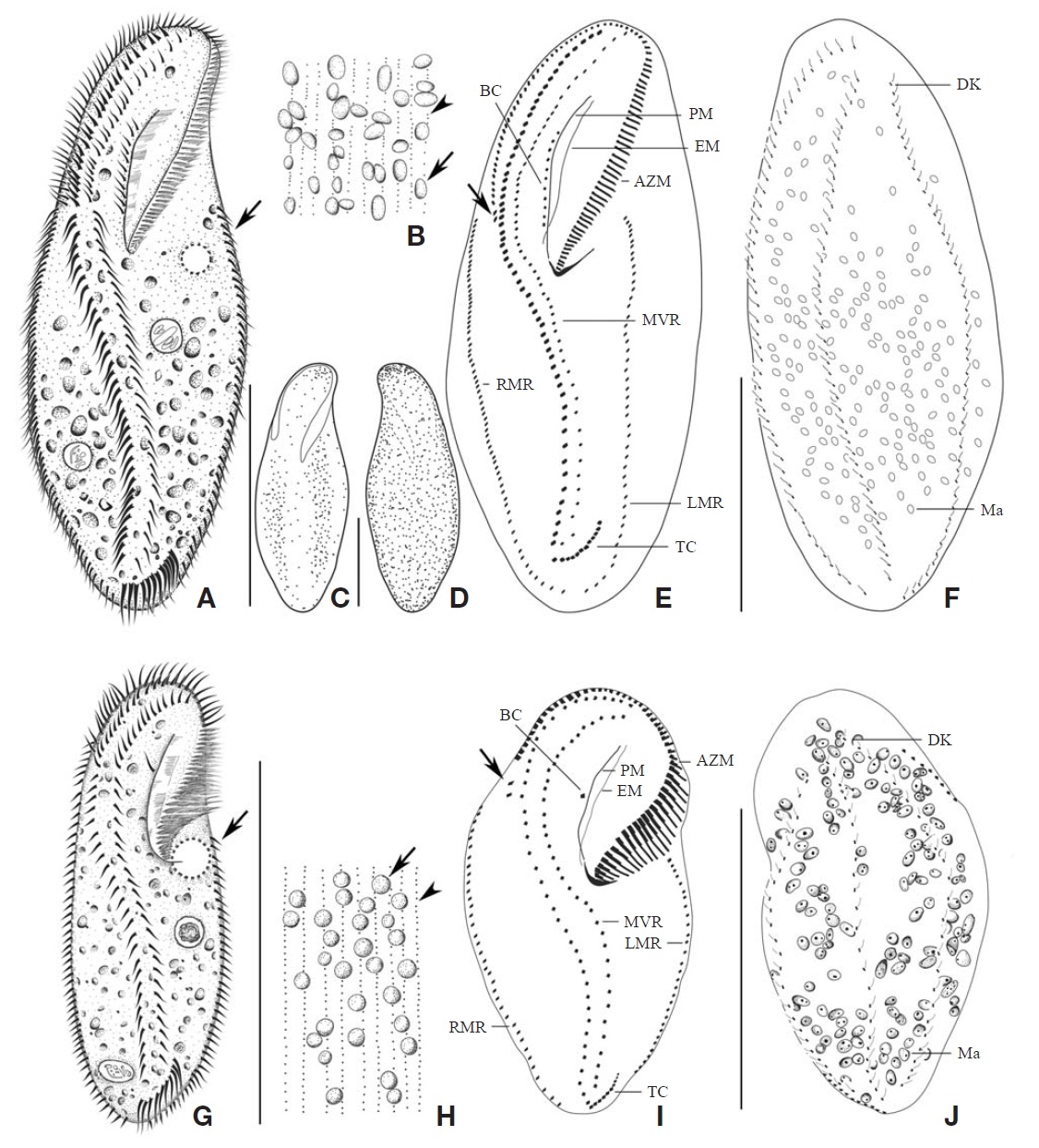
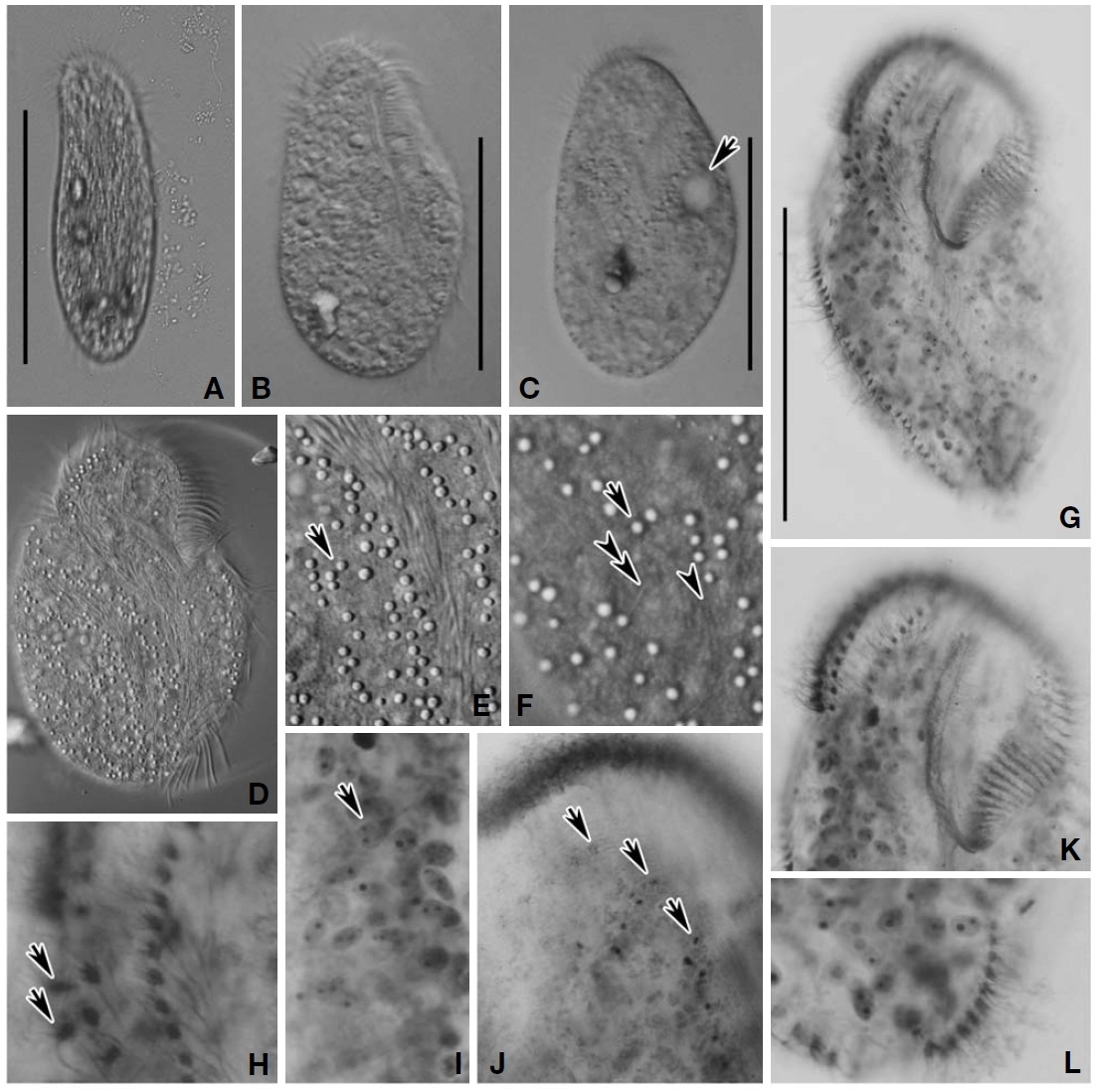
(Table 1,Figs. 1A-F, 2)
Materials examined. One population was obtained from Incheon Harbor on the 2nd of November, 2010.
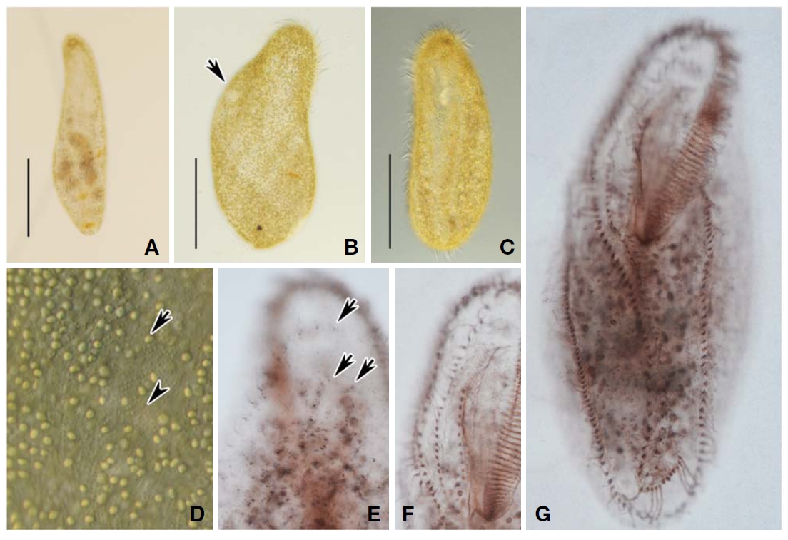
Description. Size in life usually 260×80 μm (Figs. 1A,2A). Body slightly contractile and flexible (Fig. 2B, C); fusiform shape with both ends broadly rounded; dorsoventrally flattened. Contractile vacuole located on the left side of the anterior 1/3, approximately 15 μm in diameter (Figs. 1A, 2B, arrow). Cells appeared yellow green to green in colour under low magnification. Cytoplasm opaque and colourless (Fig.2D). Macronuclei composed of about 200 nodules, each one oval shaped (Figs. 1F, 2G).
Two types of cortical granules: type I very small, ca. 0.3μm across and colourless, arranged on a line longitudinally,distributed over the entire body (Figs. 1B, 2D, arrowhead);type II ca. 2 μm across, oval shape, and yellow green in colour,densely and irregularly distributed over the entire body(Figs. 1B, arrow; C, D; 2D, arrow).
Distinct adoral zone of membranelles, about 1/3 of the cell length, formed question mark, composed of about 85 membranelles;base of the longest membranelles are about 10 μm wide (Fig.1 A, E). Paroral and endoral membranes are almost equal in length and spatially crossed at proximal end on different planes (Fig.1 E).
Most somatic cirri are relatively fine,
Distribution. China and Korea (this study).
Remarks. In terms of the infraciliature,
The Korean population of A. bergeri corresponds well with the original description by Li et al. (2008) and the ranges of characteristics in the morphometric comparisons are highly overlapped (Table 1). However, the body size of the Korean population is slightly smaller than that of the Chinese
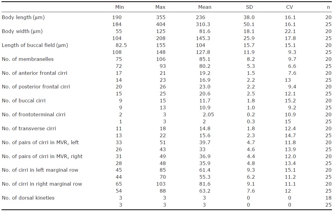
[Table 1.] Morphometric characterization of Apokeronopsis bergeri

Morphometric characterization of Apokeronopsis bergeri
population
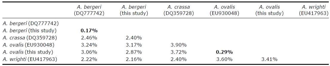
The SSU rRNA gene sequence of A. bergeri is 1,748 bp in length (GenBank accession no: JF718644). Inter-specific pairwise distances between A. bergeri and congeners are more than 2%, however the intra-specific distance within A.bergeri is 0.17% (Table 3).
1*Apokeronopsis ovalis (Kahl, 1932)
(Table 2,Figs. 1G-J, 3)
Materials examined. One population was obtained from Baengyeongdo Island on the 16th of November, 2010.
Description. Size in life usually 160×55 μm (Figs. 1G, 3A-D). Body slightly contractile and flexible (Fig.3 B-D);shape in oval form; dorsoventrally flattened. Contractile vacuole located on the left side of the anterior one third of body, about 10 μm in diameter (Figs. 1G, 3C, arrow). Cells appeared colourless to grayish in colour under low magnification.Cytoplasm opaque and colourless (Fig.3 A-D). Macronuclei usually 150 nodules, each one oval in shape (Figs. 1J;3I, arrow).
Two types of cortical granules: type I very small, about 0.2 μm across and colourless, arranged on a line longitudinally,distributed over the entire body (Figs. 1H, arrowhead;3F, arrowhead); type II granules 1.5 μm in diameter, round shape, and mostly colourless and only a few yellow greencoloured,densely and irregularly distributed over the entire body (Figs. 1H, arrow; 3E, F, arrow)
Distinct adoral zone of membranelles, about 1/3 of cell length, formed question mark, consisting of about 62 membranelles;base of the longest membranelles is about 15 μm wide (Fig.1 I). Paroral and endoral membranes are almost equal in length and spatially crossed on the proximal end(Fig.1 I).
Most somatic cirri are relatively fine, about 10 μm in length.Bicorona of frontal cirri slightly enlarged, composed of 30 cirri, connecting to the midventral complex with an inconspicuous gap. Midventral complex consisted of about 52 cirri, terminating near transverse cirri. Invariably, two frontoterminal cirri located on the distal end of the adoral zone(Fig.1I, arrow). Buccal cirri consisting of 1-2 arranged in a line beside the paroral membrane (Figs. 1I, 3K). Transverse cirri J-shaped, and composed of 9-14 (Figs. 1I, 3L). Left marginal row terminating on the posterior dorsal side, composed of 30-47 cirri; right marginal row commencing on the anterior dorsal side, consisting of 38-55 cirri. Dorsal kineties usually composed of three (Figs. 1J; 3J, arrows); cilia about 5 μm in length (Figs. 1J; 3F, double-arrowhead).
Distribution. Germany, China, and Korea (this study).
Remarks. In comparison with the congeners,
The population of Apokeronopsis ovalis in this study corresponds well with the description of combining authors Shao et al. (2008). A morphometric comparison among the Korean and two Chinese populations (Shao et al., 2008) is shown in Table 2, and most features are consistent among three populations.Shao et al. (2008) described the Chinese populations having one to five buccal cirri (vs. 1-2 in Korean), and are brown to dark brown in colour (vs. colourless to grayish in
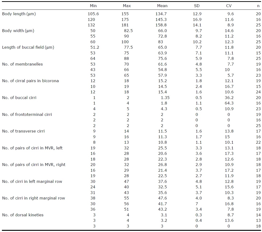
[Table 2.] Morphometric characterization of Apokeronopsis ovalis

Morphometric characterization of Apokeronopsis ovalis
Pairwise distance of SSU rRNA gene sequences among Apokeronopsis congeners were calculated using the Kimura twoparameter distance
Korean). Shao et al. (2008) suggested that the body colour of
The SSU rRNA gene sequence of A. ovalis is 1,751 bp in length (GenBank accession no: JF718645). Inter-specific pairwise distances between A. ovalis and congeners are more than 2%, however, the intra-specific distance within A. ovalis is 0.29% (Table 3).
Korean name: 1*박각모하모충속 (신칭), 2*황색박각모하모충 (신칭) Korean name: 1*난형박각모하모충 (신칭)