Aetideid copepods are relatively large-sized marine calanoids with most found in the deep sea between the mesopelagic and bathypelagic depths, even though a few species are known to be epipelagic (Boxshall and Halsey, 2004). Copepods in this group are mixed feeders and their mouthparts are well adapted for seizing and filtration (Arashkevich, 1969).
The family Aetideidae consists of 213 known species in 30 genera worldwide (Razouls et al., 2010) whereas only three species have been reported from Korea:
In this study, the following three aetideid species are reported from the East Sea of Korea, based on the samples collected by vertical towing of a plankton net from the surface level up to 300 m depth:
The sequences of the mitochondrial cytochrome c oxidase subunit 1 (CO1) for these three species are also provided as supplementary data for their molecular characteristics.
The samples were collected by vertical tows (0-300 m) using a plankton net (45 cm mouth diameter, 330㎛ mesh) at three sites in the East Sea. The specimens were preserved in 95% ethanol immediately after collection. The appendages of the specimens were dissected in glycerol on a cavity slide glass under a stereomicroscope (Olympus SZX-7; Olympus, Tokyo, Japan) and observed using a microscope (Leica DM 2500; Leica Microsystems, Wetzlar, Germany). All drawings and measurements were made using a drawing tube. Voucher specimens were deposited in the Department of Biological Sciences, Inha University, South Korea.
The Roman and Arabic numerals in the armature formula represent spines and setae, respectively.
Single antennas were removed from each specimen for genomic DNA extraction. The other body parts were used for the morphological observation. The genomic DNA was extracted using a RED Extract-N-Amp Tissue PCR kit (Sigma, St.Louis, MO, USA) according to the manufacturer’s instructions. Mitochondrial CO1 was amplified by PCR using universal primers of LCO1490 and HCO2198 (Folmer et al., 1994). PCR amplification was carried out under the following conditions: 3 min at 94℃, 35 cycles of 95℃ for 15 sec, 48℃ for 30 sec, and 72℃ for 90 sec, with a final 72℃ extension reaction for 7 min. The PCR products were gel-purified using a QIAquick® Gel Extraction Kit (Qiagen, Valencia, CA, USA) and sequenced with an ABI PRISM® 3700 DNA Analyzer using a BigDye Terminator Cycle Sequencing Ready Reaction Kit (Applied Biosystems, Foster City, CA, USA).
Order Calanoida Sars, 1903
Family Aetideidae Giesbrecht, 1893
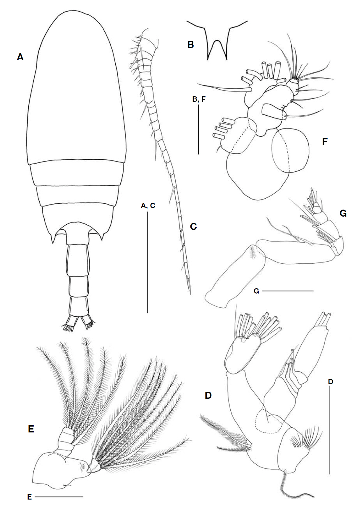
1*Genus Aetideus Brady, 1883
2*
Aetideus acutus Farran, 1929: 228, fig. 5; Tanaka, 1957: 36, fig. 25; Bradford and Jillett, 1980: 14, fig. 5.
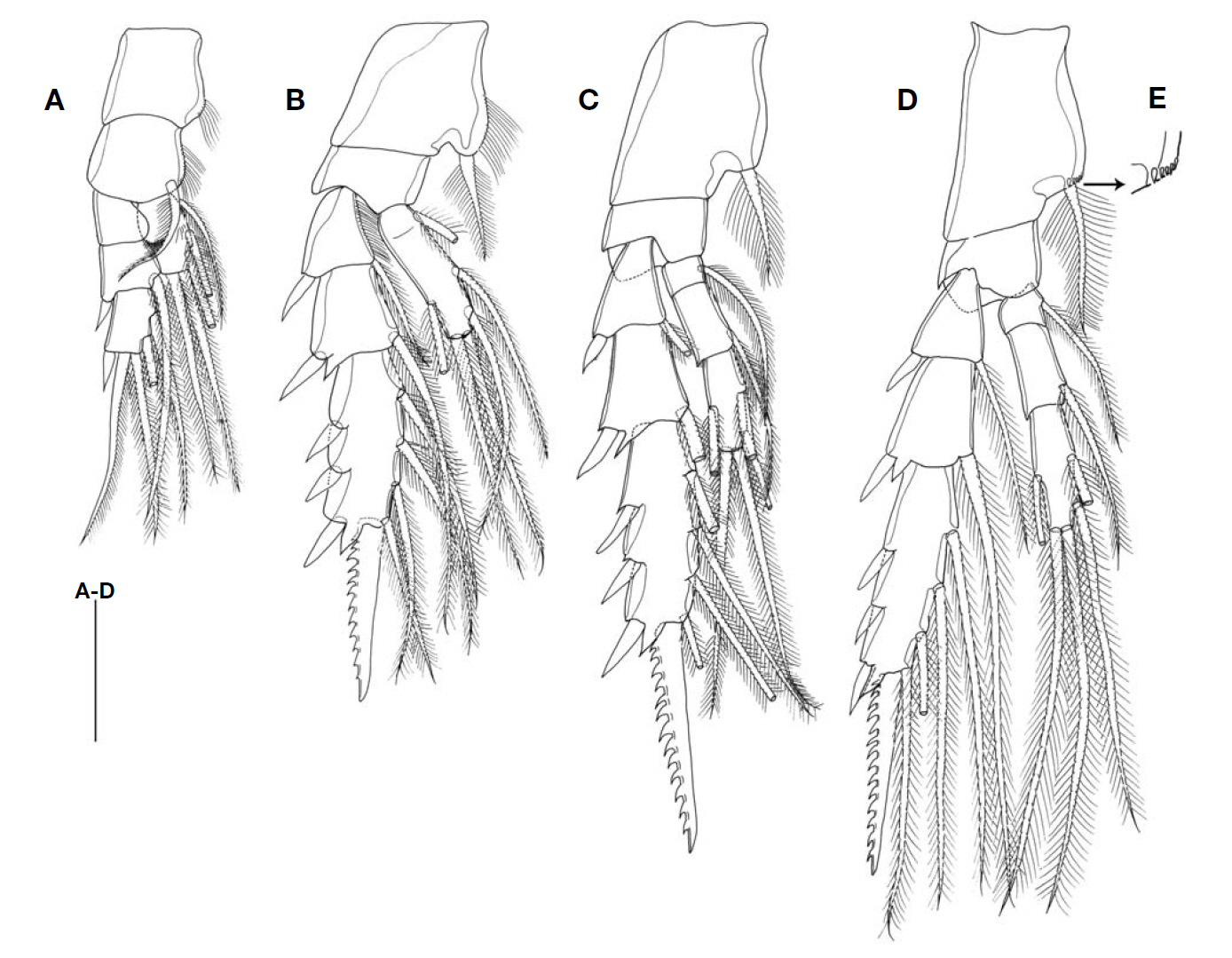
Leg 2 (Fig.3 B). Coxa with inner hairs and 1 inner seta. Basis unarmed. Endopod incompletely 1-segmented, with 6 setae. Exopod 3-segmented; first and second segments with 1 inner seta and 1 outer spine respectively; distal segment with 3 spines, 4 setae and 1 serrated spine bearing 12 teeth on outer edge.
Leg 3 (Fig.3 C). Coxa with 1 inner seta. Endopod 3-segmented; first segment with 1 inner seta; second and third segments with 1 inner seta, 5 setae, respectively. Exopod 3-segmented; distal segment with 3 spines, 4 setae and 1 serrated spine bearing 13 outer teeth.
Leg 4 (Fig.3 D) similar to leg 3 except for coxa with 1 inner seta and 5 small spines (Fig.3 E) and distal segment of exopod with 14 outer teeth. Armature formula of swimmimg legs 1-4 as follows:
Leg 1 coxa 0-0 basis 0-1 exp 0-0; Ⅰ-1; I,2,2 enp 0,2,3
Leg 2 coxa 0-1 basis 0-0 exp Ⅰ-1; Ⅰ-1; Ⅲ,Ⅰ,4 enp 1,2,3
Leg 3 coxa 0-1 basis 0-0 exp Ⅰ-1; Ⅰ-1; Ⅲ,I,4
enp 0-1; 0-1; 1,2,2
Leg 4 coxa 0-1 basis 0-0 exp Ⅰ-1; Ⅰ-1; Ⅲ,Ⅰ,4
enp 0-1; 0-1; 1,2,2
Not found.
East Sea (Korea), New Zealand, South Atlantic, Izu region (Japan), and tropical Pacific Ocean.
This species is quite rare and with only a few specimens have been reported (Chen and Zhang, 1965; Tanaka and Omori, 1970; Kim, 1985). Only a single female specimen was found.
The genus
of the rostrum is not protruded from the top of the head in the female of
The Korean specimen is congruent with that of the original description with minor differences: the last pedigerous somite of the present specimen has a process that reaches the 2/3 region of the second abdominal somite, but that of the type specimen is longer than the Korean specimen and extends beyond the second abdominal somite.
The CO1 sequence of the Korean specimen showed 99% similarity to that of the previously known sequence of
1*Genus Bradyidius Giesbrecht, 1897
>
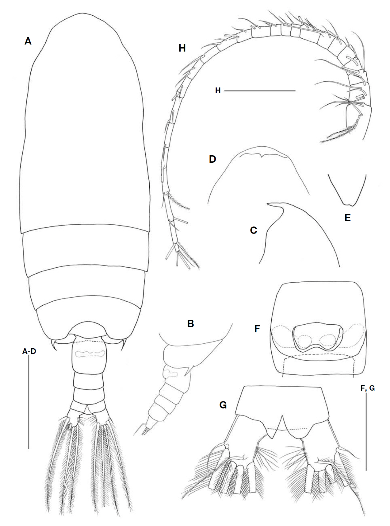
2*Bradyidius angustus (Tanaka, 1957) (Figs. 4, 5)
Undinopsis angustus Tanaka, 1957: 45, fig. 29.
Bradyidius angustus: Markhaseva, 1996: 70, fig. 49.
1♂, East Sea, 36°05′N, 131°40′E, Jun 2009.
Body as Fig.4 A. Body length 1.57 mm. Prosome about 2.3 times as long as urosome. Cephalosome and first pedigerous somite fused forming cephalothorax.Posterior corners of last pedigerous somite extending to distal margin of first abdominal somite. Rostrum (Fig.4 B) bifurcate and weakly divergent. Caudal rami about 1.3 times longer than width.
Antennule (Fig.4 C). 22-segmented, reaching posterior
border of genital somite.
Antenna (Fig.4 D). Coxa with 1 long seta and hairs; basis with 2 setae. Endopod 2-segmented; first segment unarmed; second segment with 6 inner and 6 outer setae. Exopod 7-segmented; first segment unarmed; second to sixth segments with 1 seta each; distal segment with 3 setae.
Mandible (Fig.4 E). Basis with 2 short setae. Endopodal segment with 8 setae. Exopod indistinctly 4-segmented; first segment unarmed; second and third segments with 1 seta, respectively; distal segment with 3 plumose setae.
Maxillule (Fig.4 F). Arthrite unarmed. Coxal endite degenerate. Coxal epipodite with 4 large and 1 minute setae. Basal endites with 1, 2 setae respectively. Endopod with 9 setae. Exopod with 7 setae.
Maxilla strongly reduced in size and structure.
Maxilliped (Fig.4 G). Coxa with 1 distal seta. Basis with 2 medial setae. Endopod 6-segmented; first segment with 1 seta; second to sixth segments with 4, 3, 2+1, 2, and 4 setae respectively.
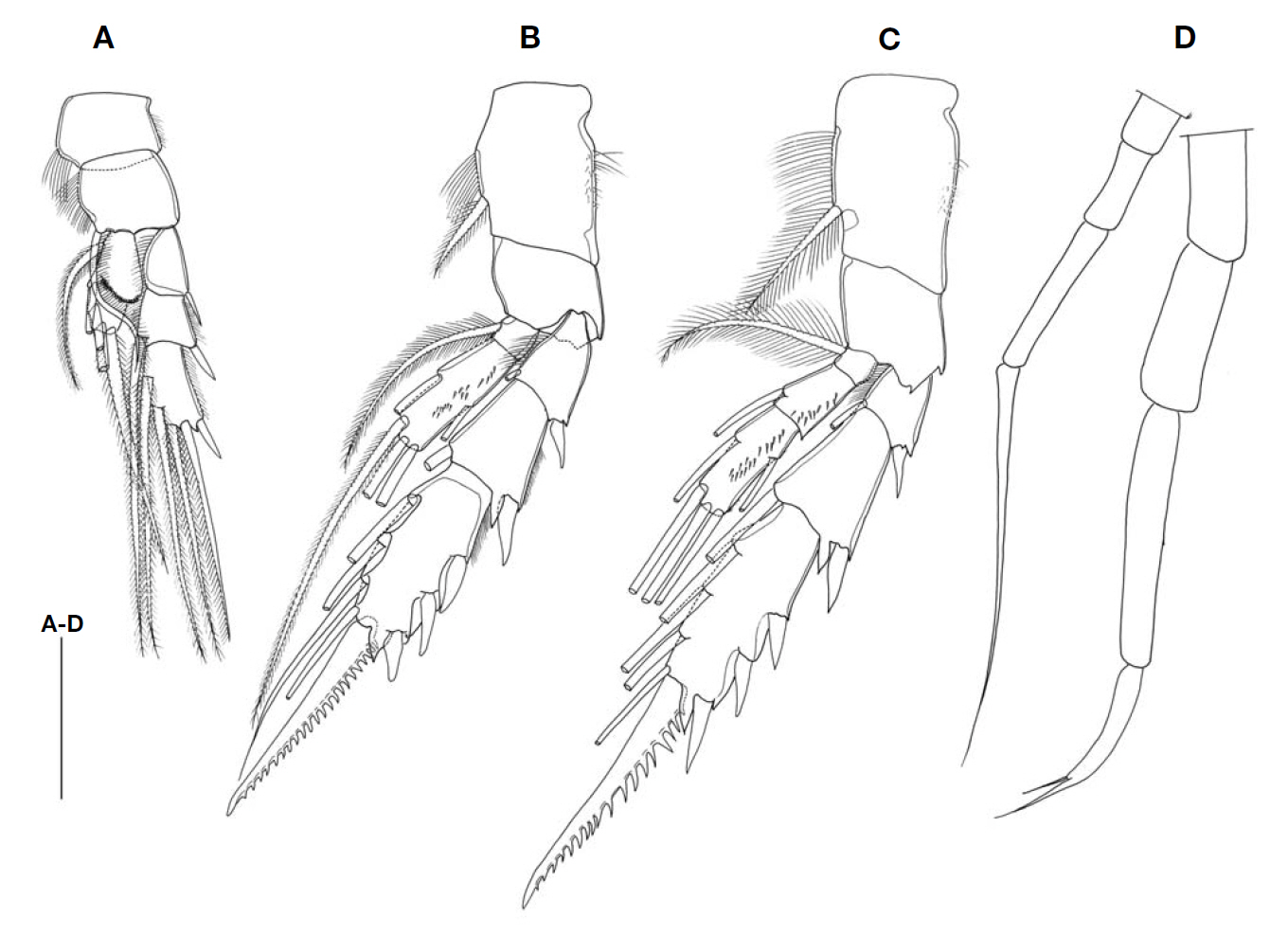
Leg 1 (Fig.5 A). Coxa with hairs along outer margin and distal part of inner margin. Basis with hairs and 1 distal seta on inner margin. Endopod 1-segmented; outer margin swollen with 1 row of spinules and 5 setae. Exopod 3-segmented; first segment with inner hairs and 1 outer spine; second segment with 1 inner seta and 1 outer spine; distal segment with 4 setae and 1 outer spine.
Leg 2 (Fig.5 B). Coxa with hairs on both margins and 1 inner seta. Basis unarmed. Endopod 2-segmented; first segment with 1 inner seta; second segment with minute spinules on posterior surface and 5 setae. Exopod 3-segmented; first and second segments with 1 inner seta and 1 outer spine, respectively; distal segment with 3 spines, 4 setae, and 1 serrated spine bearing 22 teeth on outer edge.
Leg 3 (Fig.5 C). Coxa and basis same as in leg 2. Endopod 3-segmented; first segment with 1 inner seta; second and third segments with 1 inner seta and 5 setae, respectively and minute spinules on posterior surface. Exopod 3-segmented; first and second segments with 1 inner seta and 1 outer spine; distal segment with 3 spines, 4 setae, and 1 serrated spine with 17 outer teeth. Armature formula of swimming legs 1-4 as follows:
Leg 1 coxa 0-0 basis 0-1 exp Ⅰ-0; Ⅰ-1; Ⅰ,1,3 enp 0,2,3
Leg 2 coxa 0-1 basis 0-0 exp Ⅰ-1; Ⅰ-1; Ⅲ,Ⅰ,4 enp 0-1; 1,2,2
Leg 3 coxa 0-1 basis 0-0 exp Ⅰ-1; Ⅰ-1; Ⅲ,Ⅰ,4
enp 0-1; 0-1; 1,2,2
Leg 4, missing.
Leg 5 (Fig.5 D) uniramous and asymmetrical; right and left exopod without seta and spine; third segment of right exopod spiniform, about 2.4 times longer than second segment; third segment of left exopod forming a sharp process with 1 subdistal seta.
Not found.
The East Sea (Korea), Sagami, and Suruga (Japan).
Sixteen species have been recorded in the genus
The Korean specimen coincides well with the Japanese specimen of the original description (Tanaka, 1957). Nevertheless, the Korean and Japanese specimens show the following differences: 1) Tanaka’s specimen has a short outer spine on the first exopodal segment of leg 1, whereas the same spine of our specimen extends to the middle of the second segment; 2) according to Tanaka’s description, the serrated spines of legs 2 and 3 each have 24 and 25 outer teeth, whereas the specimen in the present study has 22 and 18, respectively; and 3) the original illustration shows that the first endopodal and exopodal segments of the antenna have a distal seta, respectively, but no seta was found in the present specimen. These differences are considered variations within the species.
The partial CO1 sequence of genus
1*Genus Gaetanus Giesbrecht, 1888
>
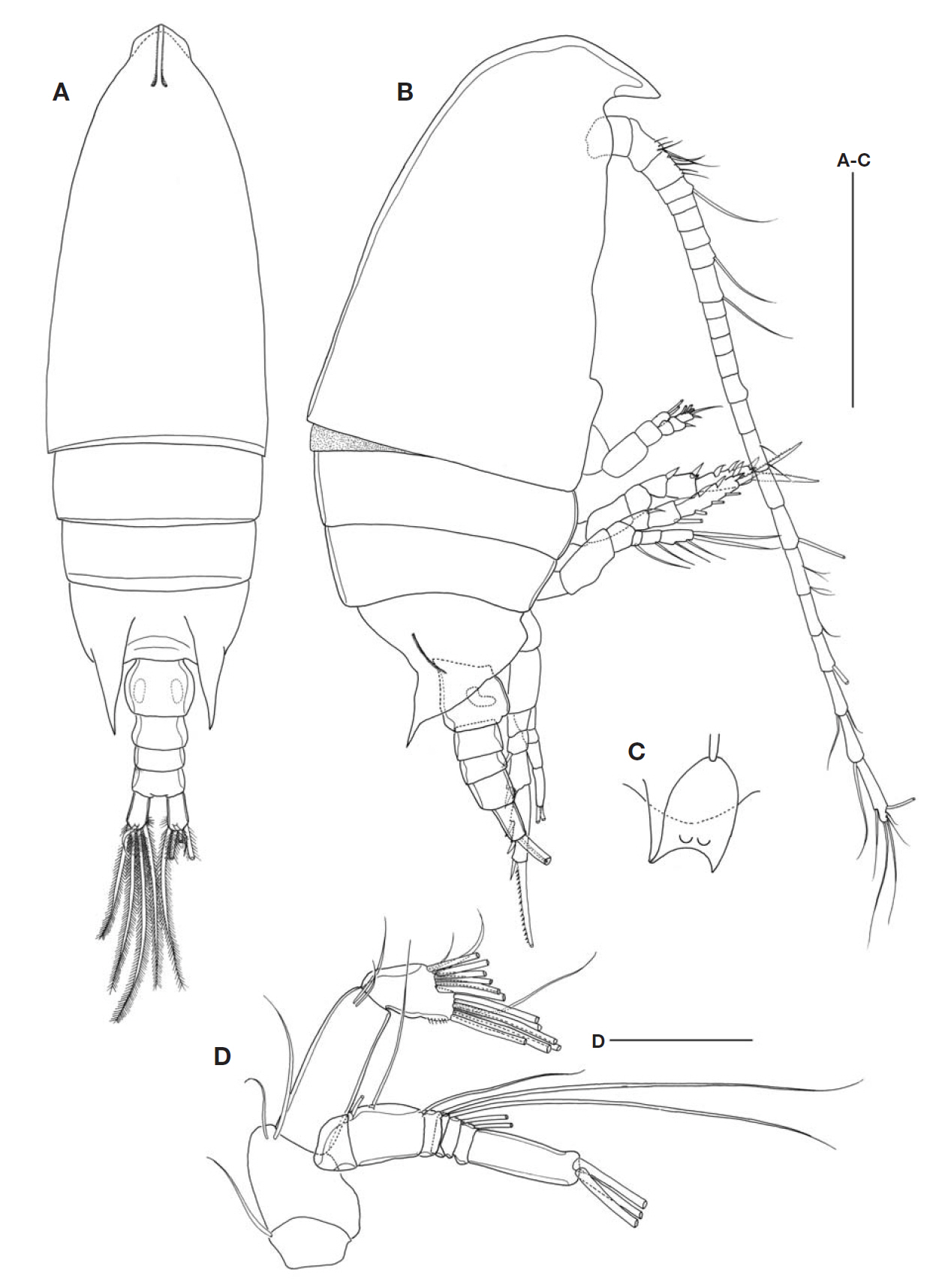
2*Gaetanus minutus (Sars, 1907) (Figs. 6-8)
Gaidius minutus Sars, 1907: 10, 1924-1925: 49, Pl. 14, figs. 14-18; Tanaka, 1957: 64, fig. 39a-f.
Gaidius variabilis Brodsky, 1950: 160, fig. 74; Tanaka and Omori, 1970: 127, fig. 6a-k.
Gaetanus moderatusTanaka, 1957: 66, fig. 40.
1♀, East Sea, 37°05′N, 131°20′E; 5♀ ♀, 36°30′N, 131°20′E, Jun 2009.
Body as Fig.6 A. Body length 4.46 mm. Prosome about 3.7 times as long as urosome. Cephalosome and first pedigerous somite fused forming cephalothorax. Posterior corners of last pedigerous somite (Fig.6 B) with short spine on each side. Rostrum (Fig.6 C, D) short and acute, but slightly notched at apex (Fig.6 E).
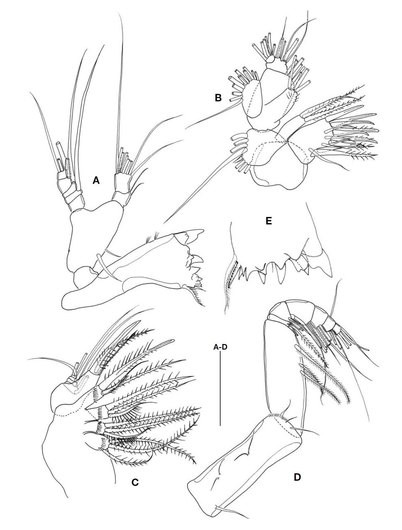
Antennule (Fig.6 H). 24-segmented, reaching end of caudal rami. Endopod of antenna (Fig.7 A) slightly shorter than exopod; coxa with 1 seta; basis with 2 setae. Endopod 2-segmented; first segment with 2 distal setae; second segment with 8 inner and 7 outer setae. Exopod 7-segmented; first segment without seta; second segment with 2 setae; third to sixth segments with 1 seta each; distal segment with 3 terminal setae and medial seta.
Mandible (Fig.7 B, C). Gnathobase with 6 large and 5 small teeth, 1 long seta and 2 rows of setules. Basis with 2 setae. Endopod 2-segmented; first segment with 1 seta; second segment with 9 setae. Exopod 5-segmented; first to fourth segments with 1 seta each; distal segment with 2 setae.
Maxillule (Fig.7 D). Arthrite with 4 posterior setae, 9 spines and 1 medial seta. Coxal epipodite with 9 setae. Coxal endite with 4 setae. Basal endites with 4+5 setae. Endopod with 14 setae. Exopod with 11 setae.
Maxilla (Fig.7 E). First and second praecoxal endites with 3 setae and row of spinules on one side. First coxal endite with 2 long setae and row of spinules on one side. Second coxal endite with 2 setae and 1 thickened seta and with a proximal border spinulose on one side. Basal endite with 3 setae, one of them thickened. Endopod with 5 setae.
Maxilliped (Fig.7 F). Coxa with 3 groups (from proximal to distal end of segment) of 2, 3 and 3 setae (distal group with small spinules). Basis with spinules on proximal edge and 3 medial setae. Endopod 6-segmented; first segment with 2 setae; second to sixth segments with 4, 4, 3, 3+1, and 4 setae, respectively.
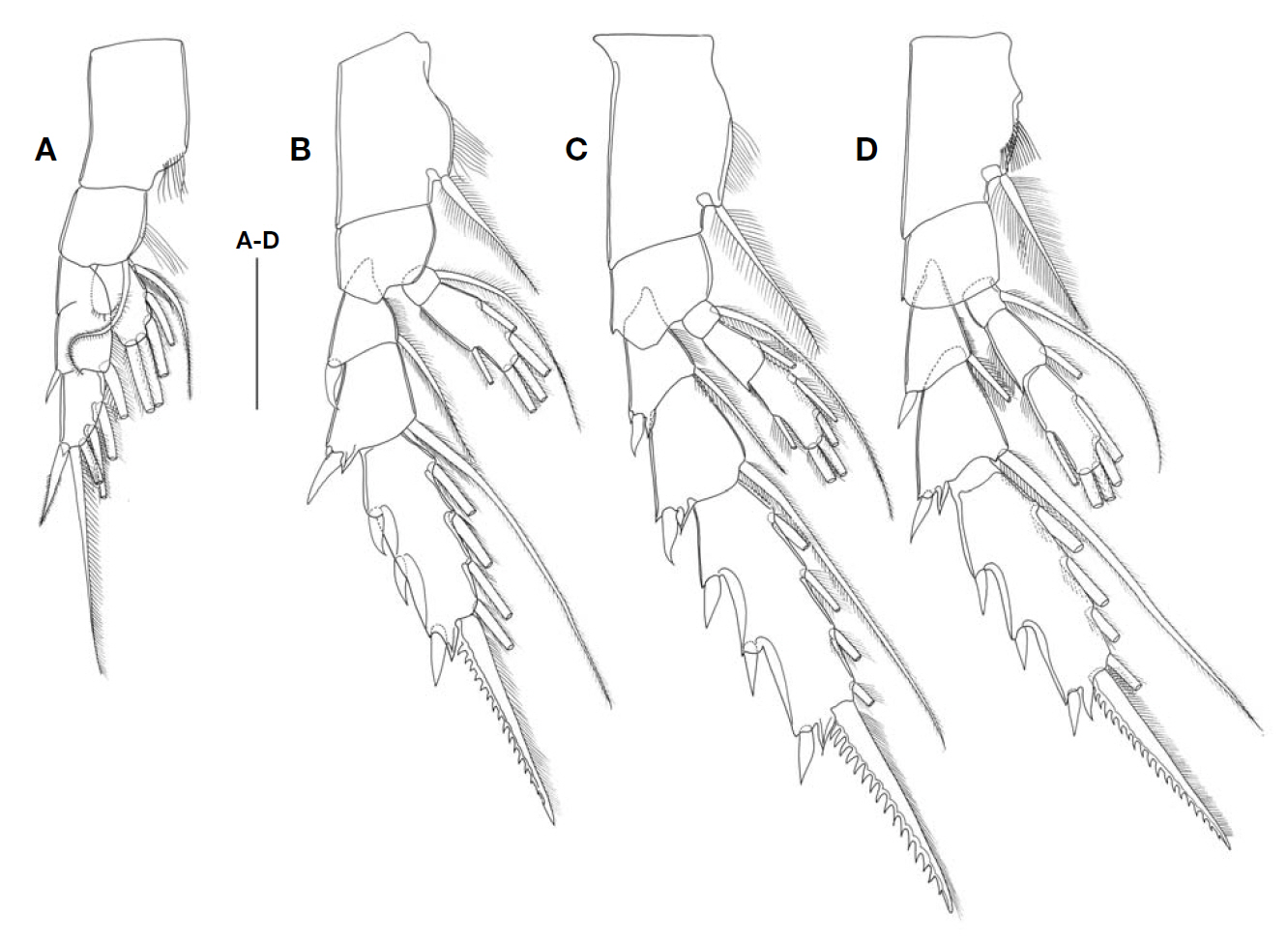
Leg 1 (Fig.8 A). Coxa with hairs on inner border. Basis with hairs on inner border and 1 inner distal seta on anterior surface. Endopod 1-segmented; outer margin swollen with 1 row of spinules. Exopod 2-segmented; first and second segments incompletely fused with 1 outer spine and 1 inner seta; third segment with 1 outer spine and 4 inner setae.
Leg 2 (Fig.8 B). Coxa with hairs on inner border and 1 inner seta. Basis unarmed. Endopod 2-segmented; first seg-
ment with 1 seta; second segment with 5 setae. Exopod 3-segmented; first and second segments with 1 outer spine and 1 inner seta, respectively; third segment with 3 spines and 1 terminal spine bearing 17 teeth on outer edge.
Leg 3 (Fig.8 C). Coxa with hairs on inner border and 1 inner seta. Basis unarmed. Endopod 3-segmented; first and second segments with 1 seta, respectively; distal segment with 5 setae. Exopod 3-segmented; first and second segments with 1 outer spine and 1 inner seta; distal segment with 3 spines, 5 setae and 1 serrated spine bearing 16 teeth on outer edge.
Leg 4 (Fig.8 D). Coxa with thin spines similar to setae. Basis unarmed. Endopod and exopod same as leg 3. Distal segment of exopod with serrated spine bearing 18 teeth on outer edge. Armature formula of swimming legs 1-4 as follows:
Leg 1 coxa 0-0 basis 0-1 exp 0-0; Ⅰ-1; Ⅰ,1,3 enp 0,2,3
enp 0-1; 0-1; 1,2,2
Leg 4 coxa 0-1 basis 0-0 exp Ⅰ-1; Ⅰ-1; Ⅲ,Ⅰ,4
enp 0-1; 0-1; 1,2,2
Leg 5 absent.
Not found.
East Sea (Korea), Sea of Okhotsk, Bering Sea, Izu (Japan), Gulf of Mexico, New Zealand, Kurile-Kamchatka and Mariana Trench.
Female
The specimen (4.46 mm) examined in this study is larger than those of other previous records (Tanaka, 1957; Tanaka and Omori, 1970; Park, 1975). The Korean specimen is congruent with the Japanese one described by Tanaka (1957). However, the present specimen shows some minor differences from Tanaka’s specimen as follows: 1) the maxilliped does not have a small lamellous process on the distal margin of the basis; 2) the terminal spines of legs 2, 3, and 4 were recorded to have 19, 17, and 20 outer teeth (Tanaka, 1957), respectively, whereas there were 15, 16, and 18, respectively, in the present specimen. These differences variations were considered to be within the species.
The CO1 sequence of the Korean specimen showed 99.8% similarity to the previous sequence of the same species (Genbank accession no: AB379980).
Korean name: 1*참수리노벌레속(신칭), 2*참수리노벌레 Korean name: 1*두갈래이마뿔노벌레속(신칭), 2*두갈래이마뿔노벌레(신칭) Korean name: 1*이마노벌레속(신칭), 2*꼬마이마노벌레(신칭)