Members of the genus
The number of morphospecies in
In this study, we describe for the first time three
>
Morphological taxonomy processes
Samples were collected from coastal areas in Korea using a polyurethane foam enveloped slide system (Xu et al., 2009)and then transferred to a laboratory. In successive steps, cultures were maintained at 17℃ in a chamber that was adjusted with a light and dark photoperiod of 12:12 hr. Rice grains were used as a food source for enrichment of bacterial growth in cultures. Living specimens were observed using phase contrast and a differential interference microscope at different magnifications. Protargol and silver nitrate impregnation(Foissner, 1991) were applied in order to reveal the cirri pattern and infraciliature. Enumeration and measurement of stained specimens were preformed under ×1,000 magnification (Leica DM2500; Leica Microsystems, Germany)and specimens were drawn with a camera lucida. The classification scheme used here was based on that of Lynn(2008) and terminologies follow those of Corliss (1979) and Lynn (2008). Abbreviations are as follows: AM, adoral membranelles;AZM, adoral zone of membranelles; BC, buccal cirrus; CC, caudal cirri; CV, contractile vacuole; DK, dorsol kineties; FC, frontal cirri; FV, food vacuole; LMC, left marginal cirri; Ma, macronuclear nodule; Mi, micronucleus;PM, paroral membrane; TC, transverse cirri; and VC, ventral cirri.
>
Molecular taxonomy processes
Isolation of each living individual ciliate was performed under a dissecting microscope using a micropipette (Leica MZ 12.5; Leica Microsystems). DNA extraction, amplification of SSU rDNA, and sequencing were performed according to Kim and Min (2009). Sequenced strands were aligned using ClustalX (Thompson et al., 1997) and BioEdit 7.0.9.0(Hall, 1999). Nucleotide diversity was calculated by MEGA 4.0 (Tamura et al., 2007) using the p-distance value.
Phylum Ciliophora Doflein, 1901
Class Spirotrichea Butschli, 1889
Subclass Hypotrichia Stein, 1859
Order Euplotida Small and Lynn, 1985
Family Uronychiidae Jankowski, 1979
1*Genus Uronychia Stein, 1859
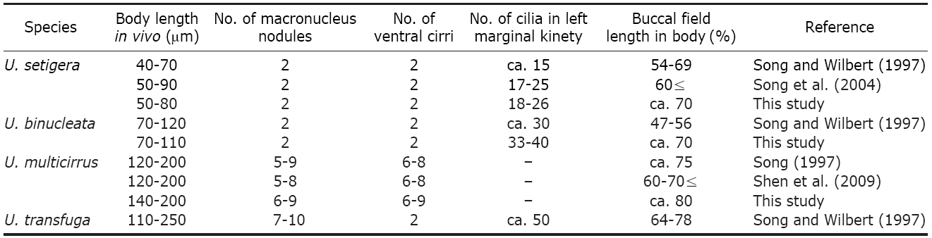
2*Uronychia setigera Calkins, 1902 (Table 1,Fig.1)
Uronychia uncinata sensu Kahl, 1932: 627, fig. S. 622, 28.
Uronychia setigera Song and Wilbert, 1997: 424, fig. 7;Song et al., 2004: 315, figs. 1-3.
Specimens collected from the coastal water of Gumjin-ri (36o22′N, 129o23′E), Gyeongsangbukdo in the East Sea of Korea on 6 Oct 2006. The raw culture maintained for up to ten days.
about 50-80 μm long
The smallest
Two Ma connected by funiculus, located on the left side of body; each nodule ellipsoid-shaped, with spherical nucleoli;Mi not recognized (Fig. 1C, D). CV recognized near the right of TC; several FV recognized after protargol impregnation.Movement typically rapid jumping aside or backwards, swimming with very fast rotation against the longitudinal axis.
AZM pattern stable in this genus; 11 AM1 on apical area of anterior region, extended dorsal surface, each membranelle consists of 3 kineties; 4 AM2 on the left of middle region,each membranelle consists of roughly 6-8 kineties; 1 BC,small cirrus-like membranelle, apart from AZM2, inconspicuous,about 2-3 ㎛ long (Fig. 1A-C, G-I). Buccal field surrounded by a horseshoe-shaped robust PM (Fig. 1B, E, F).
FC 4 in number, narrow, positioned at the most anterior area on ventral surface; 2 small VC, on right margin; 5 TC,rightmost cirrus rather small; 3 enlarged LMC, positioned on left of ventral region; 3 enormous CC emerged on dorsal surface (Fig. 1A-D, G, H). Six DK composed of dikinetids on dorsal surface; 2 DK dorsolaterally located on left margin of ventral surface, densely packed near posterior region,contained in an average of 23 cilia in the leftmost kinety;other 3 DK positioned on dorsal surface, extended to CC(Fig. 1B-D).
Three SSU rDNA sequences of Korean population were deposited in Genbank under the accession number HQ380021-HQ380023. They are 1,625 bp in length and show 99.45-99.69% similarity between Korean population and Chinese population (Genbank accession number: EF198669) of
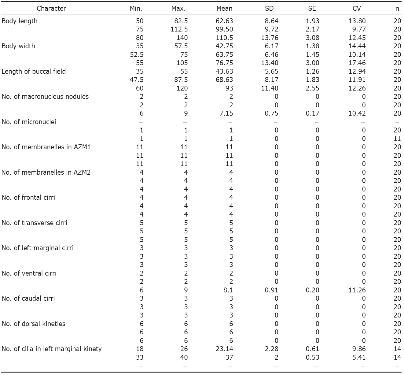
Morphometric characteristics of Uronychia setigera (1st line) U. binucleata (2nd line) and U. multicirrus (3rd line) from protargol impregnated specimens
The Korean population is congruent with the Chinese population(Song and Wilbert, 1997; Song et al., 2004) in most respects, including body shape and size, the number of dikinetids in the leftmost kinety and nuclei, and a spur on the left margin with the population by Song and Wilbert (1997) and Song et al. (2004) (Table 2).
Germany, China, and Korea (this study).
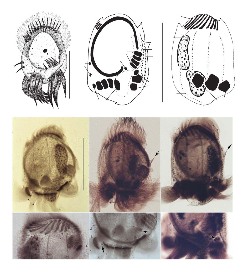
1*Uronychia binucleata Young, 1922 (Table 1,Fig. 2)
Uronychia binucleata Young, 1922: 360, figs. 4-6; Song and
Wilbert, 1997: 427, fig. 8.
Specimens collected from the public waterfront (37o26′N, 126o35′E), Incheon in the Yellow Sea of Korea on 19-26 Jun 2006. The raw culture maintained for up to one week.
Size about 70-110 μm long
ly rectangular shaped, 2 Ma, 1 Mi, 2 spurs on the posterior region,11 AM1, 4 AM2, 1 BC, 4 FC, 3 LMC, 2 VC, 5 TC, 3 CC, 6 DK, approximately 37 cilia in the leftmost kinety.
Body size about 70-110×55-75 ㎛ in vivo,oval to slightly rectangular shaped; slightly convex both right and left margins. Dorso-ventrally flattened, ratio of about 2 : 3; buccal area deeply caved, extended about 70% of body length (Fig. 2A, B, D). Two conspicuous spurs appeared on posterior region, visible after protargol staining (Fig. 2B, G);lateral spurs inconspicuous. On posterior portion, 2 depressions on ventral side, TC and LMC emerged; 1 depression on dorsal side, CC appeared (Fig. 2A-C). Cytoplasm grayish to dark gray colored, with numerous granules and large particles.
Two Ma connected by funiculus, located on left of body;each nodule irregular sausage-shaped, with spherical nucleoli;Mi located between Ma nodules, globular form (Fig. 2C).CV recognized near the right of TC; several FV recognized after protargol impregnation (Fig. 2D). Movement usually genus typical, like
AZM pattern stable in this genus; 11 AM1 on apical area of anterior region, extended dorsal surface, each membranelle contained with 3 kineties; 4 AM2 on left of middle region,each membranelle consists of about 6-8 kineties; 1 BC, small cirrus-like membranelle, aparted from AZM2, about 2-3 μm long (Fig. 2A-C, F). An oral area surrounded by horseshoe-shaped PM (Fig. 2B, D).
[Table 2.] Comparison of Uronychia species in present and previous specimens

Comparison of Uronychia species in present and previous specimens
shaped PM (Fig.2 B, D).
FC 4 in number, positioned at the most anterior area on ventral surface; 2 small VC, on right margin; 5 TC, rightmost cirrus rather small; 3 enlarged LMC, positioned on left side of ventral region; 3 strong CC emerged on right margin of dorsal side (Fig. 2A-C). Six DK composed of dikinetids on cell surface, genus-typical; 2 DK dorsolaterally located on left margin of ventral surface, densely packed on posterior region, included in an average of 37 cilia in the leftmost kinety (Fig. 2B, C, E, F). Silverline system on dorsal surface formed net-type (Fig. 2H).
The SSU rDNA sequence of the Korean population was deposited in Genbank under the accession number HQ380024.The sequence is 1,625 bp in length and shows 99.82% similarity between the Korean population and the Chinese population(Genbank accession number: EF198667) of
However,
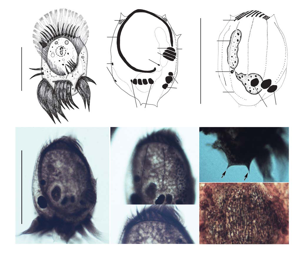
1*Uronychia multicirrus Song, 1997 (Table 1,Fig.3 )
Uronychia multicirrus Song, 1997: 279, figs. 1-5; Shen et al., 2009: 296, figs. 1-3o.
Specimens collected from the public waterfront (37o26′N, 126o35′E), Incheon in the Yellow Sea of Korea on 6-13 Aug 2007. The raw culture maintained for up to ten days.
Size about 140-200 μm long
Body size about 140-200×70-120 μm
Usually moniliform Ma connected with 6-9 nodules by funiculus, C-shaped; each nodule ellipsoidal to elongated shape, with spherical nucleoli; Mi located at the lower left corner of Ma, spherical form (Fig. 3C, G). CV recognized near the right of TC; several FV recognized after protargol impregnation (Fig. 3G). Movement usually genus typical, as
AZM pattern genus-typical; 11 AM1 on apical area of anterior region, extended dorsal surface, each membranelle consists of 3 kineties; 4 AM2 on left side of middle region;1 BC, small cirrus-like membranelle, separated from AZM2,base about 4-5 ㎛ long (Fig. 3A, B, H). A buccal field surrounded by horseshoe-shaped mighty PM (Fig. 3A, G).
FC 4 in number, positioned at the most frontal area on ventral surface; 6-9 VC, on right border of cell; 5 TC, rightmost cirrus rather small; 3 enlarged LMC, positioned on left of ventral region; 3 strong CC emerged on right margin of dorsal side (Fig. 3A-C, G-I). Six DK on cell surface, genus-typical,composed of dikinetids; 2 DK dorsolaterally located on left margin of ventral surface (Fig. 3B, C, J). Silverline system on dorsal side formed net-type (Fig. 3K).
The SSU rDNA sequence of the Korean population was deposited in Genbank under the accession number HQ380025. The sequence is 1,624 bp in length and shows 99.88% similarity between the Korean population and the Chinese population(Genbank accession number: EU267929) of
China and Korea (this study).
Korean name: 1*말총충속 (신칭), 2*강모말총충 (신칭) Korean name: 1*쌍핵말총충 (신칭) Korean name: 1*다극모말총충(신칭)