검색 전체 메뉴
PDF
맨 위로
OA 학술지
Terahertz Pulse Imaging of Micro-metastatic Lymph Nodes in Early-stage Cervical Cancer Patients
  • 비영리 CC BY-NC
  • 비영리 CC BY-NC
ABSTRACT
Terahertz Pulse Imaging of Micro-metastatic Lymph Nodes in Early-stage Cervical Cancer Patients
KEYWORD
Terahertz imaging , Cervical cancer , Lymph node metastasis , (110.6795) Terahertz imaging , (170.3880) Medical and biological imaging , (300.6495)Spectroscopy , terahertz
  • I. INTRODUCTION

    Cervical cancer is the second most common malignancy in women worldwide and is the only major gynecological malignancy clinically staged according to the International Federation of Obstetrics and Gynecology (FIGO) recommendations[1]. The prognostic factors identified in cervical cancer patients are tumor size, depth of stromal invasion,presence of lymphovascular tumor emboli, histologic type,and invasion of surgical margins [2, 3]. Lymph node metastasis is also closely associated with poor prognosis and decreased survival rate [4]; moreover, this metastasis is an indicator for adjuvant radiotherapy [5].

    Computed tomography (CT), magnetic resonance imaging(MRI), and positron emission tomography-CT (PET-CT)have been used to detect metastatic lymph nodes in cervical cancer patients, but their accuracies were lower than expected [6, 7]. In one study of cervical cancer patients negative for metastatic lymph nodes by MRI, the sensitivity of PET in detecting metastatic lymph nodes was only 10% [8]. This low diagnostic performance was attributed to the adoption of inaccurate size criteria and a possible substantial number of small metastatic lymph nodes. Although surgical lymph node staging is an option, it is not routinely recommended because it is a specialized procedure that increases the time and cost of the surgery, and it leads to operation-related complications [9, 10]. The accurate diagnosis of metastatic lymph nodes is of clinical importance, the lack of sensitive diagnostic methods indicates the need for additional imaging modalities.

    In recent years, terahertz (THz) imaging technology has shown great promise and has increasingly been performed for cancer diagnosis and treatment [11-13]. Although THz imaging may improve the diagnosis of metastatic lymph nodes in cervical cancer patients, it has not yet been evaluated in this setting. Therefore, we report a pilot study for THz imaging to detect lymph node micro-metastases in earlystage cervical cancer patients, which are usually undiagnosed by conventional imaging methods.

    II. MATERIALS AND METHODS

       2.1. Sample Preparation

    Two patients, aged 45 and 50 years, with FIGO stage IB1 and squamous cell carcinoma cervical cancer, respectively,underwent conventional lymphadenectomy combined with radical hysterectomy. Dissected lymph nodes were embedded in paraffin for histologic diagnosis and THz evaluation.Five paraffin-embedded metastatic lymph nodes from the two patients (three from one patient, two from the other)were sliced at 5-mm intervals in the longitudinal plane,and 4-㎛ sections were stained with hematoxylin and eosin. Because of the retrospective design of this study,our institutional review boards did not require approval or informed consent for the review of the patients’ specimens.

       2.2. Image Acquisition and Measurement

    All lymph node specimens were imaged using a THz time-domain spectroscopy (THz TDS) system operating in reflection mode [12]. The THz pulse was generated by an InAs wafer pumped by a Ti:Sapphire laser with a center wavelength of 790 nm, a pulse width of 100 fs, and a repetition rate of 80 MHz. The THz pulse was focused on the specimen at a normal incidence by using an off-axis parabolic mirror,and the reflected THz pulse was detected by a photoconductive antenna fabricated on a low-temperature grown GaAs. The entire THz TDS system was enclosed in a chamber and continuously purged with dry air to eliminate THz absorption by water vapor in the air. The bandwidth of the imaging setup ranged from 0.2 to 2.0 THz with a signalto-noise ratio of 2000:1. Two-dimensional THz images were obtained by raster-scanning of specimens in the x-y plane.Each scan area was ~ 20 mm × 20 mm, consisting of 40× 40 pixels with a pixel pitch of 0.5 mm, and the image quality was enhanced by using a spline interpolation.

    THz images were obtained using the normalized peak-topeak amplitude of reflected THz pulses at each pixel of the specimen, E(x, y)/E0, where E0 is the minimum peakto-peak amplitude of THz pulses within the specimen. After Fourier transformation of the measured THz pulses, the frequency dependent reflection coefficient at each interface between the specimen and air, r(x, y, ?), was calculated as

    image

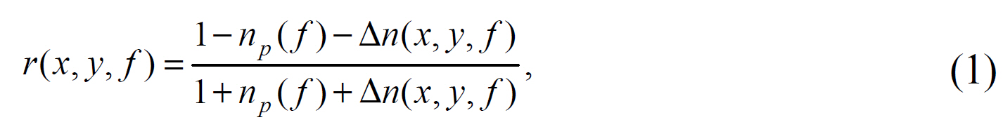
    where ?is frequency, np(?) is the refractive index of paraffin, and Δ(x, y, f) = n(x, y, f) - np(?) is the difference in refractive index between the lymph node and paraffin. Equation (1) can be rearranged as

    image

    where γ(x, y, ?) is the reflection coefficient of the lymph node, normalized by the reflection coefficient of paraffin rp(?),

    image

    Because the frequency dependence of np(? ) is negligible in the THz frequency range used in experiments [14], np(f ) is 1.5 and γ(x, y, ? ) ? E(x, y)/E0. THz images were obtained by mapping the relative refractive index using equation (2)and evaluated by comparing the size and shape of the region of contrast with the size and shape of the tumor region identified by histopathologic examination of the corresponding stained lymph node slices under an optical microscope.

    III. EXPERIMENTAL RESULTS AND DISCUSSION

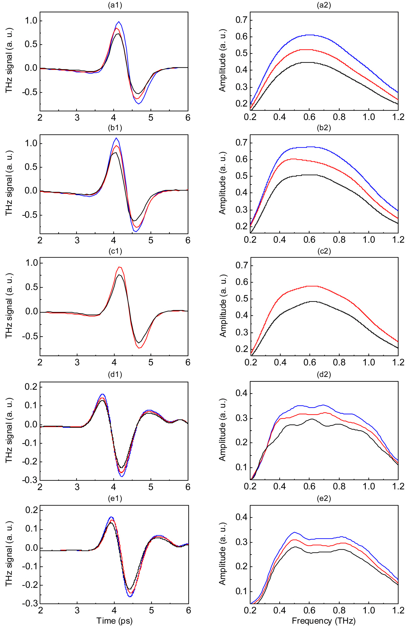
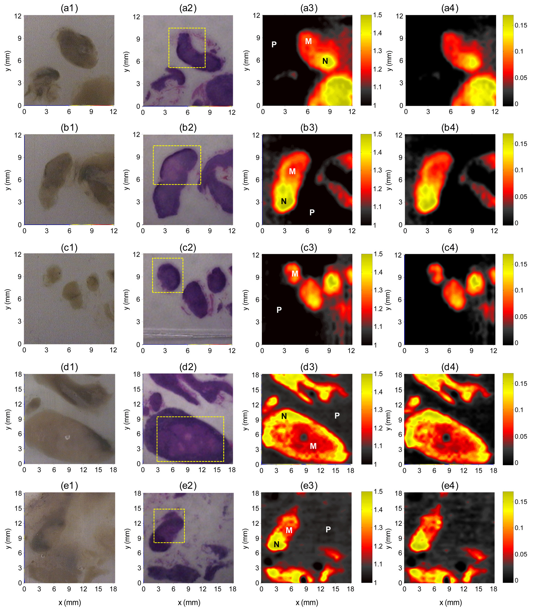
    Figure 1 shows reflected THz pulses and spectral amplitudes of lymph node specimens at the selected pixels indicated in Fig. 2. For all specimens, the reflected peak amplitudes were smaller for the metastatic than for the non-metastatic portions of lymph nodes, as was the spectral amplitude in the THz frequency range. These reductions in peak amplitudes in the metastatic region were primarily due to changes in the refractive index, as shown in Fig. 2.

    The THz images clearly differentiated metastatic regions from non-metastatic regions, even for 3-mm metastatic foci(Fig. 2) and showed good correlations with histopathologic findings. This differentiation, however, was not evident in the optical images. THz images were generated on a color scale by mapping two image parameters: E(x, y)/E0 and Δn(x, y, ? )/np. The metastatic foci ranged in size from 3 to 12 mm, with a mean size of 6 ± 0.5 mm. There were no significant differences in contrast between two THz images by using peak-to-peak amplitudes and refractive index changes,demonstrating that THz images can also determine the optical properties, such as refractive index, of lymph node specimens.We observed that the refractive index decreased in regions of metastatic lymph nodes. The mean refractive indices of the non-metastatic and metastatic portions of lymph nodes were 1.84 ± 0.04 and 1.76 ± 0.03, respectively. The index difference between non-metastatic and metastatic portions

    of lymph nodes was 4.7 ± 0.5%.

    Lymph node metastasis is an important factor influencing prognosis and therapeutic strategy in patients with cervical cancer. Although CT and MRI have been used to detect metastatic lymph nodes, including measurements of size,shape, presence of necrosis, and enhancement characteristics,these methods cannot efficiently differentiate malignant from hyperplastic lymph nodes [15-17]. Many of these metastatic

    lymph nodes are smaller than 1 cm, the usual diagnostic size criteria for metastatic lymph nodes, thereby reducing the diagnostic accuracy of CT and MRI [18]. Although PET scanning diagnoses metastatic lymph nodes by assessing the abnormal accumulation of FDG, PET is expensive and less widely available, and has lower spatial resolution. The pooled sensitivities of CT, MRI, and PET were found to be only 52%, 38%, and 54%, respectively, in region- or nodebased comparisons, which are too low to replace surgical staging [19, 20]. Thus, there are as-of-yet unmet clinical needs for the accurate imaging diagnosis of metastatic lymph nodes and detection of small metastatic nodes, i.e., diagnoses that better reflect the actual histological processes.

    Our pilot study showed that THz images delineated metastatic foci in lymph nodes to an extent similar to those of histopathologic methods. Although the overall correlation of metastatic foci and THz images were good, peak reflection amplitudes at the edges of the lymph nodes were lower because of averaging with the adjacent paraffin, which has much lower reflected peak amplitudes. This edge effect may be responsible for the false positive findings in benign portions of the lymph nodes. We found that the smallest metastatic focus delineated on THz imaging was approximately 3 mm in its largest dimension. Although these were specimen images,we believe that our findings are meaningful as a pilot study for future investigations of this imaging modality in the diagnosis of micro-metastatic lymph nodes, which are usually undiagnosed by conventional imaging methods.

    Earlier studies have suggested that water content is a major contributory factor for contrast in THz images since tumors generally contain a higher water content than normal tissue and liquid water has high attenuation for THz waves[21-23]. Although several other studies have used paraffinembedded tissues [24-26], the fundamental mechanisms underlying the contrast in THz images of the dehydrated paraffin-embedded samples that differentiate tumor from normal tissue are not clear at this stage. However, the different contrasts for tumors and normal tissues in THz images of dehydrated paraffin-embedded samples may be due to other components, including cell density, proteins, and DNA.Thus, paraffin-embedded tissue samples may be useful for assessing cancer biology using THz imaging and for investigating THz contrast mechanisms, other than the effects of water.

    IV. CONCLUSION

    We have shown here that THz imaging can clearly delineate the metastatic portions of the lymph nodes as small as 3 mm. Two THz images by using peak-to-peak amplitudes and refractive index changes were almost identical, demonstrating that THz images can also determine refractive indices of lymph node specimens. THz imaging is a promising technique for the detection of micro-metastatic lymph nodes that cannot be detected by conventional imaging methods,but technical improvements are needed prior to its clinical application.

참고문헌
  • 1. Parkin D. M, Bray F, Ferlay J, Pisani P 2001 Estimating the world cancer burden: Globocan 2000 [Int. J. Cancer] Vol.94 P.153-156 google cross ref
  • 2. Juretzka M. M, Jensen K. C, Longacre T. A, Teng N. N, Husain A 2004 Detection of pelvic lymph node micrometastasis in stage IA2-IB2 cervical cancer by immunohistochemical analysis [Gynecol. Oncol.] Vol.93 P.107-111 google cross ref
  • 3. Marchiole P, Buenerd A, Benchaib M, Nezhat K, Dargent D, Mathevet P 2005 Clinical significance of lympho vascular space involvement and lymph node micrometastases in early-stage cervical cancer: a retrospective case-control surgico-pathological study [Gynecol. Oncol.] Vol.97 P.727-732 google cross ref
  • 4. Takeshima N, Yanoh K, Tabata T, Nagai K, Hirai Y, Hasumi K 1999 Assessment of the revised international federation of gynecology and obstetrics staging for early invasive squamous cervical cancer [Gynecol. Oncol.] Vol.74 P.165-169 google cross ref
  • 5. Tanaka Y, Sawada S, Murata T 1984 Relationship between lymph node metastases and prognosis in patients irradiated postoperatively for carcinoma of the uterine cervix [Acta Radiol. Oncol.] Vol.23 P.455-459 google
  • 6. Choi H. J, Roh J. W, Seo S. S, Lee S, Kim J. Y, Kim S. K, Kang K. W, Lee J. S, Jeong J. Y, Park S. Y 2006 Comparison of the accuracy of magnetic resonance imaging and positron emission tomography/computed tomography in the presurgical detection of lymph node metastases in patients with uterine cervical carcinoma: a prospectivestudy [Cancer] Vol.106 P.914-922 google cross ref
  • 7. Williams A. D, Cousins C, Soutter W. P, Mubashar M, Peters A. M, Dina R, Fuchsel F, McIndoe G. A, deSouza N. M 2001 Detection of pelvic lymph node metastases in gynecologic malignancy: a comparison of CT MR imaging and positron emission tomography [AJR Am. J. Roentgenol.] Vol.177 P.343-348 google
  • 8. Chou H. H, Chang T. C, Yen T. C, Ng K. K, Hsueh S, Ma S. Y, Chang C. J, Huang H. J, Chao A, Wu T. I, Jung S. M, Wu Y. C, Lin C. T, Huang K. G, Lai C. H 2006 Low value of [18F]-fluoro-2-deoxy-D-glucose positron emission tomography in primary staging of early-stage cervical cancer before radical hysterectomy [J. Clin. Oncol.] Vol.24 P.123-128 google cross ref
  • 9. Nagell J. R. van, Roddick Jr. J. W, Lowin Jr. D. M 1971 The staging of cervical cancer: inevitable iscrepancies between clinical staging and pathologic findinges [Am. J. Obstet. Gynecol.] Vol.110 P.973-978 google
  • 10. Scheidler J, Hricak H, Yu K. K, Subak L, Segal M. R 1997 Radiological evaluation of lymph node metastases in patients with cervical cancer. A meta-analysis [JAMA] Vol.278 P.1096-1101 google cross ref
  • 11. Ashworth P. C, Pickwell-MacPherson E, Provenzano E, Pinder S. E, Purushotham A. D, Pepper M, Wallace V. P 2009 Terahertz pulsed spectroscopy of freshly excised human breast cancer [Opt Express] Vol.17 P.12444-12454 google cross ref
  • 12. Fitzgerald A. J, Wallace V. P, Jimenez-Linan M, Bobrow L, Pye R. J, Purushotham A. D, Arnone D. D 2006 Terahertz pulsed imaging of human breast tumors [Radiology] Vol.239 P.533-540 google cross ref
  • 13. Wallace V. P, Fitzgerald A. J, Shankar S, Flanagan N, Pye R, Cluff J, Arnone D. D 2004 Terahertz pulsed imaging of basal cell carcinoma ex vivo and in vivo [Br. J. Dermatol.] Vol.151 P.424-432 google cross ref
  • 14. Enatsu T, Kitahara H, Takano K, Nagashima T “Terahertz spectroscopic imaging of paraffin-embedded livercancer samples” P.557-558 google
  • 15. King A. D, Tse G. M, Ahuja A. T, Yuen E. H, Vlantis A. C, To E. W, Hasselt A. C. van 2004 Necrosis in metastatic neck nodes: diagnostic accuracy of CT MR imaging and US [Radiology] Vol.230 P.720-726 google cross ref
  • 16. Brekel M. W. van den, Stel H. V, Castelijns J. A, Nauta J. J, Waal I. van der, Valk J, Meyer C. J, Snow G. B 1990 Cervical lymph node metastasis: assessment of radiologic criteria [Radiology] Vol.177 P.379-384 google
  • 17. Mack M. G, Rieger J, Baghi M, Bisdas S, Vogl T. J 2008 Cervical lymph nodes [Eur. J. Radiol.] Vol.66 P.493-500 google cross ref
  • 18. Choi H. J, Kim S. H, Seo S. S, Kang S, Lee S, Kim J. Y, Kim Y. H, Lee J. S, Chung H. H, Lee J. H, Park S. Y 2006 MRI for pretreatment lymph node staging in uterine cervical cancer [AJR Am. J. Roentgenol.] Vol.187 P.538-543 google cross ref
  • 19. Choi H. J, Ju W, Myung S. K, Kim Y 2010 Diagnostic performance of computer tomography magnetic resonance imaging and positron emission tomography or positron emission tomography/computer tomography for detection of metastatic lymph nodes in patients with cervical cancer: meta-analysis [Cancer Sci.] Vol.101 P.1471-1479 google cross ref
  • 20. Hricak H, Gatsonis C, Chi D. S, Amendola M. A, Brandt K, Schwartz L. H, Koelliker S, Siegelman E. S, Brown J. J, McGhee R. B, Iyer Jr. R, Vitellas K. M, Snyder B, Long 3rd H. J, Fiorica J. V, Mitchell D. G 2005 Role of imaging in pretreatment evaluation of early invasive cervical cancer: results of the intergroup study American college of radiology imaging network 6651-gynecologic oncology group 183 [J. Clin. Oncol.] Vol.23 P.9329-9337 google cross ref
  • 21. Bruehlmeier M, Roelcke U, Blauenstein P, Missimer J, Schubiger P. A, Locher J. T, Pellikka R, Ametamey S. M 2003 Measurement of the extracellular space in brain tumors using 76Br-bromide and PET [J. Nucl. Med.] Vol.44 P.1210-1218 google
  • 22. Chen J. H, Avram H. E, Crooks L. E, Arakawa M, Kaufman L, Brito A. C 1992 In vivo relaxation times and hydrogen density at 0.063-4.85 T in rats with implanted mammary adenocarcinomas [Radiology] Vol.184 P.427-434 google
  • 23. Ross K. F, Gordon R. E 1982 Water in malignant tissue measured by cell refractometry and nuclear magnetic resonance [J. Microsc.] Vol.128 P.7-21 google
  • 24. Knobloch P, Schildknecht C, Kleine-Ostmann T, Koch M, Hoffmann S, Hofmann M, Rehberg E, Sperling M, Donhuijsen K, Hein G, Pierz K 2002 Medical THz imaging: an investigation of histo-pathological samples [Phys. Med. Biol.] Vol.47 P.3875-3884 google cross ref
  • 25. Loffler T, Siebert K, Czasch S, Bauer T, Roskos H. G 2002 Visualization and classification in biomedical terahertzpulsed imaging [Phys. Med. Biol.] Vol.47 P.3847-3852 google cross ref
  • 26. Berry E, Handley J. W, Fitzgerald A. J, Merchant W. J, Boyle R. D, Zinov'ev N. N, Miles R. E, Chamberlain J. M, Smith M. A 2004 Multispectral classification techniques for terahertz pulsed imaging: an example in histopathology [Med. Eng. Phys.] Vol.26 P.423-430 google cross ref
OAK XML 통계
이미지 / 테이블
  • [ FIG. 1. ]  Reflected THz pulses (left) and THz spectral amplitudes (right) of paraffin (black), non-metastatic (blue), and metastatic (red)portions of five lymph nodes at the selected pixels indicated in Fig. 2.
    Reflected THz pulses (left) and THz spectral amplitudes (right) of paraffin (black), non-metastatic (blue), and metastatic (red)portions of five lymph nodes at the selected pixels indicated in Fig. 2.
  • [ FIG. 2. ]  Optical (column 1), histopathologic (column 2), and THz images (columns 3 and 4) of five paraffin-embedded metastaticlymph nodes from two patients. The THz images were obtained using E(x, y)/E0 (column 3) and Δn(x, y, f)/np (column 4). Selectedpixels are indicated in column 3 (P: paraffin, M: metastatic portion, N: non-metastatic portion). The dashed boxes indicate themetastatic portion identified by histopathologic examination. The THz images showed good correlations with histopathologic results.
    Optical (column 1), histopathologic (column 2), and THz images (columns 3 and 4) of five paraffin-embedded metastaticlymph nodes from two patients. The THz images were obtained using E(x, y)/E0 (column 3) and Δn(x, y, f)/np (column 4). Selectedpixels are indicated in column 3 (P: paraffin, M: metastatic portion, N: non-metastatic portion). The dashed boxes indicate themetastatic portion identified by histopathologic examination. The THz images showed good correlations with histopathologic results.
(우)06579 서울시 서초구 반포대로 201(반포동)
Tel. 02-537-6389 | Fax. 02-590-0571 | 문의 : oak2014@korea.kr
Copyright(c) National Library of Korea. All rights reserved.