Red algal parasites are characterized by little or no pigmentation, relatively small size, hemispherical cushion-like pustules on the surface of their host, and the establishment of secondary pit connections between penetrating parasite cells and medullary cells of the host (Setchell 1918, Apt and Schlech 1998). Approximately 100 species of parasitic red algae represent a diversity of taxonomic groups and are fairly widespread geographically (Kraft and Abbott 2002). Among them, the greatest concentration of parasites is found in the Rhodomelaceae, for which approximately 30-35 species in 18 genera have been described (Goff 1982, Kraft and Abbott 2002).
At least three red algal parasites belonging to the tribe Laurencieae have been reported from the flora of Korea (Lee and Kang 2001). These include Janczewskia gardneri Setchell et Guernsey and J. morimotoiTokida, found on Laurencia sp. and Benzaitenia yenoshimensis Yendo, found on Chondria sp. Parasites on species of the Rhodomelaceae are relatively common but reports on members of the tribe Pterosiphonieae being parasitized are uncommon, with only three genera reported in the literature: Leachiella Kugrens parasitizing Pterosiphonia Falkenberg in the eastern Pacific from Washington to California (Kugrens 1982), Levringiella Kylin parasitizing Pterosiphonia on the North American Pacific coast and Juan Fernandez Islands, and Stromatocarpus Falkenberg parasitizing Tayloriella Kylin on the southwestern coast of South Africa.
In a collection of algae from Busan, Korea, we noticed a parasitic alga on Symphyocladia latiuscula (Harvey) Yamada, belonging to the tribe Pterosiphonieae. A careful examination revealed that this alga represented a new example of parasitic algae in the Rhodomelaceae. Here, we report on this newly discovered parasitic species on the basis of its vegetative and reproductive morphology on the south coast of Korea.
Plants were collected by the author (M. S. Kim) on 21 February 2008 in Molundae, Busan, Korea, growing on Symphyocladia latiuscula. Plants were fixed in 4% formalin/seawater. For anatomical studies, sections were cut by hand and stained with 1% aqueous aniline blue acidified by addition of 1 N HCl. Photographs were taken with a Digital Sight DS-Fi1 camera (Nikon, Tokyo, Japan) attached to a microscope (ECLIPSE 80i; Nikon). Representative material has been deposited as preserved samples in the herbarium of Jeju National University (JNUB), Jeju, Korea.
Plantae minutae, laete pigmentiferae, in Symphyocladia latiuscula parasiticae aut hemiparasiticae; axes erecti polysiphoni cellulis pericentralibus 6 habentes corticem proximalem, ex pulvino centralibus cellularum orientes, per pseudoparenchymatus pulvinatus basi quae pit-connectiones secundarias cellulis hospitis formant affixi; gametophyti feminini procarpia in cellulis suprabasalibus trichoblastorum portati; procarpiis ex ramo carpogoniali 4-cellulato in cellula sustentanti cum 2 cellulato basalis sterile constantibus; cystocarpium ovatus cum ostiolum; capitula spermatangialia lanceolata portatus unus per segmentum in monosiphonius pedicelli in spiralis position cum fertilis trichoblasto; tetrasporangia tetraedice divisa, factus unus per successivus segmentum, in strictus series intra ramis.
Plants minute, lightly pigmented, parasitic or hemiparasitic on Symphyocladia latiuscula; erect polysiphonous axes of six pericentral cells with proximal cortication, arising from the central cushion of cells attached by pseudoparenchymatous pulvinate bases, which form secondary pit connections with host cells; female gametophytes producing procarps on the suprabasal cells of trichoblasts; the procarps consisting of four-celled carpogonial branches borne on supporting cells that also bear two-celled sterile basal branches; cystocarps ovate with an ostiole; lanceolate spermatangial branches borne one per segment on monosiphonous pedicels in spiral positions with fertile trichoblast; tetrasporangia, tetrahedrally divided, formed one per successive segment, in a straight series within branches.
Thalli in Symphyocladia latiuscula hemiparasitici; axes erecti polysiphonii ad 2 mm alt., 100-250 ㎛ diametro; cellulis pericentralibus 6, 100 ㎛ longis et 30 ㎛ latis in statura ex cellulis pericentralibus ad inferior parte; oblique divisa cellula apicalis, 15 ㎛ longis et 8 ㎛ latis;
procarpia in suprabasale cellula trichoblasti; cystocarpia ovatus in basibus brevibus polysiphoniis, usque ad 230 ㎛ logis et 200 ㎛ diametro; spermatangia in trichoblastis semel dichotomis alter dimidium axem spermatangialem faciens, 110 ㎛ longis et 30 ㎛ diametro; tetrasporangia tetraedice divisa, 30 ㎛ longis et 20 ㎛ latis.
Thalli hemiparasitic on S. latiuscula (Fig. 1); erect polysiphonous axes to 2 mm high, 120-280 ㎛ in diameter; six pericentral cells, 100 ㎛ long and 30 ㎛ wide, of a pericentral cell in the lower part; obliquely divided apical cells, 15 ㎛ long and 8 ㎛ wide; procarps on suprabasal cell of trichoblast; cystocarps ovate on short polysiphonous bases, up to 230 ㎛ long and 200 ㎛ in diameter; spermatangia on once-dichotomous trichoblasts, one half becoming the spermatangial axis, 110 ㎛ long and 35 ㎛ in diameter; tetrasporangia tetrahedrally divided, 30 ㎛ long and 20 ㎛ wide.
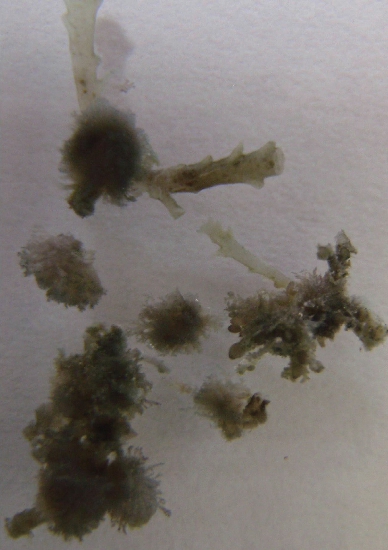
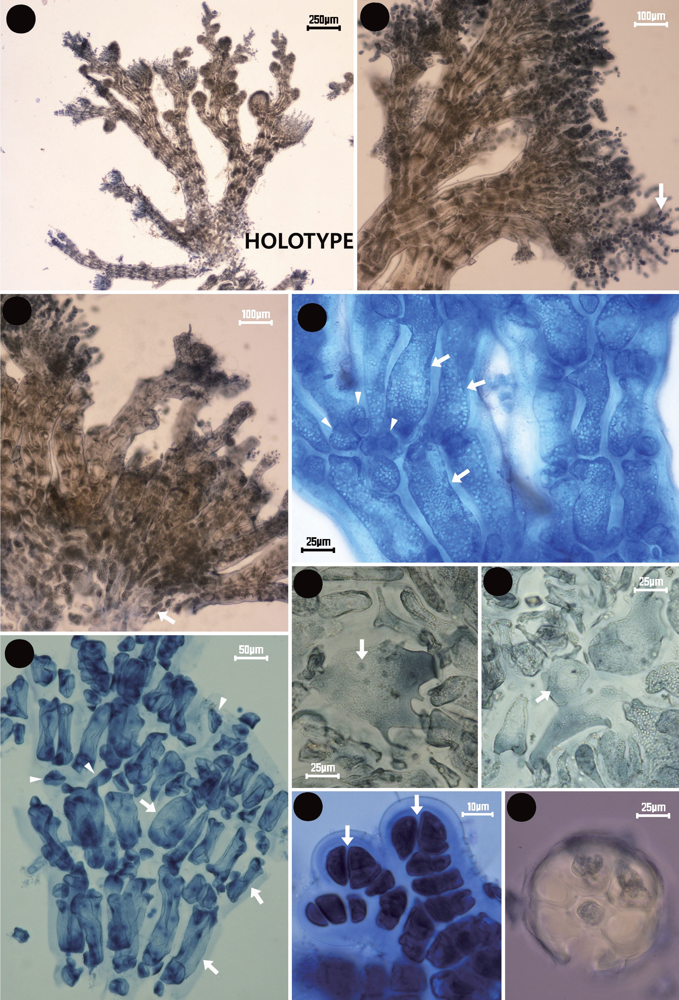
Holotype: The type collection consists of ten microscope slides, the holotype (JNU-MSK30201) female gametophyte(Fig. 2A) plus female, spermatangial and tetrasporangial isotypes on slide # JNU-MSK30202~30210, all plants parasitic on Symphyocladia latiuscula, collected on 21 February 2008 by the author in Busan, Korea.
Type locality: Molundae, Dadaepo, Busan, Korea (35°02´28.41˝N, 128°58´38.09˝E).
Korean name: 보라색우무털
Distribution: Although the host is recorded widely throughout northeast Asian coastal areas, the parasite is only known from one collection at the type locality.
Habitat:The type collection was parasitic on S. latiuscula. The host is growing on a rock at the low tide mark near the subtidal zone on the southeastern coast of Korea. Habitat is characteristic of sheltered rocky sites.
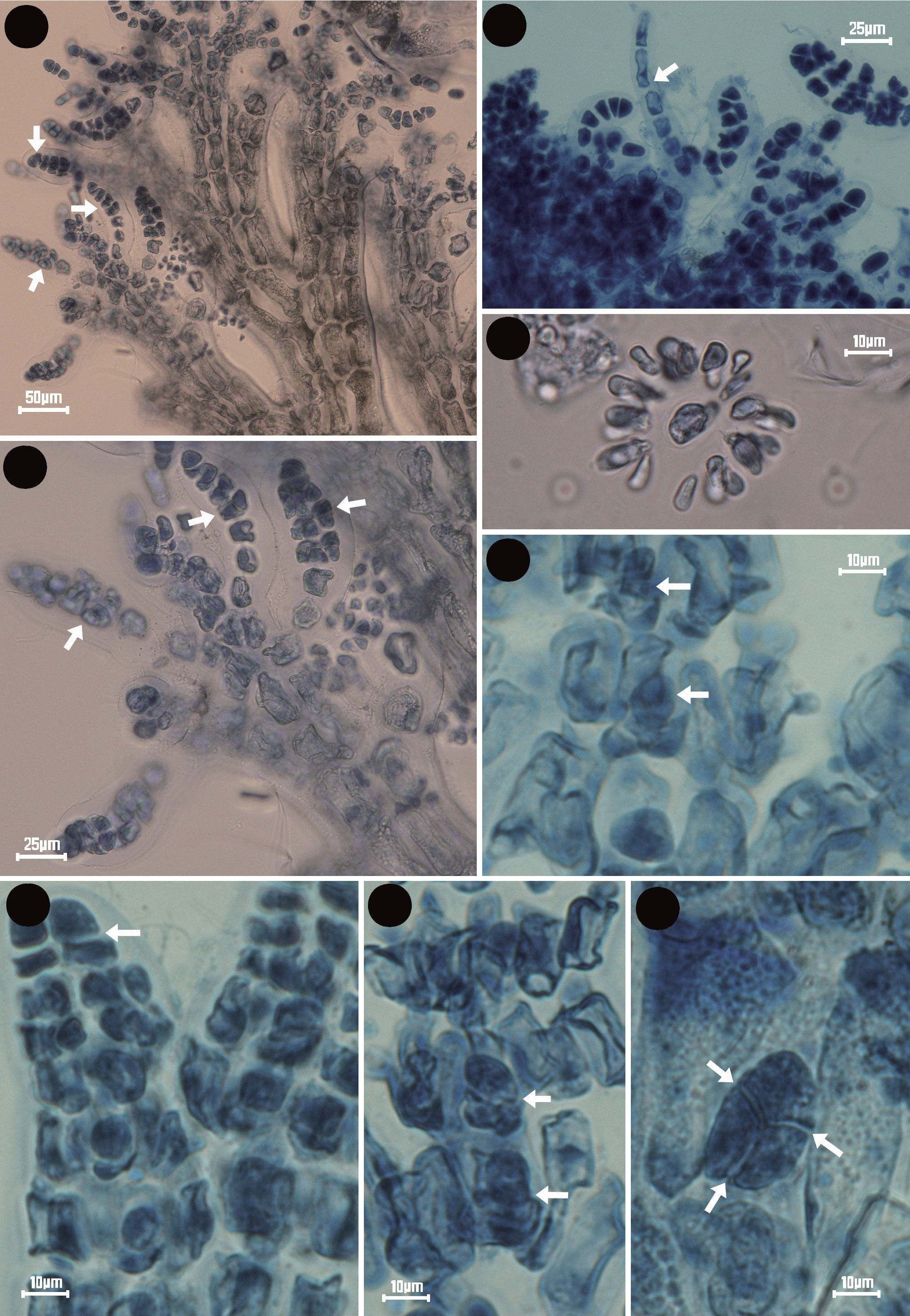
Vegetative structure: Thalli are attached to the host through a pseudoparenchymatous pulvinate cell 100 ㎛ in diameter that establishes several pit-connections to the central axial and adjacent pericentral cells of the host at the node between successive segments (Fig. 2C, F & G). All the material collected was reproductive (Fig. 2A & B). The corticated axes have six pericentral cells (Fig. 2D, E & I). Apical cells divide obliquely (Fig. 2H). Trichoblasts, when deciduous, leave prominent round basal cells, especially on male plants.
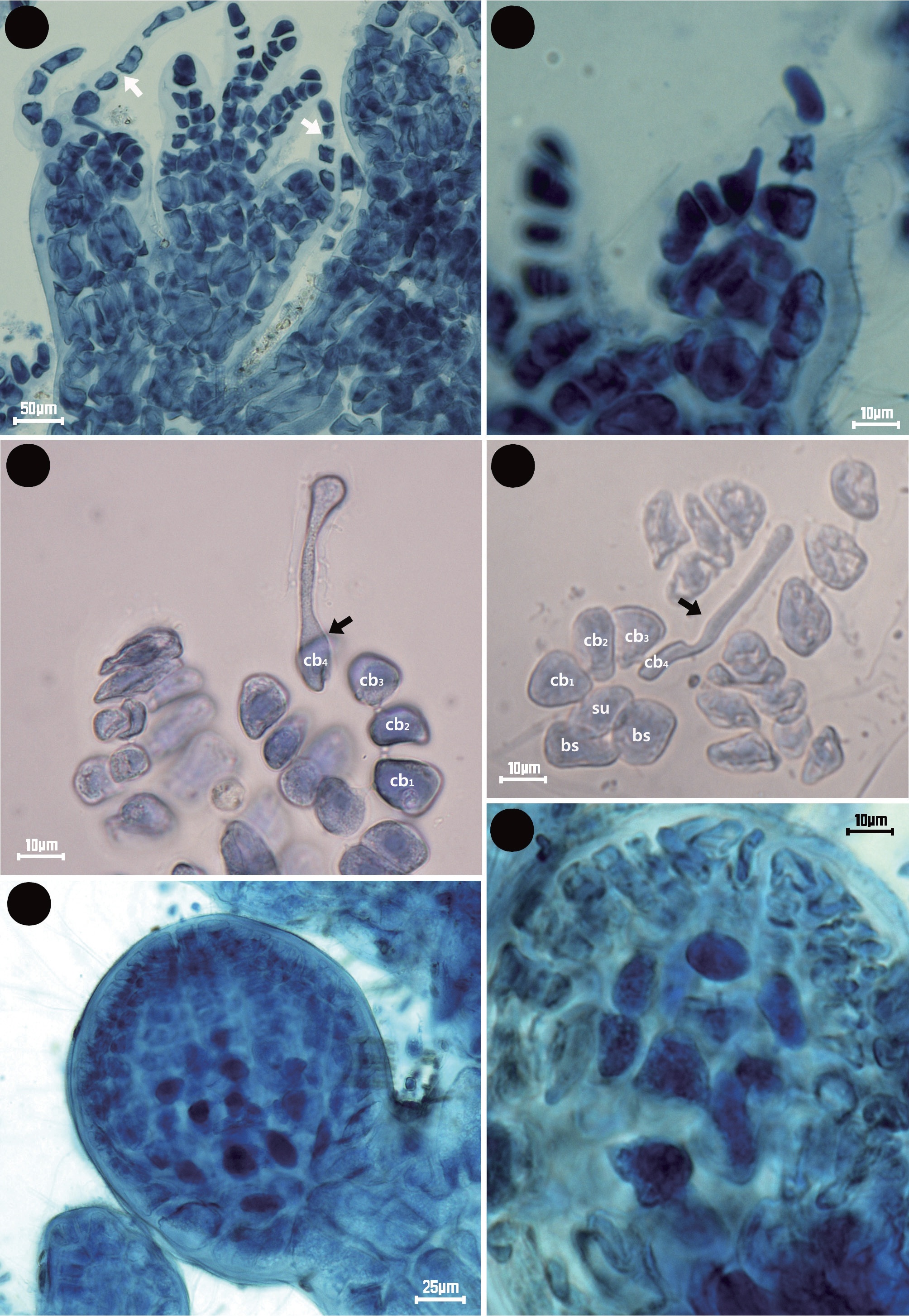
Carpogonial branches and carposporophyte: Carpogonial branches with protruding trichogynes are borne on fruiting heads only 60-80 ㎛ in length (Fig. 3A). Procarps consist of a four-celled carpogonial branch with two sterile cell groups borne on the supporting cell and surrounded very early by filaments of a globose pericarp (Fig. 3B & C). The supporting cell is one of a cluster of six pericentral cells of similar size and shape to the pyriform apical cell (Fig. 3D). The mature cystocarpic plants have rudimentary filamentous cortication growing acropetally and basipetally from pericentral cells. The cystocarps are ovate with an ostiole and approximately 300 ㎛ long and 200 ㎛ in diameter (Fig. 3E & F).
Spermatangia:The species is dioecious, the male gametophytes producing deciduous trichoblasts that leave round basal cells (Fig. 4A & B), as well as persistent fertile trichoblasts (Fig. 4C & D). Spermatangial branches are borne one per each successive segment on monosiphonous pedicels in spiral positions with fertile trichoblast which is being once dichotomous and composed of lanceolate spermatangial branches.
Tetrasporangia: Transversely divided apical cells in tetrasporangial axes result in a central filament of linearly aligned cells (Fig. 4E, F & G). Tetrahedrally divided sporangia are produced within the main branches (Fig. 4H). A single tetrasporangium is formed per successive segment in straight series, with two cover cells and mature tetrasporangia reaching 20 × 30 ㎛ in size.
The red algal parasite genus Symphyocolax is newly described from the south coast of Korea based on morphological evidence. The parasite on S. latiuscula was found infecting only one plant of the relatively common host (Choi and Lee 1991). The parasite was fertile, producing young female, tetrasporangiate, and male reproductive structures. Thus it is possible to compare this species with other similar genera in reproductive as well as structural characteristics. The most notable diagnostic features of Symphyocolax are the polysiphonous upright axes with many branches attached by pseudoparenchymatous pulvinate tissue, six pericentral cells, and having many corticated cells. Although the new genus resembles Levringiella Kylin and Meridiocolax J. Morrill in appearance, Symphyocolax is distinguishable from Levringiella by its many polysiphonous branches and corticated cells, and from Meridiocolax by the six pericentral cells and a pulvinate base.
The new genus Symphyocolax has little pigmentation in polysiphonous upright axis, although the general form of the parasite is colorless pustules which are small and compact with a cluster habit growing on mostly distal parts of host branches (Norris 1988). In recognizing the parasite genus, vegetative characters such as the type of attachment at the base, the presence or absence of cortical cell and trichoblasts, and the number of pericentral cells are considered important diagnostic characters (Noble and Kraft 1983, Norris 1988, Apt and Schlech 1998). Tetrasporangial cleavage and arrangement are also valuable characters at the genus level, although gametangial reproductive structures are of less taxonomic value (Morrill 1976, Stegenga et al. 1997). Most Rhodomelaceae produce spermatangia in dense heads borne on forks or basal cells of trichoblasts and procarps consisting of four-celled carpogonial branches borne on a supporting cell additionally bearing a single sterile cell (Kraft and Abbott 2002).
Pseudoparenchymatous pulvinate tissue has been reported in the genus Levringiella in both the North American Pacific and Juan Fernandez Islands of the South Pacific (Kylin 1956, Abbott and Hollenberg 1976). In his discussion of the genus Levringiella, Kylin (1956) used that character to separate it from the genus Stromatocarpus Falkenberg attached by rhizoidal filaments to the
host cells. Because the new genus Symphyocolax has no rhizoidal filaments penetrating the host, the presence of a pulvinate base to the erect branches is useful for separating the genus. The number of pericentral cells has been shown to be a valid character for delimiting some genera of parasites, as is the presence or absence of cortical cells and trichoblasts (Kylin 1956, Kraft and Abbott 2002). Symphyocolax has six pericentral cells with many cortical cells at the lower parts of main branches, but most genera of the family Rhodomelaceae have four pericentral cells without cortication. On the other hand, the genus Neotenophycus Kraft et Abbott has three pericentral cells (Kraft and Abbott 2002), Ululania Apt et Schlech has five pericentral cells with outer cortical layers (Apt and Schlech 1998), and Chamaethamnion Falkenberg also has five pericentral cells with cortication (Norris 1988).
An additional character of importance in separating genera in the Rhodomelaceae is whether or not the tetrasporangia are produced in straight or spiral arrangements. Morrill (1976) used tetrasporangial type on bipartite versus ternate as a generically diagnostic character. The new genus Symphyocolax formed tetrasporangia as one per segment in a straight series within determinate branches and divided tetrahedrally. Chamaethamnion and Tylocolax are the only two parasites in the Rhodomelaceae that are known to produce two tetrasporangia in each stichidial segment (Kylin 1956). Noble and Kraft (1983) discussed the tetrasporangia of two genera, Meridiocolax and Stromatocarpus, in which transversely divided apical cells in tetrasporangial axes of Meridiocolax result in a central filament of linearly aligned cells, in contrast to Stromatocarpus in which the divisions are oblique and the cells aligned in a zigzag fashion.
Kraft and Abbott (2002) mentioned that at least 35 species in 17 or 18 genera of parasites are attributed to the Rhodomelaceae. Among these, three parasitic genera, Levringiella Kylin, Meridiocolax J. Morrill, and StromatocarpusFalkenberg, are very similar to the new genus Symphyocolax (Table 1). Although they occur on hosts belonging to the Pterosiphonieae (Levringiella and Stromatocarpus) and Polysiphonieae (Meridiocolax), the host of the new genus Symphyocolax belongs to the Pterosiphonieae.
Levringiella, a little-known parasite of Pterosiphonia, forms dense monopodial polysiphonous branches from pseudoparenchymatous pulvinate tissue and develops all reproductive structures on most segments in distal regions of erect branchlets (Guiry and Guiry 2010). In addition, carpogonial branches are unknown and spermatangia with short monosiphonous pedicels are formed continuously on most segments of upright axes. As pointed out by Abbott and Hollenberg (1976) on Levringiella gardneri (Setchell) Kylin from the North American Pacific, the new genus Symphyocolax differs from this species in the shape of the pericentral cells, which are longer rather than shorter, and in having six pericentral cells with cortical cells that are produced in the proximal parts of main branches rather than five to seven pericentral cells without cortical cells, as in the type species. The shape of the cystocarp in the new genus Symphyocolax is ovate rather than spheroidal. Symphyocolax and Levringiella are the two parasites in the Rhodomelaceae that are known to produce pseudoparenchymatous pulvinate tissue at the base. These two genera, from the type of attachment point of view, seem to be closely related generic status, but this issue requires more extensive research.
Meridiocolax was erected as the type species Meridiocolax narcissus Morrill to delimit parasites of Polysiphonia ferulacea Suhr from the Caribbean Sea and the western Atlantic Ocean (Morrill 1976). Noble and Kraft (1983), in reporting characters of Meridiocolax, noted that the ecorticate axes have four pericentral cells and the lower axial cells give rise to rhizoidal filaments from their bases, which anchor the thallus to the host, forming secondary connections to both adjacent host and parasite cells. The new genus Symphyocolax, however, has six pericentral cells with many cortical cells from pulvinate tissue. According to the reproductive features of Meridiocolax, some species have unusual characters, such as several male capitula on a single trichoblast, bisporangia, and transversely divided apical cells in tetrasporophytes (Noble and Kraft 1983). However, the male gametophytes of the new genus Symphyocolax produce only one spermatangial branch on a deciduous trichoblast, which leaves round basal cells as well as persistent fertile trichoblasts. Although the apical cells in tetrasporangial axes of the new genus Symphyocolax are also divided transversely, it has no bisporangia in the tetrasporophyte.
Moreover, in virtually all other features of vegetative and reproductive structure, Symphyocolax and Stromatocarpus are very dissimilar, which strongly suggests that the two genera are not closely related. Stromatocarpus, a little-known parasite of Tayloriella and Placophora from the southwest coast of South Africa, forms four pericentral cells that obliquely divide at the apical cell. In addition, they lack corticated cells in both vegetative and reproductive filaments. Other significant differences between Symphyocolax and Stromatocarpus are the type of attachment at the base and the arrangement of tetrasporangia.
Approximately 80% of red algal parasites are termed ‘adelphoparasites,’ meaning that they are very closely related to their hosts taxonomically. Consequently, they are members of the same tribe in the large family Rhodomelaceae based on morphological/reproductive features (Goff 1982, Zuccarello et al. 2004). A smaller number of red algal parasites, on the other hand, are not closely related to their hosts and have been termed ‘alloparasites.’This term has not been precisely defined but is usually applied to parasites that are found on hosts from a different tribe or family than the parasite (Goff 1982). The taxonomic position of the new genus Symphyocolax has been difficult to identify because of relatively sparse material in addition to the rather incomplete generic descriptions of Symphyocladia (Kim et al. 2010). Nevertheless, features of pericentral cell formation, procarp, and particularly spermatangial trichoblasts clearly ally Symphyocolax with the tribe Polysiphonieae of the Rho domelaceae, although the host, S. latiuscula, is a member of the tribe Pterosiphonieae (Hommersand 1963).