검색 전체 메뉴
PDF
맨 위로
OA 학술지
Characterization of Fibrinolytic Proteases from Gloydius blomhoffii siniticus Venom
  • 비영리 CC BY-NC
  • 비영리 CC BY-NC
ABSTRACT
Characterization of Fibrinolytic Proteases from Gloydius blomhoffii siniticus Venom
KEYWORD
Gloydius blomhoffii siniticus , snake venom , fibrinolytic protease
  • I. Introduction

    The hemotoxic venoms of rattlesnakes and vipers usually affect the circulation system1). Some of these toxins, both enzymattic and non-enzymatic, lead to the spectacular changes in haemostasis and to the frequent hemorrhage seen after snake bite. The enzyme proteins are acetylcholinesterases, ADPases, phospholipases, hialuronidases, and haemostatic proteases. The non-enzymic proteins are disintegrin and bradykinin-potentiating peptides. The activities of these toxins include coagulation and anticoagulation of blood, platelet-activation, anti-platelet function, fibrinolytic activation, and hemorrhage2- 6).

    Fibrinolytic and fibrinogenolytic activities have been described in venom from the following snake families: Viperidae, Elapidae, and Crotalidae7). Venom fibrin(ogen)olytic proteases have been classified into metalloproteases and serine proteases7, 8). These fibrin(ogen)olytic proteases differ in their mechanism of action and they target different amino acid sequences in fibrin(ogen).

    The fibrin(ogen)olytic proteases have drawn particular attentions as potential therapeutic agents to remove plasma fibrinogen from the circulation for treatment of acute isochaemic stroke and to dissolve blood clot for treatment of occlusive thrombi7-9). Fibrolase from copperhead snake venom degrades both α and β chains of fibrin and showed some promise as a thrombolytic agent. Alfimeprase, a recombinant fibrinolytic enzyme derived from fibrolase has been being developed as a clinical agent.

    Gloydius blomhoffii with the common name of mamushi, known as salmusa in Korea, is a venomous pit viper found in China, Japan, and Korea. There are four subspecies including G. b. blomhoffii, G. b. brevicaudus, G. b. dubitatus, and G. b. siniticus10). G. b. siniticus with the common name of Yangtze mamushi is located in China from Shandong south to Chiang Jing Basin11).

    Terada and his colleagues12-14) purified and characterized several metalloproteases from Chinese mamushi (Agkistroden halys brevicaudus) venom. A high molecular weight metalloprotease H6 (brevilysin H6) was purified from the venom of Chinese mamushi. The molecular weight is 60 kDa and it has Zn+ in the active site. Addition of Ca2+ ion enhanced both the proteolytic activity and the thermal stability. Brevilysin H6 degrades preferentially Aα chain of fibrinogen and slowly Bβ chain and has weak hemorrhagic activity12). The molecular weights of two low molecular weight metalloproteases, brevilysin L4 and brevilysin L6, were estimated to be 22 and 21.5 kDa by SDS-PAGE13, 14). Brevilysin L6 and brevilysin L4 only attack Aα-chain of fibrinogen. The velocity of degradation of fibrinogen by brevilysin L6 is much lower than that by brevilysin L4.

    The molecular weight of halystase, a serine protease, purified from Japanese common viper (Agkistroden halys blomhoffii) was estimated to be 38 kDa under reducing condition of SDS-PAGE15). Halystase cleaves Bβ-chain of fibrinogen and slowly Aα-chain and hydrolyzes kininogen to produce bradykinin. A heterogenous two-chain fibrinolytic enzyme, brevinase, was purified from the venom of Agkistroden blomhoffii brevicaudus, a Korean snake16). The venom enzyme belongs to serine proteinase, which cleaves preferentially Bβ-chain of fibrinogen and slowly Aα-chain, and consists with two polypeptides of 16.5 kDa and 17 kDa of which the N-terminal amino acid sequences show homology to the N-terminal and the internal amino acid sequences of single-chain fibrinolytic enzymes from other snake venoms, respectively.

    The objectives of this study were to identify fibinolytic proteases from Yangtze mamushi (G. b. siniticus) venom and to characterize a major fibrinolytic protease as a potential therapeutic agent for pharmacopuncture treatment of the circulation-related diseases.

    II. Materials and Methods

       1. Snake venom

    The snake venom from G. b. siniticus in this study was purchased in China.

       2. Isolation of fibrinolytic enzyme

    The venom powder (2 g) was dissolved and dialyzed in 50 mM Tris-HCl, pH 7.6, which was then centrifuged at 10,000 g for 30 min using the refrigerated centrifuge of model VS-15CF (Vision Scientific, Korea) to remove insoluble substances. The venom solution was applied to a column (2.5 cm × 8 cm) of Q-Sepharose (GE, USA) equilibrated with 50 mM Tris-HCl, pH 7.6. The column was eluted with 200 ml of 50 mM Tris-HCl, pH 7.6 and then with a concentration gradient from 150ml of 50 mM Tris-HCl, pH 7.6 to 150ml of 50 mM Tris-HCl, pH 7.6, 0.3 M NaCl. The column was subsequently eluted with 50ml of 50 mM Tris-HCl, pH 7.6, 0.3 M NaCl. The flow rate was 21 ml per hour and the fraction volume was 7 ml. The fractions with fibrinolytic activity were combined and concentrated by using polyethylene glycol and then applied to a column (2.5 cm × 109 cm) of Sephadex G-75 (GE, USA). The column was eluted with 50 mM Tris-HCl, pH 7.6, 0.15 M NaCl. The flow rate was 14 ml per hour and the fraction volume was 7 ml. All the isolation steps were performed at refrigeration temperature. Absorbance at 280 nm and fibrinolytic activity in the fibrin plate assay of the fractions from the chromatography were determined. The fractions with fibrinolytic activity were subjected to SDS-PAGE analysis.

       3. Fibrin plate assay

    Fibrinolytic activity was determined on fibrin plates according to the following procedure17). An aliquot (200 μl) of thrombin solution (10 U/ml) in 100 mM Tris-HCl, pH 7.8 was added to 9 ml of 0.1% fibrinogen in the same buffer. The mixed solution was added into petri dish with the diameter of 9 cm and incubated at 37C for 1 hr, until it was converted into fibrin gel. An aliquot (10 μl) of the venom fraction was placed on the fibrin gel and then incubated at 37C for 18 hr. Two diameters (r1 and r2) at a right angle of hydrolysis zone were measured. The fibrinolysis area was calculated following the formula of 0.785r1 × r2. Relative fibrinolysis (%) was percentage of the fibrinolysis area of treated fibrinolytic protease to that of control without treatment.

       4. SDS-PAGE

    Sodium dodecyl sulfate-polyacrylamide gel electrophoresis (SDS-PAGE) was performed according to the procedure described by Laemmli18). The molecular weight markers (Bio-Rad, USA) for SDS-PAGE consisted of myosin (200 kDa), β-galactosidase (116.25 kDa), phosphorylase b (97.4 kDa), bovine serum albumin (66.2 kDa), ovalbumin (45.0 kDa), carbonic anhydrase (31.0 kDa), trypsin inhibitor(21.5 kDa), lysozyme(14.4 kDa), and aprotinin (6.5 kDa).

       5. Hydrolysis of proteins

    An aliquote (200 μl) of 2% proteins in 50 mM Tris-HCl, pH 7.5, 0.15 M NaCl, 10mM CaCl2 was added with 50 μl of the fibrinolytic protease (2 μg/ml) in the same buffer and then the mixture was incubated at 37C for 6 hour. An aliquote (10 μl) of the mixture was taken at specified times during the incubation and added with equal amount of the sample buffers for SDS-PAGE each time. The mixtures were heated at 100C for 5 min and analyzed in SDS-PAGE.

       6. Protein determination

    Protein concentration was determined using BCA Protein Assay Reagent (Pierce, USA). Bovine serum albumin was used for calibration curve.

    III. Results

       1. Isolation of fibrinolytic enzymes from the snake venom

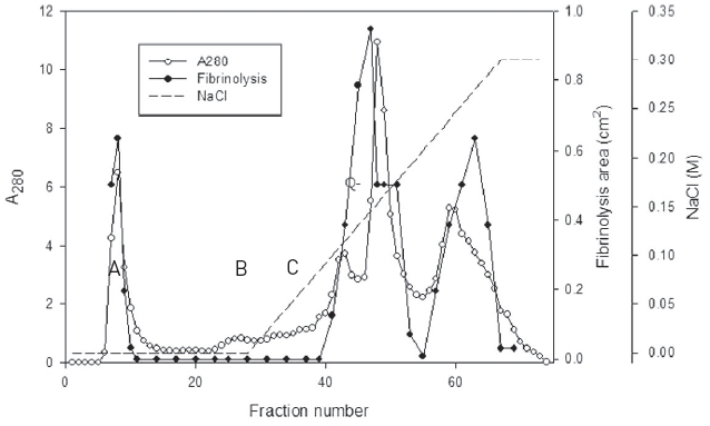
    The snake venom was separated into three major peaks of A, B, and C by using chromatography on the column of Q-Sepharose which was eluted using 50 mM Tris-HCl, pH 7.6 and then the same buffer with the concentration gradient up to 0.3 M sodium chloride (Fig. 1).

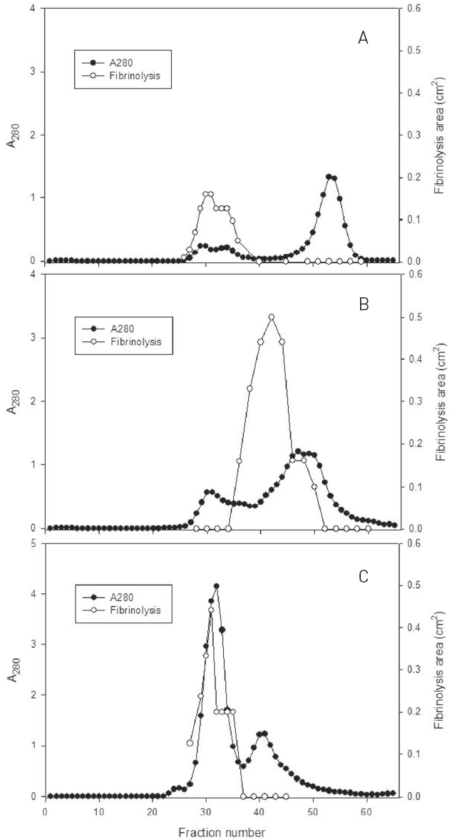
    Fibrinolytic activities of the fractions of the three peaks were closely related with protein concentration. The fractions of three major peaks showing fibrinolytic activity were combined, concentrated, and subjected to chromatography using a column of Sephadex G-75 (Fig. 2).

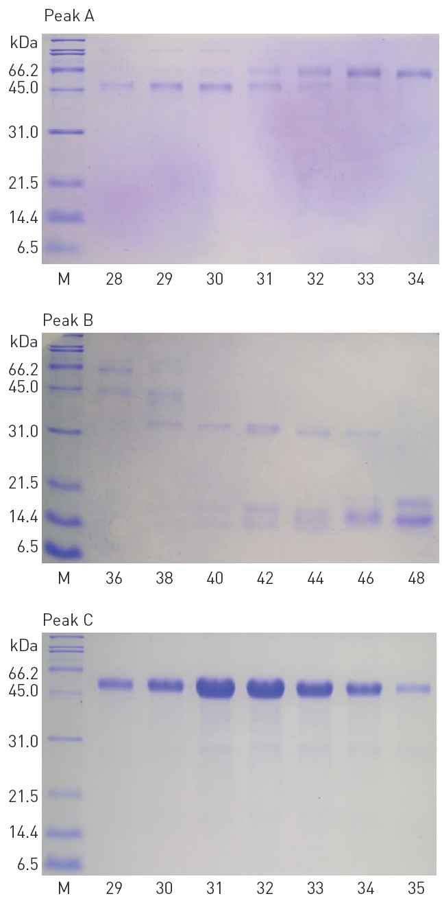
    The fractions 28-34 from the peak A, 36-48 from the peak B, and 29-35 from the peak C, showed significant fibrinolytic activities. These fibrinolytic fractions were subjected to SDS-PAGE analysis (Fig. 3).

       2. SDS-PAGE analysis of the fibrinolytic fractions

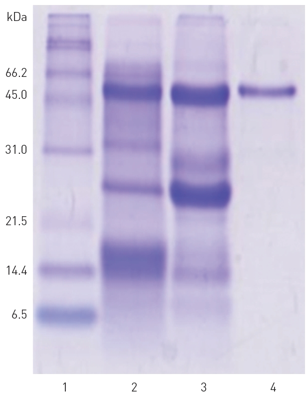
    The polypeptide patterns of the fibrinolytic fractions which showed strong fibrinolytic activity are shown in (Fig. 3). The fractions 28-30 and 33-34 contained two polypeptides of 46 and 59 kDa, respectively. The fraction 42 showing the strongest fibrinolytic activity in the peak B contained three polypeptides of 32, 18, and 15 kDa. All the frac-

    tions 29-35 from the peak C contained a major polypeptide of 54 kDa. These results suggested that these polypeptides were potential fibrinolytic proteases and the fibrinolytic protease of 54 kDa was a predominant fibrinolytic protease in the venom.

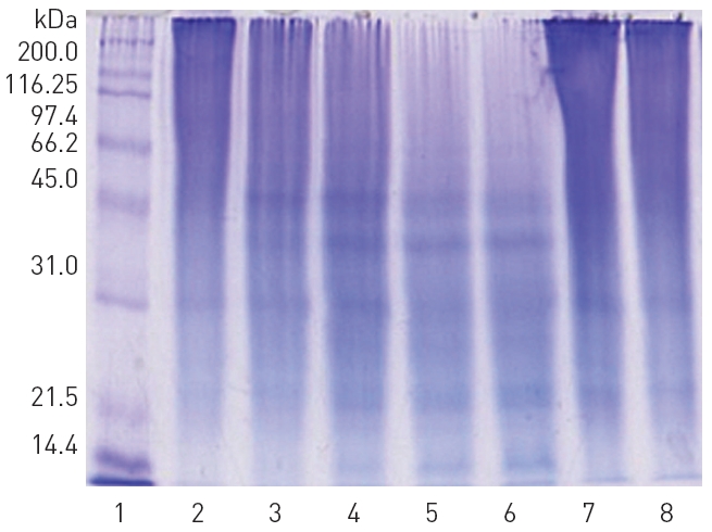
    The fibrinolytic fractions 29-35 from the peak C obtained from the Sephadex G-75 column chromatography (Fig. 2) were combined together, concentrated in dialysis tubing using polyethylene glycol, and then subjected to chromatography on the same column of Sephadex G-75 again to purify the

    major fibrinolytic protease (results not shown). SDS-PAGE in Fig. 4 confirmed that the purified fibrinolytic protease of 54 kDa (lane 4) was one of the major polypeptides in the snake venom (lane 2) as well as in the combined fibrinolytic fraction of

    peak C (lane 3) obtained after Q-Sepharose column chromatography. The amount of the purified fibrinolytic protease obtained from 2 g of the snake venom was 81 mg.

       3. Characterization of the fibrinolytic protease

    The fibrinolytic protease was diluted to the protein concentrations of 0.5, 0.4, 0.3, 0.2, 0.1, and 0.05 mg/ml and 10 μl of the diluted fibrinolytic protease was used in fibrin plate assay as shown in (Fig. 5). The fibrinolysis zone sizes which indicated fibrinolytic activity were very weak at the protein concentration of 0.05 and 0.1 mg/ml. As the protein concentration increased from 0.2 to 0.5 mg/ml, the size of the fibrinolysis zone gradually increased. The protein concentration of 0.45 mg/ml was used

    in the following study to determine the effects of protease inhibitors and salts on the fibrinolytic activity.

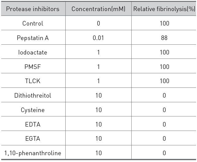
    The effects of protease inhibitors on the fibrinolytic activity were determined as shown in (Table 1).

    The reducing compounds, such as dithiothreitol and cysteine, which reduce disulfide linkage to sulfhydryl group of proteins completely destroyed the fibrinolytic activity, which suggested that the fibrinolytic protease contained disulfide linkage which is necessary for the activity. Metal chelators, such as EDTA, EGTA, and 1,10-phenanthroline, also destroyed the fibrinolytic activity, which indicated that the fibrinolytic protease was a metalloprotease which required metal ion for the activity. Serine protease inhibitors, PMSF and TLCK, a cysteine protease inhibitor, iodoacetate, and an acid protease inhibitor, pepstatin A showed no inhibition on the activity. These results indicated that the fibrinolytic protease was a metalloprotease which contained disulfide linkage.

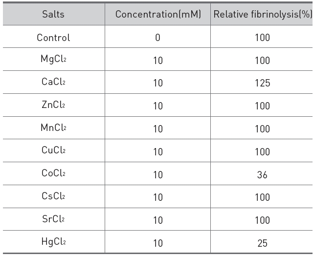
    The effects of salts on the fibrinolytic activity of the protease were shown in (Table 2).

    Addition of calcium chloride to the fibrinolytic protease increased the fibrinolytic activity and

    [Table 1.] Effects of protease inhibitors on the fibrinolytic protease from G. b. siniticus.

    label

    Effects of protease inhibitors on the fibrinolytic protease from G. b. siniticus.

    [Table 2.] Effects of salts on the fibrinolytic protease from G. b. siniticus.

    label

    Effects of salts on the fibrinolytic protease from G. b. siniticus.

    made the fibrinolysis zone clearer. Addition of cobalt(II) chloride and mercuric chloride partially inhibited the fibrinolytic activity.

       4. Hydrolysis of proteins by the fibrinolytic protease

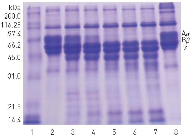
    The fibrinolytic protease cleaved preferentially Aα-chain and slowly Bβ-chain of fibrinogen but γ-chain was unaffected as shown in (Fig. 6).

    When 10 mM calcium choride was omitted from the reaction mixture, hydrolysis of Aα-chain was retarded and Bβ-chain and γ- chain were unaffected (data not shown). The fibrinolytic protease hydrolyzed high molecular weight polypeptides of gelatin into low molecular weight peptides. However, plasmin did not hydrolyze gelatin (Fig. 7). The fibrinolytic protease did not hydrolyze bovine serum albumin (results not shown). Since gelatin is heat-denatured collagen, the fibrinolytic protease may also hydrolyze collagen which is a major protein in extracellular basal membrane and connective tissue.

    These results showed that the fibrinolytic protease purified from G. b. siniticus venom was a major polypeptide of 54 kDa and α-fibri(ogen)olytic metalloprotease which require Ca2+ ion and disulfide linkage for the fibrinolytic activity. Hydrolysis of fibrin, fibrinogen, and gelatin may enable the fibrinolytic protease to cause hemorrhage in the body.

    IV. Discussion

    Fibrin(ogen)olytic enzymes have been purified from venoms of Chinese mamush (Agkistroden halys brevicaudus)12-14), Japanese common viper (Agkistroden halys blomhoffii)15), and Korean snake (Agkistroden blomhoffii brevicaudus)16). The molecular weights of metalloproteases, brevilysin H6, brevilysin L4, and brevilysin L6, from Chinese mamush venom were 60, 22, and 21.5, respectively12-14). The molecular weight of halystase, a serine protease, purified from Japanese common viper was estimated to be 38 kDa. The molecular weights of brevinase, a serine protease and heterogenous two-chain fibrinolytic enzyme, from Korean snake venom were 17 and 16.5 kDa under reducing condition of SDS-PAGE16). The enzyme was shown as a polypeptide band with the molecular weight of 26.5 kDa under non-reducing condition of SDS-PAGE.

    The molecular weights of polypeptides from the fibrinolytic fractions obtained from G. b. sinister in this study were 59, 54, 46, 32, 18, and 15 kDa. The two polypeptides of 18 and 15 kDa under reducing condition of SDS-PAGE were converted into a polypeptide of 25 kDa under non-reducing condition of SDS-PAGE (results not shown). These results suggested that the protein consisting with the two polypeptides of 18 and 15 kDa might be the fibrinolytic enzyme which is similar with brevinase.

    The molecular weight of brevilysine H5 from Chinese mamushi (Agkistroden halys brevicaudus) is 60 kDa12-14). Addition of Ca2+ ion enhanced both the proteolytic activity and the thermal stability. Brevilysin H6 degrades preferentially Aα chain of fibrinogen and slowly Bβ chain12). The fibrinolytic protease of 54 kDa purified from G. b. sinister in this study had similar characteristics as brevilysine H5 has. The protease was inhibited by metal chelators, such as EDTA, EGTA, and 1,10-phenanthroline and had a cleavage pattern of fibrinogen, indicating that it was a α-fibrin(ogen)olytic metalloprotease, which was similar with brevilysine H5.

    However, the protease purified in this study hydrolyzed gelatin, suggesting that it might destroy the connective tissue and induce hemorrhage, if it would be introduced into the body. Snake venom metalloproteases are enzymes with multiple domains whose main toxic effects are due to disruption of the hemostatic system8). They exert their hemorrhagic effects by degradation of proteins such as laminin, fibronectin, collagen type IV, and proteoglycansfrom the endothelial basal membrane20, 21) and by cleavage of fibrinogen, thereby making the protein unclottable and so enhancing the hemorrhagic state4). These effects are thought to be the major mechanism of hemorrhage.

    Fibrin(ogen)olytic non-hemorrhagic snake venom metalloproteases, such as fibrolase from Agkistrodon contortrix contortric, were explored as thrombolytic agents by thrombus dissolution or by preventing its formation7, 8). Advances have been made in the use of fibrolase for clinical treatment of occulusive thrombi. The direct fibrinolytic activity of fibrolase has been effective in dissolving blood clot with no adverse effects on blood pressure or heart rate. Snake venom metalloproteases present promising advantages for use of thrombolysis, since they are not affected by blood serine protease inhibitors. Fibrin(ogen)olytic non-hemorrhagic metalloproteases can also be applied to clinical pharmapuncture treatment of occulusive thrombi by dissolving blood clot directly.

참고문헌
  • 1. Markland FS 1998 Snake venoms and the hemostatic system [Toxicon] Vol.36 P.1749-1800 google
  • 2. Bjarnason JB, Fox JW 1994 Hemorrhagic metalloproteinases from snake venoms [Pharmacol] Vol.62 P.325-372 google
  • 3. Marsh NA 1994 Snake venom affecting the haemostatic mechanism - a consideration of their mechanisms practical applications and biological significance [Blood Coagul. Fibrinolysis] Vol.5 P.399-410 google
  • 4. Kamiguti AS, Hay CRM, Theakston RDG, Zuzel M 1996 Insights into the mechanism of haemorrhage caused by snake venom metalloproteinases [Toxicon] Vol.34 P.627-42 google
  • 5. Suhr S-M, Kim D-S 1996 Identification of the snake venom substance that induces apoptosis [Biochem. Biophys. Res. Commun] Vol.224 P.134-139 google
  • 6. Matsui T, Fujimura Y, Titani K 2000 Snake venom proteases affecting hemostasis and thrombosis [Biochim. Biophys. Acta] Vol.1477 P.146-156 google
  • 7. Swenson S, Markland Jr FS 2005 Snake venom fibrin(ogen)olytic enzymes [Toxicon] Vol.45 P.1021-1039 google
  • 8. Ramos OHP 2006 Snake venom metalloproteases - structure and function of catalytic and disintegrin domains [Comp. Biochem. Physiol. Part C] Vol.142 P.328-346 google
  • 9. Marsh N 2005 Practical applications of snake venom toxins in haemostasis [Toxicon] Vol.45 P.1171-1181 google
  • 10. McDiarmid R, Campbell JA, Toure T 1999 Snake species of the world: A taxonomic and geographic reference google
  • 11. Mehrtens JM 1987 Living snakes of the world in color google
  • 12. Terada S, Kimoto E, Kawasaki H, Hao WX, Li HW 1981 Purification of a metalloprotease from Chinese Mamushi (Agkistrodon halys brevicaudus) venom [Fukuoka Univ. Sci. Rep] Vol.21 P.147-153 google
  • 13. Terada S, Kimoto E, Kawasaki H, Hao WX, Li HW 1981 Purification of a metalloprotease from Chinese Mamushi (Agkistrodon halys brevicaudus) venom [Fukuoka Univ. Sci. Rep] Vol.21 P.147-153 google
  • 14. Terada S, Hori J, Fujimura S, Kimoto E 1999 Purification and amino acid sequence of Brevilysin L6 a non-hemorrhagic metalloprotease from Agkistrodon halys brevicaudus venom [J. Biochem] Vol.125 P.64-69 google
  • 15. Matsui T, Sakurai Y, Fujimura Y, Hayashi I, Oh-Ishi S, Suzuki M, Hamako J, Yamamoto Y, Yamazaki J, Kinoshita M, Titani K 1998 Purification and amino sequence of halystase from snake venom of Agkistrodon halys brevicaudus a serine protease that cleaves specifically fibrinogen and kininogen [Eur. J. Biochem] Vol.252 P.69-575 google
  • 16. Lee J-W, Seu J-H, Rhee I-K, Jin I, Kawamura Y, Park W 1999 Purificiation and characterization of brevinase a heterogenous two-chain fibrinolytic enzyme from the venom of Korean snake [Agkistroden blomhoffii brevicaudus. Biochem. Biophys. Res. Commun] Vol.260 P.665-670 google
  • 17. Astrup T, Mullertz S 1952 The fibrin plate method for estimating of fibrinolytic activity. Archs. [Biochem. Biophys] Vol.40 P.346-351 google
  • 18. Laemmli UK 1970 Cleavage of structural proteins during the assembly of the head of bacteriophage T-4 [Nature] Vol.227 P.680-685 google
  • 19. Baramova EN, Shannon JD, Bjarnason JB, Fox JW 1989 Degradation of extracellular matrix proteins by hemorrhagic metalloproteinases [Arch. Biochem. Biophys] Vol.275 P.63-71 google
  • 20. Shannon JD, Baramova EN, Bjarnason JB, Fox JW 1989 Amino acid sequence of a Croatalus atrox venom metalloproteinase which cleaves type IV collagen and gelatin [J. Biol. Chem] Vol.264 P.11575-11583 google
  • 21. Johnson EK, Ownby CL 1993 Isolation of hemorrhagic toxin from the venom of Agkistrodon contortrix laticinetus (broad-banded copperhead) and pathogenesis of the hemorrhage induced by the toxin in mice [Int. J. Biochem] Vol.25 P.267-278 google
OAK XML 통계
이미지 / 테이블
  • [ Fig. 1. ]  Chromatography of Gloydius blomhoffii siniticus venom on a column (2.5 × 8 cm) of Q-Sepharose equilibrated with 50 mM Tris-HCl pH 7.6 using a gradient elution to 0.3M NaCl.
    Chromatography of Gloydius blomhoffii siniticus venom on a column (2.5 × 8 cm) of Q-Sepharose equilibrated with 50 mM Tris-HCl pH 7.6 using a gradient elution to 0.3M NaCl.
  • [ Fig. 2. ]  Chromatography of the fibrinolytic fractions from the three peaks A B and C shown in Fig. 1 on a column (2.5 × 109 cm) of Sephadex G-75 equilibrated with 50 mM Tris-HCl pH 7.6 0.15M NaCl.
    Chromatography of the fibrinolytic fractions from the three peaks A B and C shown in Fig. 1 on a column (2.5 × 109 cm) of Sephadex G-75 equilibrated with 50 mM Tris-HCl pH 7.6 0.15M NaCl.
  • [ Fig. 3. ]  SDS-PAGE of the fibrinolytic fractions obtained from Sephadex G-75 column chromatography (Fig. 2) of the peaks A B and C which are obtained from Q-Sepharose column chromatography (Fig. 1) of G. b. siniticus venom. M Molecular weight markers; The lane numbers correspond to the fraction numbers of the chromatography in Fig. 2. Molecular weight markers are listed in SDS-PAGE of Materials and Methods.
    SDS-PAGE of the fibrinolytic fractions obtained from Sephadex G-75 column chromatography (Fig. 2) of the peaks A B and C which are obtained from Q-Sepharose column chromatography (Fig. 1) of G. b. siniticus venom. M Molecular weight markers; The lane numbers correspond to the fraction numbers of the chromatography in Fig. 2. Molecular weight markers are listed in SDS-PAGE of Materials and Methods.
  • [ Fig. 4. ]  SDS-PAGE of the fibrinolytic preparations from G. b. siniticus venom. 1 Molecular weight markers; 2 crude snake venom before chromatography procedure; 3 combined fibrinolytic fraction of peak C after Q-Sepharose column chromatography; 4 purified fibrinolytic protease of 54 kDa. Molecular weight markers are listed in SDS-PAGE of Materials and Methods.
    SDS-PAGE of the fibrinolytic preparations from G. b. siniticus venom. 1 Molecular weight markers; 2 crude snake venom before chromatography procedure; 3 combined fibrinolytic fraction of peak C after Q-Sepharose column chromatography; 4 purified fibrinolytic protease of 54 kDa. Molecular weight markers are listed in SDS-PAGE of Materials and Methods.
  • [ Fig. 5. ]  Fibrin plate assay of the fibrinolytic protease at the protein concentrations of 0.5 (A) 0.4 (B) 0.3 (C) 0.2 (D) 0.1 (E) and 0.05 (F) mg/ml.
    Fibrin plate assay of the fibrinolytic protease at the protein concentrations of 0.5 (A) 0.4 (B) 0.3 (C) 0.2 (D) 0.1 (E) and 0.05 (F) mg/ml.
  • [ Table 1. ]  Effects of protease inhibitors on the fibrinolytic protease from G. b. siniticus.
    Effects of protease inhibitors on the fibrinolytic protease from G. b. siniticus.
  • [ Table 2. ]  Effects of salts on the fibrinolytic protease from G. b. siniticus.
    Effects of salts on the fibrinolytic protease from G. b. siniticus.
  • [ Fig. 6. ]  Hydrolysis of fibrinogen by the fibrinolytic protease. 1 Standard molecular weight marker; 2 3 4 5 6 and 7 fibrinogen taken at 0 30 60 120 240 and 360 min after incubating with the fibrinolytic protease at 37?C; 8 fibrinogen incubated for 360 min without the fibrinolytic protease.
    Hydrolysis of fibrinogen by the fibrinolytic protease. 1 Standard molecular weight marker; 2 3 4 5 6 and 7 fibrinogen taken at 0 30 60 120 240 and 360 min after incubating with the fibrinolytic protease at 37?C; 8 fibrinogen incubated for 360 min without the fibrinolytic protease.
  • [ Fig. 7. ]  Hydrolysis of gelatin by the fibrinolytic protease. 1 Standard molecular weight marker; 2 3 4 5 and 6 gelatin taken at 0 60 120 240 and 360 min after incubating with the fibrinolytic protease at 37?C; 7 gelain incubated for 360 min without the fibrinolytic protease; gelatin incubated for 360 min with plasmin (2 μg/ml).
    Hydrolysis of gelatin by the fibrinolytic protease. 1 Standard molecular weight marker; 2 3 4 5 and 6 gelatin taken at 0 60 120 240 and 360 min after incubating with the fibrinolytic protease at 37?C; 7 gelain incubated for 360 min without the fibrinolytic protease; gelatin incubated for 360 min with plasmin (2 μg/ml).
(우)06579 서울시 서초구 반포대로 201(반포동)
Tel. 02-537-6389 | Fax. 02-590-0571 | 문의 : oak2014@korea.kr
Copyright(c) National Library of Korea. All rights reserved.