검색 전체 메뉴
PDF
맨 위로
OA 학술지
Factors Affecting Primary Cultures of Abalone Haliotis discus hannai Ovary-dissociated Cells and General Culture Aspects
  • 비영리 CC BY-NC
  • 비영리 CC BY-NC
ABSTRACT
Factors Affecting Primary Cultures of Abalone Haliotis discus hannai Ovary-dissociated Cells and General Culture Aspects
KEYWORD
Haliotis discus hannai , Ovary , Primary culture , Medium supplement
  • Introduction

    Abalones which belong to mollusc are important in aquaculture, and their cell lines can be used as in vitro models for biological and pathological research on marine molluscs (van der Merwe, 2010; Pichon et al., 2013; Yoshino et al., 2013). In particular, in vitro cultures of germline stem cells such as ovarian or spermatogonial stem cells have more utility than somatic cells due to their greater developmental competence; they can be applied to the production of transgenic animals and the preservation of species through cryopreservation (Guan et al., 2006; Zou et al., 2009; Lacerda et al., 2010; Wong et al., 2013). Therefore, developing cell culture techniques for germline stem cells in abalone is important to future biotechnological applications of this organism, and thus we must establish general guidelines for abalone cell culture.

    To date, considerable effort has been put into culturing abalone cells, including hemocytes (Lebel et al., 1996; Auzoux-Bordenave et al., 2007; Farcy et al., 2007; van der Merwe et al., 2010; Gaume et al., 2012; Latire et al., 2012) and tissue-dissociated cells derived from the gills (Suwattana et al., 2010; Gaume et al., 2012; Pichon et al., 2013), heart (Suwattana et al., 2010), larvae (Naganuma et al., 1994; van der Merwe et al., 2010), mantle (Poncet et al., 2000; Sud et al., 2001; Poncet et al., 2002; Suja and Dharmaraj, 2005; Auzoux-Bordenave et al., 2007; Suja et al., 2007), and radula (Kim et al., 2014a). However, despite these efforts, culture outcomes thus far have been generally unsatisfactory. The factors involved in cell survival and growth in vitro, and the conditions required to induce stable cultures in vitro, remain unknown. Consequently, cultures can generally only be maintained for less than 84 days (Naganuma et al., 1994), aside from an exceptional case of 370 days (Suja et al., 2005).

    For these reasons, we conducted primary cultures of cell populations derived from Pacific abalone Haliotis discus hannai ovaries to determine optimal culture conditions. We determined which components of the culture medium are involved in in vitro cell survival and growth, and assessed the feasibility of culturing abalone ovary-dissociated cells by evaluating their general culture characteristics. Based on initial culture conditions from our previous reports, which showed the importance of media salinity and growth factor addition in primary cultures of radula-derived abalone cell populations (Kim et al., 2014a), we carried out primary cultures of H. discus hannai ovary-dissociated cells and evaluated the effects of media supplements. We also examined general culture characteristics such as cell morphology, time required for subculture, and cell death according to culture period and passage number.

    Materials and Methods

      >  Animals

    Pacific abalones were obtained from the Genetics & Breeding Research Center of the National Fisheries Research and Development Institute (Geoje, Korea). To obtain mature ovaries, abalones with swollen, dark green ovaries were selected and transported directly to the laboratory. The ovaries contained many late vitellogenic oocytes with distinct nuclei and nucleoli, and were classified as stage III of maturity (Najmudeen, 2007). Thirty abalones were sacrificed for this study, with an average shell length and total weight of 9.85 ± 0.36 cm and 99.07 ± 9.07 g, respectively.

      >  Hemolymph preparation

    Prior to dissecting the abalones for tissue collection, we collected hemolymph from the body cavities around the heart using 3-mL syringes as a medium supplement. Hemocytes were separated by centrifugation at 3,500 g for 15 min, and the supernatant hemolymph was kept at 4℃ overnight. The next day, after additional centrifugation at 3,500 g for 30 min, the supernatant was inactivated at 56°C for 30 min, filtered with a 0.1-μm syringe filter (Sartorius Stedim Biotech, Göttingen, Germany), and stored at –20℃ until use.

      >  Tissue dissociation and cell collection

    Abalones were disinfected in 70% ethanol (SK Chemicals, Sungnam, Korea) for 2 min, and the swollen, dark green ovaries surrounding the hepatopancreas were dissected with sterile scissors and tweezers. Two abalones were sacrificed for each replicate. Ovaries were placed in 35-mm petri dishes (SPL life Sciences, Pocheon, Korea) and rinsed five times with washing solution (WS) consisting of 0.2 μm filtered seawater supplemented with a 1% (v/v) mixed solution of penicillin and streptomycin (P/S; Gibco, Grand Island, NY, USA) and 2.5 μg/mL amphotericin B (Mediatech, Manassas, VA, USA). For cell dissociation, ovaries were finely minced with a surgical blade in an enzymatic solution consisting of Leibovitz’s L-15 medium (L15; Gibco), adjusted to 35 psu by dissolving Red Sea salt (Red Sea, Houston, TX, USA), supplemented with 0.05% trypsin–EDTA (Gibco) and 500 U/mL collagenase type I (Worthington Biochemical Corporation, Lakewood Township, NJ, USA), and incubated for 30 min at 23℃. After enzyme inactivation via the addition of L15 adjusted to 35 psu containing 10% (v/v) fetal bovine serum (FBS; Gibco) and 1% (v/v) P/S, all tissue derivatives were filtered on a 40-μm cell strainer (SPL Life Sciences) and centrifuged at 400 × g for 4 min. After removing the supernatant, retrieved cells were resuspended in L15 adjusted to 35 psu containing 1% (v/v) P/S. Viable cells were counted with a hemocytometer (Paul Marienfeld GmbH and Co. KG, Lauda-Königshofen, Germany) after Trypan Blue (Gibco) staining.

      >  Culture media and supplements

    Basal culture medium was L15 supplemented with 1% (v/v) P/S, 1% (v/v) nonessential amino acids (Gibco), 100 μM β-mercaptoethanol (Gibco), and 2 nM sodium selenite (Sigma-Aldrich, St. Louis, MO, USA). To enhance cell proliferation, seven supplements consisting of 15% (v/v) FBS (Gibco), 15% (v/v) hemolymph, 10 ng/mL basic fibroblast growth factor (bFGF; Gibco), 25 ng/mL epidermal growth factor (EGF; Sigma-Aldrich), 1000 U/mL leukemia inhibitory factor (LIF; Millipore, Billerica, MA, USA), 50 μg/mL abalone embryo extract (AEE), and 50 μg/mL medaka embryo extract (MEE) were added to the basal culture medium. The final salinity of all culture media was adjusted to 35 psu. MEE was prepared according to the method reported by Lee et al. (2013). Briefly, collected blastula embryos of Oryzias dancena were homogenized, processed in three freeze–thaw cycles, and centrifuged twice at 3,500 g for 30 min at 4℃ and 18,000 g for 30 min at 4℃. Of the resulting three layers, the middle layer, which contained the embryo extract, was retrieved, sterilized with a 0.1-μm syringe filter (Sartorius Stedim Biotech), and stored at ‒20℃ until use. For AEE preparation, sperm and eggs from mature H. discus hannai males and females that had been induced to spawn by sequential exposure to air and UV-irradiated seawater were artificially fertilized. When embryos reached the blastula stage, embryo extract was obtained and stored using the same method as for MEE preparation.

      >  Cell culture

    Dissociated cells were seeded in 0.1% gelatin (Sigma- Aldrich)-coated 48-well culture plates (Becton Dickinson, Franklin Lakes, NJ, USA) filled with 800 μL of culture medium at 2 × 10⁵ cells/well density; 200 μL of mineral oil (Sigma-Aldrich) was used to cover the medium to prevent its evaporation. Cells were cultured in an 18℃ incubator in an air atmosphere. After 3 days of culture, cells were washed twice with WS and filled with fresh culture medium. Thereafter, half the media were replaced every 4 or 5 days. When cells reached 100% confluency and cell sheets were formed, we conducted subculturing. For the subculture, cells were washed twice with 1% (v/v) P/S-containing calcium magnesium-free artificial seawater (CMFAS) consisting of 26.2 g/L NaCl, 0.67 g/L KCl, 4.62 g/L Na2SO4, 0.21 g/L NaHCO3, and 0.37 g/L Na2∙EDTA and trypsinized with CMFAS containing 0.05% trypsin–EDTA. After detachment of cells, L15 adjusted to 35 psu containing 10% (v/v) FBS and 1% (v/v) P/S were added for trypsin inactivation, and the cell suspension was centrifuged at 400 g for 4 min. The collected cells were plated at different ratios according to the subculture. Until passage 2, cells were subcultured at a 1:1 ratio in a 48-well culture plate, while from passage 2 to 3, cells were split at a 1:2 ratio in a 48-well culture plate. From passage 3 to 4, cells cultured in two wells of a 48-well culture plate were subcultured together in one well of a 24-well culture plate (SPL life Sciences) and from passage 4 to 5, cells were split at a 1:2 ratio in a 24-well culture plate. In all subcultures, 50% (v/v) conditioned media were used. Initial cell attachment, survival, and cell morphology during maintenance were visually investigated under an inverted microscope (TS100-F; Nikon, Tokyo, Japan).

      >  Hoechst 33342 staining

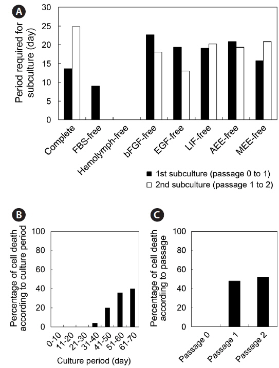
    To investigate the state of the nuclei in cultured cells, we conducted nucleus staining. Live cells after initial attachment (passage 0) and the fourth subculture (passage 4) were washed twice with WS, and fresh media containing Hoechst 33342 (Molecular Probes, Eugene, OR, USA) at a final concentration of 5 μg/mL were added. After incubation for 30 min, cells were washed twice with WS and culture wells filled with fresh culture medium. Stained nuclei were visualized under an inverted microscope equipped with a fluorescent lamp (Nikon).

      >  Experimental design and statistical analysis

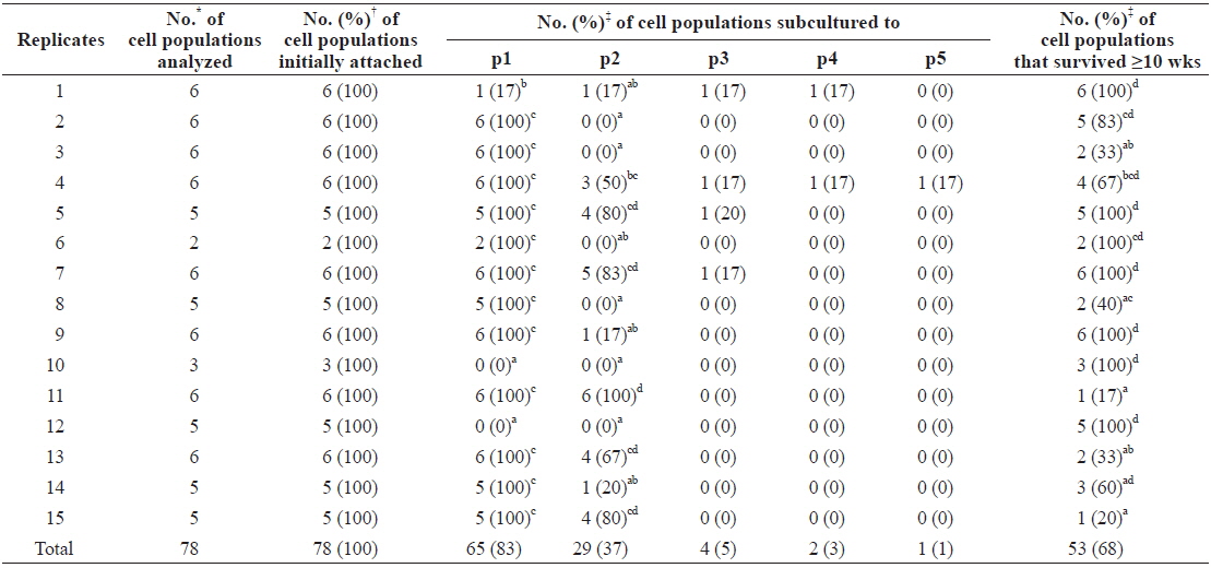
    To investigate the effects of seven medium supplements consisting of FBS, hemolymph, bFGF, EGF, LIF, AEE, and MEE, the composition of the culture medium was varied by removing one supplement from culture media containing all seven supplements. That is, we produced a total of eight culture media including one complete medium (containing all seven supplements) and one each without FBS, hemolymph, bFGF, EGF, LIF, AEE, or MEE, and cultured H. discus hannai ovary-dissociated cells in each. Each single supplement-deprived medium was designated as the “supplement name”-free medium. We used a generalized linear model (PROC-GLM) in Statistical Analysis System software (SAS Institute, Cary, NC, USA) to analyze the numerical data. When a significant main effect was detected, treatments were subsequently compared by either the least squares or Duncan’s method. Significant differences among treatments were defined as P < 0.05.

    Results

      >  Initial cell attachment of ovary-dissociated cell populations

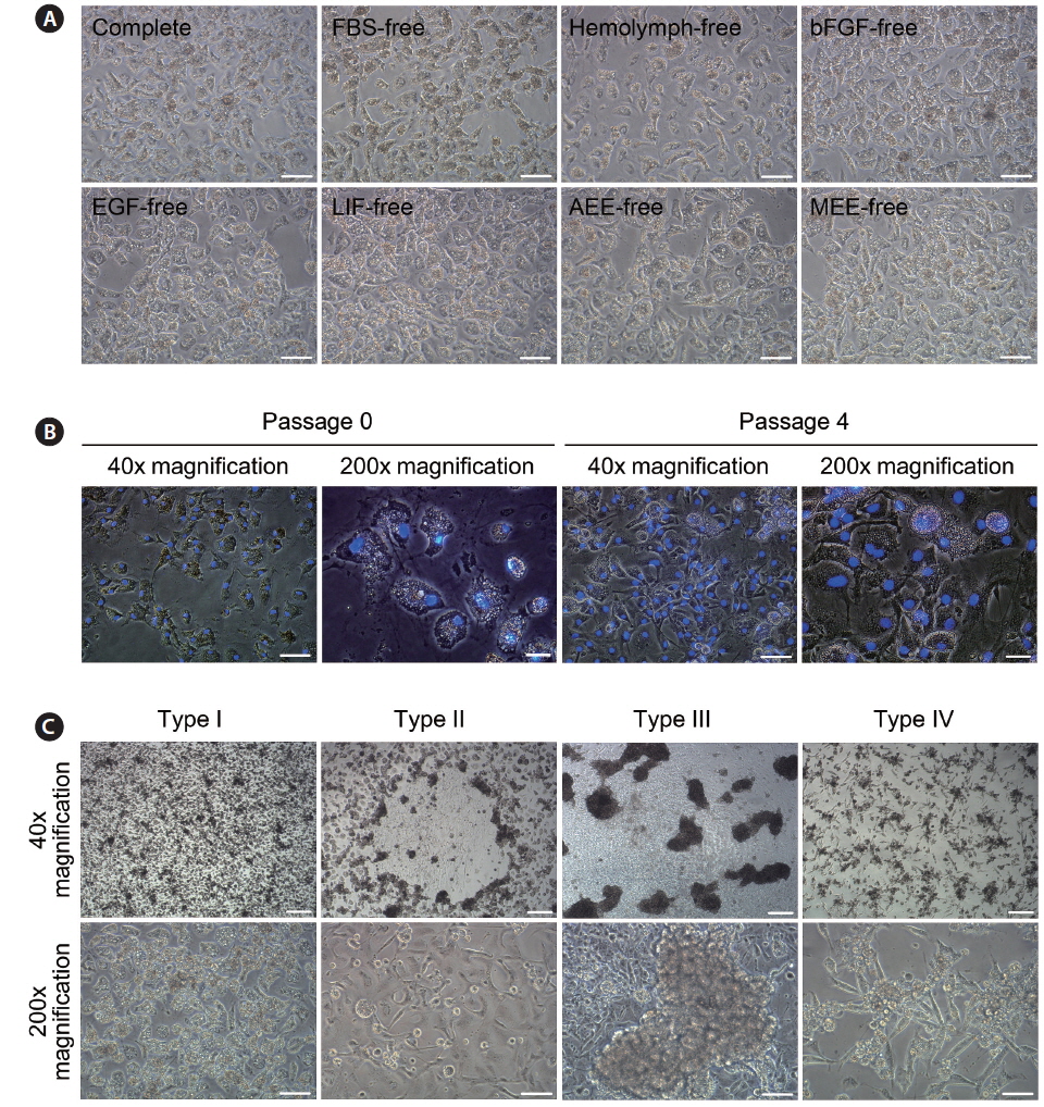
    On day 3 of culture after initial cell seeding, we identified initial cell attachment. All ovary-dissociated cell populations cultured in these experiments showed successful initial cell attachment regardless of medium composition (total attachment in all 97 trials = 100%; Table 1). Moreover, the rate of initial cell attachment was an average of 70% in all cell populations and reached a maximum of 100% when scored visually. The morphologies of initially attached cells were very similar in all cell populations regardless of treatment, and all displayed thick, irregular forms harboring one nucleus with two or three nucleoli and many granules within their cytoplasm (Fig. 1A and 1B).

    [Table 1.] Effects of medium supplements on primary culture of Haliotis discus hannai ovary-dissociated cells

    label

    Effects of medium supplements on primary culture of Haliotis discus hannai ovary-dissociated cells

      >  Effects of medium supplements on primary culture and in vitro maintenance

    In contrast to the results for initial cell attachment, we detected significant differences in the growth of initially attached cells among treatments. As shown in Table 1, cell populations cultured in FBS-free or hemolymph-free media showed significantly lower rates in the first subculture. Only 18% (2 of 11) and 0% (0 of 8) of the initially attached cell populations cultured in FBS-free and hemolymph-free media, respectively, survived beyond the first subculture, while 79%–93% of cell populations cultured in the other six groups did so (0%–18% vs. 79%–93%, P < 0.0001). Of all cell populations cultured, 60% (58 of 97) survived more than 10 weeks, and cell populations cultured in MEE-free media showed the highest survival rate at 86%. No cell populations cultured in hemolymph-free media survived past 10 weeks, while those cultured in FBS-free media survived better than those in hemolymph-free media (45% vs. 0%, P < 0.05) despite their similar survival in the first subculture. Only one cell population cultured in MEE-free medium was subcultured to passage 5, for a total culture period of 170 days (Table 1). Morphological inspection assisted by Hoechst 33342 staining confirmed that after the fourth subculture, cells still possessed one nucleus containing two or three nucleoli with no abnormal appearance (Fig. 1B).

      >  Effects of individual differences on primary culture outcome

    To examine the effects of individual differences among abalones on culture outcomes, we rearranged the data from Table 1 according to the replicates (Table 2). In this reallocation, data from cell populations cultured in FBS-free and hemolymph-free media were excluded as they differed significantly from the other six groups. Results showed that 65 of 78 (83%) cell populations and all cell populations in 12 of 15 replicates grew and survived beyond the first subculture, while significantly different results were detected in cell populations from the other three replicates. These did not grow enough to progress to subculturing, with the exception of one cell population (0%–17% vs. 100%, P < 0.05; Table 2). Subsequently, 37% (29 of 78) of the cell populations were subcultured twice but additional cell populations from four different replicates stopped their growth, and they did not progress to the second subculture. Sixty-eight percent (53 of 78) of cell populations survived beyond 10 weeks, but significant differences were detected among replicates. No significant data correlation was detected between the subculture results and survival after 10 or more weeks.

    [Table 2.] Effects of individual difference of abalones used on primary culture outcome

    label

    Effects of individual difference of abalones used on primary culture outcome

      >  Cell morphology during culture

    Throughout the culture of H. discus hannai ovary-dissociated cells, we observed different cultured cell morphologies, which we arbitrarily classified into four types (types I–IV; Fig. 1C). Almost all initially attached cells corresponded to type I, which showed thick and irregular shapes harboring many granules within their cytoplasm. In contrast, the other three cell types appeared during the maintenance period. Type II cells were flattened and had fewer granules within their cytoplasm than type I cells, while type III cells formed large cell aggregates. Type IV cells displayed fibroblast-like morphology, with a thick, elongated shape. In most cases, two or three cell types coexisted in a cell population.

      >  Subculture period and cell death in early-phase cultures

    The period after initial cell seeding until first subculture varied from 7 to 65 days according to the cell populations analyzed, and the average period was 18.4 days. Likewise, the period between the first and second subculture varied from 4 to 43 days, with an average of 19.5 days (Fig. 2A). For analysis of early-phase cell deaths, the cell populations that died before 10 weeks of culture were subjected to these analyses but those cultured in FBS-free and hemolymph-free media were excluded. No cell death took place during the first 30 days of culture; the first deaths occurred on day 31, and 32% (25 of 78) of cell populations died prior to day 70. Cells deaths were concentrated between days 41 and 70, and increased over time until day 70 (Fig. 2B). In terms of passage number, cells deaths did not occur in passage 0, and their frequencies in passages 1 and 2 were 48% and 52%, respectively (Fig. 2C).

    Discussion

    In this study, we investigated the effects of medium supplements on cultures of H. discus hannai ovary-dissociated cells and described the general properties of these cultures during in vitro maintenance.

    We demonstrated that initial attachment of freshly isolated cell populations is not affected by either a lack of specific medium supplements or by individual differences, showing that all cell populations cultured attached at an average rate of 70% regardless of treatment. In a previous study, we revealed that supplements added to media containing FBS and hemolymph improved the initial attachment of abalone cells (Kim et al., 2014a). Since the same factors used in this previous report were added in this study, we assumed that similar effects on cell attachment would be induced, even if one factor was omitted in each experimental group. Another possibility, based on previous reports that gonad extract enhances the growth performance of oyster heart cells (Chen and Wen, 1999) and abalone mantle cells (Suja et al., 2007), is that ovary tissue extracts, which might be extracted during the tissue dissociation procedure, might positively affect cell attachment. Otherwise, H. discus hannai ovary-dissociated cells may simply have an exceptional ability to attach to substrata.

    The growth of cell populations cultured with FBS-free or hemolymph-free media was significantly lower than that of other groups, indicating that FBS and hemolymph contain critical components for the stable growth and maintenance of H. discus hannai ovary-dissociated cells. Indeed, FBS is known to be an important component of cell cultures and is used in almost all animal cell cultures (Freshney, 2010). Additionally, some studies have used hemolymph in several invertebrate cell cultures as a medium component (Srivatsan and Peretz, 1997; Chen and Wen, 1999; Khurad et al., 2009; Kim et al., 2014a) and occasionally in mammalian cell cultures as a substitute for FBS (Choi et al., 2005; Rhee et al., 2013). Several studies that attempted in vitro cultures of molluscan cells also support our results. For example, Domart-Coulon et al. (1994) and Chen and Wen (1999), which examined cultures of Crassostrea gigas heart-derived cells, demonstrated that FBS and/or hemolymph promoted cell growth and viability. In addition, others have reported that the insulin contained in FBS can increase in vitro incorporation of amino acids in the mantle collar of Helisoma duryi (Sevala et al., 1993) and that the molluscan insulin-like peptide, acetylcholinesterase, or nerve growth factor-like molecules within molluscan hemolymph may promote neurite outgrowth and synaptogenesis in neuronal cell cultures of Aplysia californica (Ghirardi et al., 1996; Srivatsan and Peretz, 1997; Hyland et al., 2014). Here, we showed that the removal of other medium supplements, including bFGF, EGF, LIF, AEE, and MEE, did not affect cell growth and maintenance, which can be explained by the overlap of signals provided by each supplement (Andrae et al., 2008; Klingseisen et al., 2009). We propose that additional experiments in which two or more supplements are removed are conducted to more clearly determine the critical components necessary for primary cultures of H. discus hannai ovary-dissociated cells.

    The growth and maintenance of cell populations cultured in several replicates were significantly lower, suggesting that individual differences play a major role in the outcome of culturing—although, as we used abalone pairs as single replicates, the term “individual difference” may be inappropriate. Regardless, the presence of individual differences is unsurprising and is a well-known phenomenon in biological experiments (d’Agostino, 1998). This should be carefully considered in invertebrate cell culturing, a field that is still under development, to reduce experimental error in future studies.

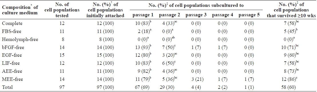
    During maintenance of cell populations, we investigated general culturing aspects such as which cell morphologies appeared, the time required for subculturing, and the cell death rate during the early culture phase. We identified four distinct morphologies among the cultured cells, which may reflect different cell types. Alternately, cell populations initially consisting of one cell type may change their cell morphology during culture according to the culturing environment. Whatever the case, our data provide fundamental information on the morphologies that may appear during the culture of H. discus hannai ovary-dissociated cells. Although high variation was observed, the average period required for subculture was longer than for mammalian and fish species whose cell culture systems are well established (Freshney, 2010; Lee et al., 2013; Kim et al., 2014b), indicating very poor growth performance in the cells cultured in this study. This suggests that the optimization of cell culturing systems for abalone should be prioritized, and that data from the general culturing conditions we examined can provide valuable information in this regard.

    Because abalone ovarian tissues consist of germline cells and various types of somatic cells (Wetakan et al., 2001; Awaji and Hamano, 2004), the cell populations cultured and maintained in this study need to be characterized to identify their cellular origin. The cell populations derived from germline cells can be used directly in biotechnological applications. On the other hand, somatic cells can be used as feeder cells for culturing ovary-derived germline stem cells. Molecular characterization of the cells cultured in this study should be conducted in further studies.

    In conclusion, primary cultures of H. discus hannai ovary-dissociated cells can be significantly affected by two medium supplements, FBS and hemolymph, and by individual variation. Our results can contribute to the development of universal conditions for the primary culture of abalone cells and further suggest the feasibility of culturing ovary-derived germline stem cells in this species.

참고문헌
  • 1. Andrae J, Gallini R, Betsholtz C 2008 Role of platelet-derived growth factors in physiology and medicine [Genes Dev] Vol.22 P.1276-1312 google cross ref
  • 2. Auzoux-Bordenave S, Fouchereau-Peron M, Helleouet MN, Doumenc D 2007 CGRP regulates the activity of mantle cells and hemocytes in abalone primary cell cultures (Haliotis tuberculata) [J Shellfish Res] Vol.26 P.887-894 google cross ref
  • 3. Awaji M, Hamano K 2004 Gonad formation, sex differentiation and gonad maturation processes in artificially produced juveniles of the abalone, Haliotis discus hannai [Aquaculture] Vol.239 P.397-411 google cross ref
  • 4. Chen SN, Wen CM 1999 Establishment of cell lines derived from oyster, Crassostrea gigas Thunberg and hard clam, Meretrix lusoria Roding [Meth Cell Sci] Vol.21 P.183-192 google cross ref
  • 5. Choi SS, Rhee WJ, Park TH 2005 Beneficial effect of silkworm hemolymph on a CHO cell system: Inhibition of apoptosis and increase of EPO production [Biotechnol Bioeng] Vol.91 P.793-800 google cross ref
  • 6. d’Agostino RB 1998 Tutorial in biostatistics: propensity score methods for bias reduction in the comparison of a treatment to a nonrandomized control group [Stat Med] Vol.17 P.2265-2281 google cross ref
  • 7. Domart-Coulon I, Doumence D, Auzous-Bordenave S, Le Fichant Y 1994 Identification of media supplements that improve the viability of primary cell cultures of Crassostrea gigas oysters [Cytotechnology] Vol.16 P.109-120 google cross ref
  • 8. Farcy E, Serpentini A, Fievet B, Lebel JM 2007 Identification of cDNAs encoding HSP70 and HSP90 in the abalone Haliotis tuberculata: Transcriptional induction in response to thermal stress in hemocyte primary culture [Comp Biochem Physiol B Biochem Mol Biol] Vol.146 P.540-550 google cross ref
  • 9. Freshney RI 2010 Culture of Animal Cells: A Manual of Basic Technique and Specialized Applications google
  • 10. Gaume B, Bourgougnon N, Auzoux-Bordenave S, Roig B, Le Bot B, Bedoux G 2012 In vitro effects of triclosan and methyl-triclosan on the marine gastropod Haliotis tuberculate [Comp Biochem Physiol C Toxicol Pharmacol] Vol.156 P.87-94 google cross ref
  • 11. Ghirardi M, Casadio A, Santarelli L, Montarolo PG 1996 Aplysia hemolymph promotes neurite outgrowth and synaptogenesis of identified Helix neurons in cell culture [Invert Neurosci] Vol.2 P.41-49 google cross ref
  • 12. Guan K, Nayernia K, Maier LS, Wagner S, Dressel R, Lee JH, Nolte J, Wolf F, Li M, Engel W, Hasenfuss G 2006 Pluripotency of spermatogonial stem cells from adult mouse testis [Nature] Vol.440 P.1199-1203 google cross ref
  • 13. Hyland C, Dufresne ER, Forscher P 2014 Regeneration of Aplysia bag cell neurons is synergistically enhanced by substrate-bound hemolymph proteins and laminin [Sci Rep] Vol.4 P.4617 google
  • 14. Khurad AM, Zhang MJ, Deshmukh CG, Bahekar RS, Tiple AD, Zhang CX 2009 A new continuous cell line from larval ovaries of silkworm, Bombyx mori [In Vitro Cell Dev Biol Anim] Vol.45 P.414-419 google
  • 15. Kim MS, Nam YK, Kim DS, Gong SP 2014 Initial culture conditions for primary cell populations derived from radula tissue in abalone Haliotis discus hannai [Fish Aquat Sci] Vol.17 P.385-390 google
  • 16. Kim MS, Nam YK, Park C, Kim HW, Ahn J, Lim JM, Gong SP 2014 Establishment condition and characterization of heart-derived cell culture in Siberian sturgeon (Acipenser baerii) [In Vitro Cell Dev Biol Anim] Vol.50 P.909-917 google cross ref
  • 17. Klingseisen A, Clark IB, Gryzik T, Muller HA 2009 Differential and overlapping functions of two closely related Drosophila FGF8-like growth factors in mesoderm development [Development] Vol.136 P.2393-2402 google cross ref
  • 18. Lacerda SM, Batlouni SR, Costa GM, Segatelli TM, Quirino BR, Queiroz BM, Kalapothakis E, Franca LR 2010 A new and fast technique to generate offspring after germ cells transplantation in adult fish: the Nile tilapia (Oreochromis niloticus) model [PLoS One] Vol.5 P.e10740 google cross ref
  • 19. Latire T, Le Pabic C, Mottin E, Mottier A, Costil K, Koueta N, Lebel JM, Serpentini A 2012 Responses of primary cultured haemocytes from the marine gastropod Haliotis tuberculata under 10-day exposure to cadmium chloride [Aquat Toxicol] Vol.109 P.213-221 google cross ref
  • 20. Lebel JM, Giard W, Favrel P, Boucaud-Camou E 1996 Effects of different vertebrate growth factors on primary cultures of hemocytes from the gastropod mollusc, Haliotis tuberculata [Biol Cell] Vol.86 P.67-72 google cross ref
  • 21. Lee D, Kim MS, Nam YK, Kim DS, Gong SP 2013 Establishment and characterization of permanent cell lines from Oryzias dancena embryos [Fish Aquat Sci] Vol.16 P.177-185 google
  • 22. Naganuma T, Degnan BM, Horikoshi K, Morse DE 1994 Myogenesis in primary cell cultures from larvae of the abalone, Haliotis rufescens [Mol Mar Biol Biotechnol] Vol.3 P.131-140 google
  • 23. Najmudeen TM 2007 Gonad maturation of the tropical abalone Haliotis varia Linnaeus 1758 (Vetigastropoda: Haliotidae) [Molluscan Res] Vol.27 P.140-146 google
  • 24. Pichon D, Cudennec B, Huchette S, Djediat C, Renault T, Paillard C, Auzoux-Bordenave S 2013 Characterization of abalone Haliotis tuberculata?Vibrio harveyi interactions in gill primary cultures [Cytotechnology] Vol.65 P.759-772 google cross ref
  • 25. Poncet JM, Serpentini A, Boucaud-Camou E, Lebel JM 2002 Cryopreservation of mantle dissociated cells from Haliotis tuberculata (Gastropoda) and postthawed primary cell cultures [Cryobiology] Vol.44 P.38-45 google cross ref
  • 26. Poncet JM, Serpentini A, Thiebot B, Villers C, Bocquet J, Boucaud-Camou E, Lebel JM 2000 In vitro synthesis of proteoglycans and collagen in primary cultures of mantle cells from the nacreous mollusk, Haliotis tuberculata: a new model for study of molluscan extracellular matrix [Mar Biotechnol] Vol.2 P.387-398 google
  • 27. Rhee WJ, Park JH, Hahn JS, Park TH 2013 Anti-apoptotic mechanism of silkworm hemolymph in HeLa cell apoptosis [Process Biochem] Vol.48 P.1375-1380 google cross ref
  • 28. Sevala VM, Sevala VL, Saleuddin ASM 1993 Hemolymph insulin-like peptides (ILP) titers and the influence of ILP and mammalian insulin on the amino acid incorporation in the mantle collar In vitro in Helisoma (Mollusca) [Biol Bull] Vol.185 P.140-148 google cross ref
  • 29. Srivatsan M, Peretz B 1997 Acetylcholinesterase promotes regeneration of neurites in cultured adult neurons of Aplysia [Neuroscience] Vol.77 P.921-931 google cross ref
  • 30. Sud D, Doumenc D, Lopez E, Milet C 2001 Role of water-soluble matrix fraction, extracted from the nacre of Pinctada maxima, in the regulation of cell activity in abalone mantle cell culture (Haliotis tuberculata) [Tiss Cell] Vol.33 P.154-160 google cross ref
  • 31. Suja CP, Dharmaraj S 2005 In vitro culture of mantle tissue of the abalone Haliotis varia Linnaeus [Tiss Cell] Vol.37 P.1-10 google cross ref
  • 32. Suja CP, Sukumaran N, Dharmaraj S 2007 Effect of culture media and tissue extracts in the mantle explant culture of abalone, Haliotis varia Linnaeus [Aquaculture] Vol.271 P.516-522 google cross ref
  • 33. Suwattana D, Jirasupphachok J, Jarayabhand P, Koykul W 2010 In vitro culture of gill and heart tissues of the abalone Haliotis asinina [J Shellfish Res] Vol.29 P.651-653 google cross ref
  • 34. van der Merwe M, Auzoux-Bordenave S, Niesler C, Roodt-Wilding R 2010 Investigating the establishment of primary cell culture from different abalone (Haliotis midae) tissues [Cytotechnology] Vol.62 P.265-277 google cross ref
  • 35. Wetakan SA, Linthong V, Wanichanon C, Panasophonkul S, Meepool A, Kruatrachue M, Upatham ES, Pumthong T, Sobhon P 2001 Ultrastructure of female germ cells in Haliotis asinina Linnaeus [Invertebr Reprod Dev] Vol.39 P.67-79 google cross ref
  • 36. Wong TT, Tesfamichael A, Collodi P 2013 Production of zebrafish offspring from cultured female germline stem cells [PLoS ONE] Vol.8 P.e62660 google cross ref
  • 37. Yoshino TP, Bickham U, Bayne CJ 2013 Molluscan cells in culture: primary cell cultures and cell lines [Can J Zool] Vol.91 P.391-404 google cross ref
  • 38. Zou K, Yuan Z, Yang Z, Luo H, Sun K, Zhou L, Xiang J, Shi L, Yu Q, Zhang Y, Hou R, Wu J 2009 Production of offspring from a germline stem cell line derived from neonatal ovaries [Nat Cell Biol] Vol.11 P.631-636 google cross ref
OAK XML 통계
이미지 / 테이블
  • [ Table 1. ]  Effects of medium supplements on primary culture of Haliotis discus hannai ovary-dissociated cells
    Effects of medium supplements on primary culture of Haliotis discus hannai ovary-dissociated cells
  • [ Fig. 1. ]  Cell morphologies during primary culture of Haliotis discus hannai ovary-dissociate cells. (A) Cell morphologies at initial attachment (day 3 after initial cell seeding) of cell populations from all treatment groups. Scale bar=50 μm. (B) Nuclear staining by hoechst 33342. Initially-attached cells and the cells at passage 4 were stained by hoechst 33342 and merged pictures of two images derived from light microscopic and fluorescent images at same spots were presented. Blue fluorescent indicates nuclei within the cells. Scale bar=200 μm in 40x and 50 μm in 200x. (C) Cell morphologies of cultured cells. Four different types of cell morphologies were identified during cell culture and the representative pictures were presented. Scale bar= 200 μm in 40x and 50 μm in 200x.
    Cell morphologies during primary culture of Haliotis discus hannai ovary-dissociate cells. (A) Cell morphologies at initial attachment (day 3 after initial cell seeding) of cell populations from all treatment groups. Scale bar=50 μm. (B) Nuclear staining by hoechst 33342. Initially-attached cells and the cells at passage 4 were stained by hoechst 33342 and merged pictures of two images derived from light microscopic and fluorescent images at same spots were presented. Blue fluorescent indicates nuclei within the cells. Scale bar=200 μm in 40x and 50 μm in 200x. (C) Cell morphologies of cultured cells. Four different types of cell morphologies were identified during cell culture and the representative pictures were presented. Scale bar= 200 μm in 40x and 50 μm in 200x.
  • [ Table 2. ]  Effects of individual difference of abalones used on primary culture outcome
    Effects of individual difference of abalones used on primary culture outcome
  • [ Fig. 2. ]  General culture aspects in early phase of culture. (A) Periods required for subculture were analyzed in all cell populations from each treatment group and the average days were presented. Average days required for first and second subcultures on all cell populations cultured were 18.4 days and 19.5 days, respectively. (B) and (C) indicate the rate of cell death of cell populations in early phase of culture (within 10 weeks) according to culture period (day) and passage number, respectively. No cell death were identified within 30 days of culture and in passage 0.
    General culture aspects in early phase of culture. (A) Periods required for subculture were analyzed in all cell populations from each treatment group and the average days were presented. Average days required for first and second subcultures on all cell populations cultured were 18.4 days and 19.5 days, respectively. (B) and (C) indicate the rate of cell death of cell populations in early phase of culture (within 10 weeks) according to culture period (day) and passage number, respectively. No cell death were identified within 30 days of culture and in passage 0.
(우)06579 서울시 서초구 반포대로 201(반포동)
Tel. 02-537-6389 | Fax. 02-590-0571 | 문의 : oak2014@korea.kr
Copyright(c) National Library of Korea. All rights reserved.